

 PDF
PDF ePub
ePub Citation
 Print
Print


The most catastrophic injury to the proximal interphalangeal (PIP) joint is the fracture dislocation which involves a complete articular surface combined with metaphyseal compaction. This intraarticular fracture of the base of the middle phalanges is uncommon but it is difficult to treat because it is highly unstable refractory to standard surgical techniques.1) The purpose of the treatment is to prevent traumatic arthritis and recover range of motion by early joint exercises.
A wide spectrum of treatments has been introduced, including splinting in extreme flexion, extension block splinting, various ingenious traction devices, a force couple, volar plate interposition arthroplasty and numerous open and closed reduction techniques, but have not achieved satisfactory results. Because comminuted fragments make reduction and fixation difficult. Although pin site infection can occur, stiffness is an important complication because pin fixes joint for few weeks.1) Thus dynamic traction using spring was introduced. This system makes length of fracture site maintained by traction along axis of middle phalanx and allows for early joint exercise.
This article describes a case of neglected pilon fracture of middle phalanx treated with external fixator combined with dental orthodontic rubber band.
Case Report
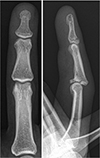
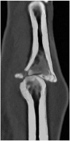
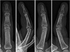
A 44-year-old man was admitted to hospital with painful swelling and limitation of motion of right ring finger PIP joint. He had symptoms after punching 17 days ago, but he could not visit a hospital because of the funeral of his relative. The physical examination showed normal blood circulation and sensation but showed tenderness and limitation of motion on the ring finger PIP joint. Radiographs showed dorsal dislocation of PIP joint and volar lip fracture which involves more than 50% of articular surface. The fracture of base of distal phalanx was observed on the radiographs (Fig. 1). Sagittal computed tomography (CT) image showed volar lip fracture which involves more than 50% of articular surface (Fig. 2).
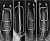
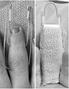
Surgery was performed using Suzuki frame under brachial plexus block. The proximal K-wire (1.1 mm in diameter) was inserted transversely through the center of the head of the proximal phalanx. The distal K-wire which was same sized in diameter was placed transversely through the center of the head of the middle phalanx. Then both pins were bent at 90° close to skin (approximately 5 mm), and hooks were created at the end of each protruding wire. Orthodontic rubber bands were linked to hooks of both K-wires. According to the CT image, it looks like the impacted fracture fragment needed to be reducted by joystick technique. However only after traction of PIP joint, we have successed to do reduction of subluxation of PIP joint and impacted fracture fragment of base of middle phalanx with a C-arm X-ray device (Fig. 3). Therefore joystick technic was no needed. The nelaton catheter was inserted at the end of the distal K-wire to prevent damage caused by the wire tip (Fig. 4).
The patient was discharged the day after the operation with oral antibiotics to prevent pin site infection. Additional aluminum finger splint was applied for 1 week. At the start of the second week, passive mobilization was started by himself in day time and kept splint at night. At the start of the third week, splint was off and active mobilization was started. The traction device was removed in the outpatient clinic without anesthesia at the end of the fourth week. The patient was observed clinically and radiologically every week for 4 weeks.
The postoperative follow-up period was 2 months. No pin site infection occurred. The total active range of motion for the PIP joint of the fractured finger was 90°, and for the distal interphalangeal joint of the fractured finger was from 0° in extension to 60° in flexion (Fig. 5). We observed radiologic evidence of union of the base of distal phalanx and reduction state of the PIP joint and fracture fragment of the base of proximal phalanx (Fig. 6). The patient was satisfied with the functional result of treatment with the traction device.
DISCUSSION
Fracture dislocation of PIP joint of finger can be divided simply into volar, dorsal, and pilon fracture dislocation. The most common injury pattern is a dorsal fracture dislocation with a volar lip fracture of a middle phalanx. Dorsal fracture dislocations result from axial load with the joint held in a slight degree of flexion. In this case, the longitudinal force causes the volar lip of the middle phalanx to shear off when it impacts the head of the proximal phalanx. Unfortunately, patients often do not seek treatment until the resultant stiffness and swelling becomes unbearable, weeks to months after the initial injury.
The patient in this case underwent surgery in 17 days after injury. In animal models, the peak of soft callus formation occurs 7 to 9 days post trauma with a peak in both type II procollagen and proteoglycan core protein extracellular markers soft callus make it difficult to satisfactory reduction.2) In this case, the reason why the reduction was successful is because the patient did not get the conservative treatment such as splint. The patient kept moving his injured finger and too much motion is known to result in delayed healing or even nonunion.3)
Treatment options are determined by classification according to fracture pattern, articular involvement and stability.4) In this case, surgical treatment was performed because the PIP joint was unstable and more than 50% of the joint surface was involved. Various operative treatment modalities, such as open reduction and internal fixation and closed reduction with K-wires have been suggested. But dissection for open reduction cause tissue trauma and fixation of damaged joint for 3 to 4 weeks with K-wire results in joint stiffness and later joint contracture. The traction device which applies ligamentotaxis maintains joint space and prevents contracture and risk of joint stiffness. Because early mobilization of damaged joint facilitates osteochondral remodeling, decreases adhesions, incidence of stiffness and joint contracture.
Several dynamic traction devices have been developed. As applied on the joint, concentric articular congruity must be achieved via a combination of longitudinal traction and translation to obtain a successful outcome. Schenck5) described a prototype distraction device and range of motion has been recovered to 87° maintaining articular congruity and joint space, but it was bulky that caused discomfort to patients. The force couple splint does not produce ligamentotaxis and cannot be used for comminuted fractures of the PIP joint. Rawes and Oni6) reported swan neck deformity as a complication of force couple splint. Duteille et al.7) used traction system device using pins and rubber bands to treat complex fractures of the PIP joint of the finger. Badia et al.8) reported a modification of the dynamic external fixator. This design does not involve rubber bands but uses longitudinal traction between the pins to induce ligamentotaxis and joint reduction. But such devices without rubber bands do not allow for effective distraction across the proximal phalangeal joint at all degrees of motion. Like this study, a recent study has shown that treatment of middle phalanx base fracture using traction device with dental rubber bands enabled satisfactory reduction and early return of daily life without joint stiffness.9)
These authors used dynamic external fixation devices using K-wire and orthodontic rubber band to treat neglected fracture and dislocation of PIP joint. It was simple and easy to apply. It allowed stable reduction state and early exercise at low cost and showed good postoperative range of motion. Surgery techniques using K-wire and rubber bands can be divided into three main ways: two K-wires, two K-wires and rubber bands, and three K-wires and rubber bands. Shin et al.10) reported that it is important to check the postoperative maximum range of motion. They also reported that adjusting the traction force more than 0.5 mm compared to the adjacent PIP joint helps to obtain the maximum range of motion. These authors confirmed the reduction and maximum range of motion with an intraoperative C-arm X-ray device.
To prevent pin track infection, the device was removed at the end of the 4th week. Because Syed et al.1) reported that retaining the device for up to 6 weeks may increase the risk of pin site infection. When drilling phalanx with K-wire, it avoided being inserted into the cancellous bone of the metaphysis because it is not strong enough to resist the torque generated at the bone wire interface.
Although this case report has limitations in the absence of the control group, it is clinically meaningful in that fractures, which was neglected for 17 days, was treated with an external fixation device. This device was difficult to active exercise because the rubber bands distracted the joint, but it was possible because the wires inserted into the bone are not fixed together. Further prospective study is needed including more cases and control group.
These authors experienced a PIP joint fracture dislocation that was neglected for 17 days in a 44-year-old man. The patient was able to get a good postoperative range of motion after surgery using the dynamic traction device and had no residual pain. The radiologic images showed good results also. Therefore, dynamic traction device is an effective treatment for neglected fracture dislocation of PIP joint of finger.

 XML Download