

 PDF
PDF ePub
ePub Citation
 Print
Print


Abstract
Purpose
This study investigated the clinical and radiological outcomes of patients undergoing provisional fixation in conjunction with locking plate fixation. Miniplates were used as the reduction plates for the surgical treatment of severe comminuted metadiaphyseal fractures with an intra-articular fracture of the distal radius.
Materials and Methods
The radial length, radial inclination, volar tilt, and radial intra-articular step-off were measured preoperatively, postoperatively, and at one year after surgery in 12 patients (eight males, four females, mean age 55.4 years old). The patients underwent volar locking plate fixation with miniplate as a reduction plate for severe comminuted metadiaphyseal fractures with an intra-articular fracture of the distal radius. Clinical evaluations were conducted using the modified Mayo wrist score (MMWS).
Results
Bone union was achieved in all cases. The mean MMWS was 81.8 points, including two excellent, three good, and seven fair cases. Radiological improvements were observed in the average radial length (preoperative, 6.4 mm; postoperative, 11.8 mm), average radial inclination (10.2° to 22.4°), average volar tilt (–4.5° to 10.6°), and average radial intra-articular step-off (4.8–0.8 mm) (all, p<0.05). Radiographic measurements obtained immediately after surgery and at the final follow-up revealed insignificant decreases in radial length (0.6 mm), radial inclination (0.4°), and volar tilt (0.9°) (all, p>0.05).
Go to : 
References
1. Kapoor H, Agarwal A, Dhaon BK. Displaced intraarticular fractures of distal radius: a comparative evaluation of results following closed reduction, external fixation and open reduction with internal fixation. Injury. 31:75–79. 2000.

2. Harley BJ, Scharfenberger A, Beaupre LA, Jomha N, Weber DW. Augmented external fixation versus percutaneous pinning and casting for unstable fractures of the distal radius–a prospective randomized trial. J Hand Surg Am. 29:815–824. 2004.
3. Gruber G, Bernhardt GA, Köhler G, Gruber K. Surgical treatment of distal radius fractures with an angle fixed bar palmar plating system: a single center study of 102 patients over a 2-year period. Arch Orthop Trauma Surg. 126:680–685. 2006.
4. Figl M, Weninger P, Liska M, Hofbauer M, Leixnering M. Volar fixed-angle plate osteosynthesis of unstable distal radius fractures: 12 months results. Arch Orthop Trauma Surg. 129:661–669. 2009.
5. Chung KC, Watt AJ, Kotsis SV, Margaliot Z, Haase SC, Kim HM. Treatment of unstable distal radial fractures with the volar locking plating system. J Bone Joint Surg Am. 88:2687–2694. 2006.
6. Downing ND, Karantana A. A revolution in the management of fractures of the distal radius? J Bone Joint Surg Br. 90:1271–1275. 2008.
7. Cho SH, Park HG, Jun DS, et al. Outcomes of severe comminuted distal radius fractures with pronator preserving approach. J Korean Fract Soc. 28:178–185. 2015.
8. Mast J, Jakob R, Ganz R. Planning and reduction technique in fracture surgery. Berlin, Heidelberg: Springer;p. 54–75. 1989.
9. Benirschke SK, Henley MB, Ott JW. Proximal one-third tibial fracture solution. Orthop Trans. 18:1055–1056. 1995.
10. Lang GJ, Cohen BE, Bosse MJ, Kellam JF. Proximal third tibial shaft fractures. Should they be nailed? Clin Orthop Relat Res. 315:64–74. 1995.
11. Archdeacon MT, Wyrick JD. Reduction plating for provisional fracture fixation. J Orthop Trauma. 20:206–211. 2006.
12. Yun HH, Lee YI, Kim KH, Yun SH. Use of auxiliary locking plates for the treatment of unstable pertrochanteric femur fractures. Orthopedics. 15:305–309. 2015.
Go to :
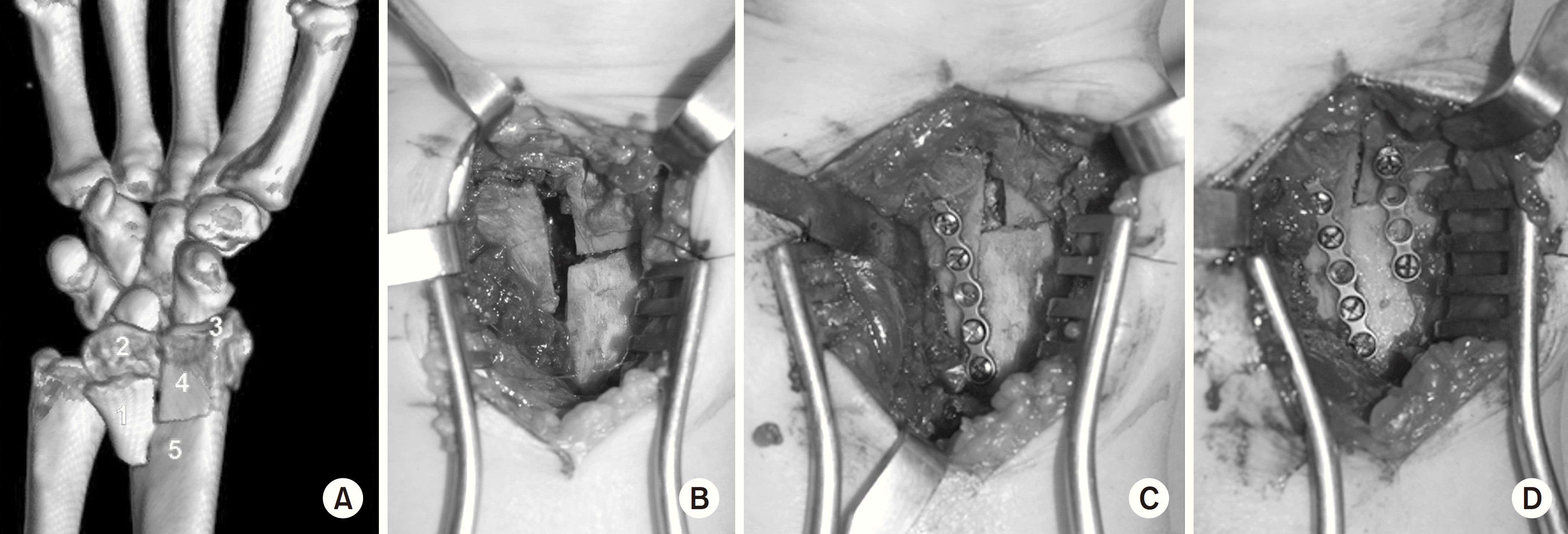 | Fig. 1.A 55-year-old male (patient No. 8) visited the emergency room with a distal radial fracture due to a 2-m fall. (A) Preoperative computed tomography allowed the numbering of each fragment. (B) Following the anterior Henry approach, (C) fragments 1 and 5 were reduced using 1.5-mm miniplates for radial height restoration, and (D) fragments 4 and 5 were reduced using 2.0-mm miniplates. |
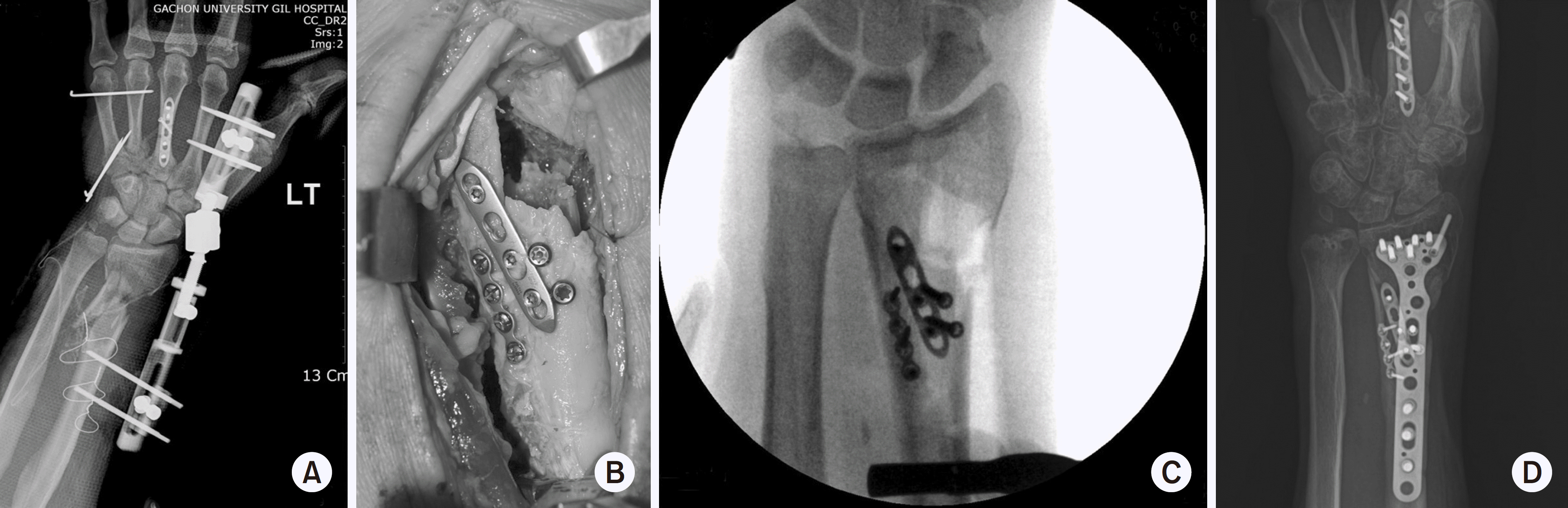
 | Fig. 2.Intraoperative image obtained using an image intensifier. (A, B) Miniplate was used as a reduction plate. (C) Volar locking plate was used as a definitive fixation plate. |
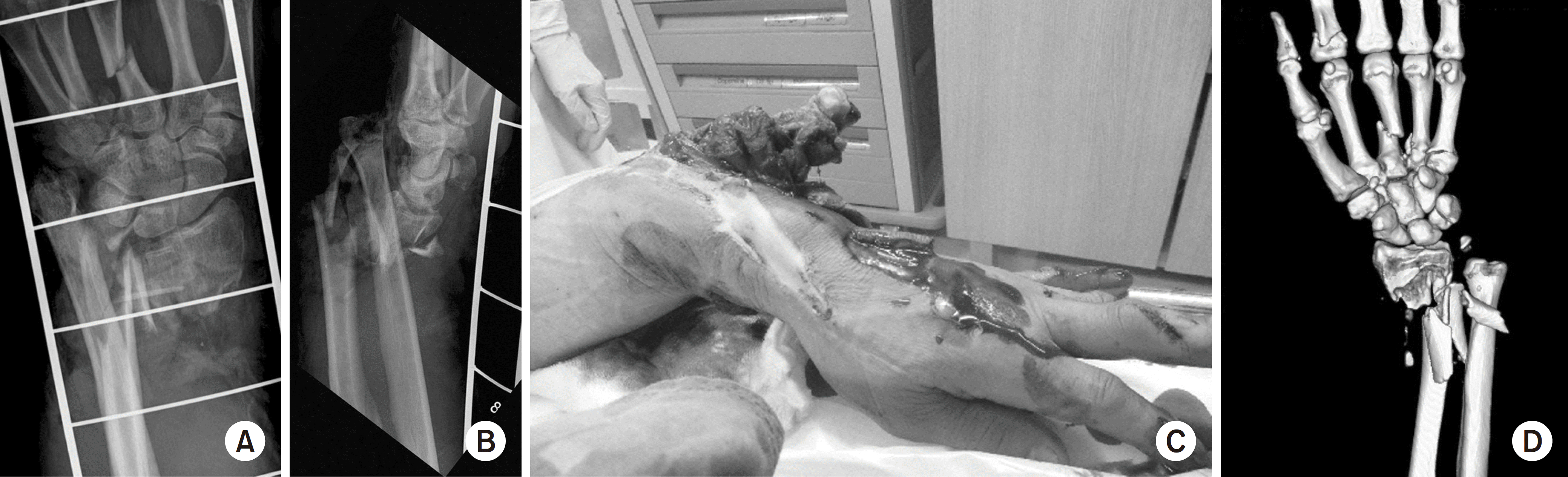 | Fig. 3.(A) Anteroposterior and (B) lateral radiographs, (C) clinical image, and (D) computed tomography image of the wrist of a 59-year-old male (patient No. 11) with a metadiaphyseal comminuted distal radial open fracture (Gustilo–Anderson classification IIIA) due to a crush injury. The associated injuries include metacarpal fractures 3, 4, and 5 and an ulnar styloid process fracture. |
 | Fig. 4.Meticulous debridement and irrigation were performed on the day of admission, and an external fixator was applied to the fracture site. (A) Two weeks later, definitive surgery was performed when the soft tissue was healed and infection is not evident. (B) Secure surgical fixation of the small metadiaphyseal comminuted fragments involves the use of a miniplate, (C) image intensifier confirmation, and (D) fixation using a definitive plate. |
Table 1.
Radiological Data for Distal Radial Fractures Treated Using Volar Locking Plates and Reduction Plates
Table 2.
Demographics, Injury Types, and Treatment Information of the 12 Patients
 XML Download