This article has been corrected. See "Corrigendum: Outcomes following Treatment of Geriatric Distal Femur Fractures with Analyzing Risk Factors for the Nonunion" in Volume 33 on page 62.
Abstract
Purpose
Many international journals have published studies on the results of distal femoral fractures in elderly people, but only a few studies have been conducted on the Korean population. The aim of this study was to determine the factors that are associated with the outcomes and prognosis of fixation of distal femur fractures using the minimally invasive plate osteosynthesis (MIPO) technique in elderly patients (age≥60) and to determine the risk factors related with the occurrence of nonunion.
Materials and Methods
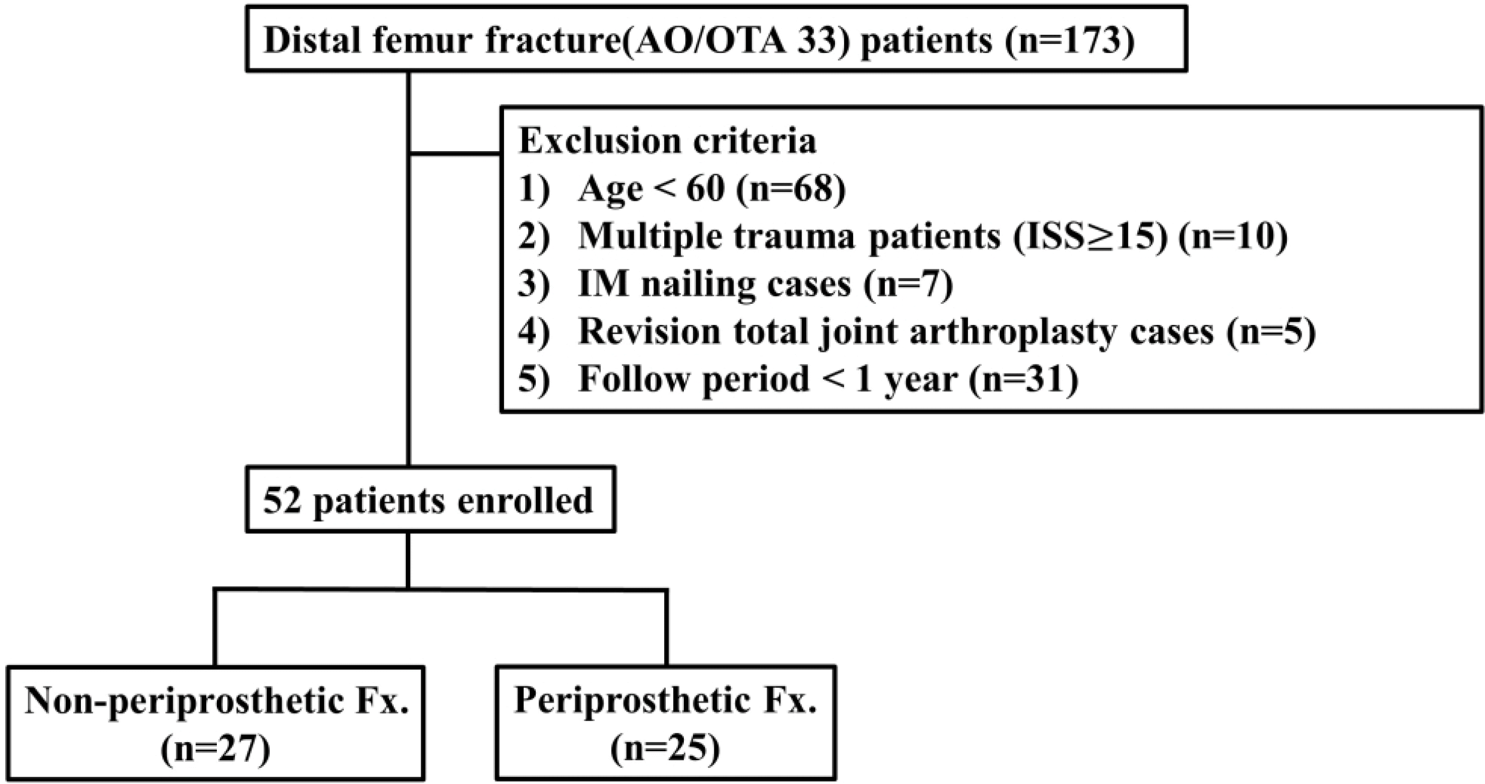
This study is a retrospective study. From January 2008 to June 2018, distal femur fracture (AO/OTA 33) patients who underwent surgical treatment (MIPO) were analyzed. A total of 52 patients were included in the study after removing 121 patients that met with the exclusion criteria. Medical records, including surgical records, were reviewed to evaluate the patients’ underlying disease, bone mineral density, the number of days delayed from surgery, complications and mortality. In addition, follow-up radiographs were used to determine bone union, delayed union and nonunion.
Results
The average time to achieve bone union was 19.95 weeks, the rate of nonunion was 20.0% (10/50) and the overall mortality was 3.8% (2/52). There were no significant differences in the clinical and radiological results of those patients with or without periprosthetic fracture. On the univariate analysis, which compared the union group vs. the nonunion group, no factors were identified as significant risk factors for nonunion. On the multiple logistic regression analysis, medical history of cancer was identified as a significant risk factor for nonunion (p=0.045).
Conclusion
The rate of nonunion is high in the Korean population of elderly people suffering from distal femur fracture, but the mortality rate appears to be low. A medical history of cancer is a significant risk factor for nonunion. Further prospective studies are required to determine other associated factors.
Go to : 
References
1. Gwathmey FW Jr, Jones-Quaidoo SM, Kahler D, Hurwitz S, Cui Q. Distal femoral fractures: current concepts. J Am Acad Orthop Surg. 18:597–607. 2010.

2. Martinet O, Cordey J, Harder Y, Maier A, Bühler M, Barraud GE. The epidemiology of fractures of the distal femur. Injury. 31(Suppl 3):C62–C63. 2000.

3. Elsoe R, Ceccotti AA, Larsen P. Population-based epidemiology and incidence of distal femur fractures. Int Orthop. 42:191–196. 2018.

4. Kregor PJ, Stannard J, Zlowodzki M, Cole PA, Alonso J. Distal femoral fracture fixation utilizing the Less Invasive Stabilization System (L.I.S.S.): the technique and early results. Injury. 32(Suppl 3):SC32–SC47. 2001.

5. Arneson TJ, Melton LJ 3rd, Lewallen DG, O'Fallon WM. Epidemiology of diaphyseal and distal femoral fractures in Rochester, Minnesota, 1965–1984. Clin Orthop Relat Res. 234:188–194. 1988.

6. Bolhofner BR, Carmen B, Clifford P. The results of open reduction and Internal fixation of distal femur fractures using a biologic (indirect) reduction technique. J Orthop Trauma. 10:372–377. 1996.

7. Grimes JP, Gregory PM, Noveck H, Butler MS, Carson JL. The effects of time-to-surgery on mortality and morbidity in patients following hip fracture. Am J Med. 112:702–709. 2002.

8. Kregor PJ, Stannard JA, Zlowodzki M, Cole PA. Treatment of distal femur fractures using the less invasive stabilization system: surgical experience and early clinical results in 103 fractures. J Orthop Trauma. 18:509–520. 2004.
9. Ebraheim NA, Martin A, Sochacki KR, Liu J. Nonunion of distal femoral fractures: a systematic review. Orthop Surg. 5:46–50. 2013.

10. Toro-Ibarguen A, Moreno-Beamud JA, Porras-Moreno MÁ, Aroca-Peinado M, León-Baltasar JL, Jorge-Mora AA. The number of locking screws predicts the risk of nonunion and reintervention in periprosthetic total knee arthroplasty fractures treated with a nail. Eur J Orthop Surg Traumatol. 25:661–664. 2015.

11. Marsh D. Concepts of fracture union, delayed union, and nonunion. Clin Orthop Relat Res. 355(Suppl):S22–S30. 1998.

12. Rodriguez EK, Boulton C, Weaver MJ, et al. Predictive factors of distal femoral fracture nonunion after lateral locked plating: a retrospective multicenter case-control study of 283 fractures. Injury. 45:554–559. 2014.

13. Streubel PN, Gardner MJ, Morshed S, Collinge CA, Gallagher B, Ricci WM. Are extreme distal periprosthetic supracondylar fractures of the femur too distal to fix using a lateral locked plate? J Bone Joint Surg Br. 92:527–534. 2010.

14. Lujan TJ, Henderson CE, Madey SM, Fitzpatrick DC, Marsh JL, Bottlang M. Locked plating of distal femur fractures leads to inconsistent and asymmetric callus formation. J Orthop Trauma. 24:156–162. 2010.

15. Myers P, Laboe P, Johnson KJ, et al. Patient mortality in geriatric distal femur fractures. J Orthop Trauma. 32:111–115. 2018.

16. Karam J, Campbell P, David M, Hunter M. Comparison of outcomes and analysis of risk factors for nonunion in locked plating of closed periprosthetic and non-periprosthetic distal femoral fractures in a retrospective cohort study. J Orthop Surg Res. 14:150. 2019.

17. Faimali M, Karuppiah SV, Hassan S, Swamy G, Badhe N, Geu-tjens G. Mortality and morbidity associated with periprosthetic fracture after total knee replacement. J Orthop Bone Disord. 2:000155. 2018.

18. Ricci WM, Streubel PN, Morshed S, Collinge CA, Nork SE, Gardner MJ. Risk factors for failure of locked plate fixation of distal femur fractures: an analysis of 335 cases. J Orthop Trauma. 28:83–89. 2014.
19. Hoffmann MF, Jones CB, Sietsema DL, Tornetta P 3rd, Koenig SJ, Maatman B. Clinical outcomes of locked plating of distal femoral fractures in a retrospective cohort. J Orthop Surg Res. 8:43. 2013.

20. Barei DP, Beingessner DM. Open distal femur fractures treated with lateral locked implants: union, secondary bone grafting, and predictive parameters. Orthopedics. 35:e843–e846. 2012.

21. Tomlinson RE, Silva MJ. Skeletal blood flow in bone repair and maintenance. Bone Res. 1:311–322. 2013.

22. Park SG, Shon OJ. Impaired bone healing metabolic and mechanical causes. J Korean Fract Soc. 30:40–51. 2017.

23. Streubel PN, Ricci WM, Wong A, Gardner MJ. Mortality after distal femur fractures in elderly patients. Clin Orthop Relat Res. 469:1188–1196. 2011.

24. Jordan RW, Chahal GS, Davies M, Srinivas K. A comparison of mortality following distal femoral fractures and hip fractures in an elderly population. Adv Orthop Surg. 2014:873785. 2014.

25. Czernichow P, Thomine JM, Ertaud A, Biga N, Froment L. [Vital prognosis in fractures of the proximal femur. Study in 506 patients of 60 years of age and over]. Rev Chir Orthop Reparatrice Appar Mot. 76:161–169. 1990; French.
26. Gregory JJ, Kostakopoulou K, Cool WP, Ford DJ. One-year outcome for elderly patients with displaced intracapsular fractures of the femoral neck managed nonoperatively. Injury. 41:1273–1276. 2010.

27. Ishimaru D, Ogawa H, Maeda M, Shimizu K. Outcomes of elderly patients with proximal femoral fractures according to positive criteria for surgical treatment. Orthopedics. 35:e353–e358. 2012.

Go to : 
 | Fig. 1.Flow chart of the study population after exclusion of patients. ISS: injury severity score, IM: intramedullary, Fx.: fracture. |
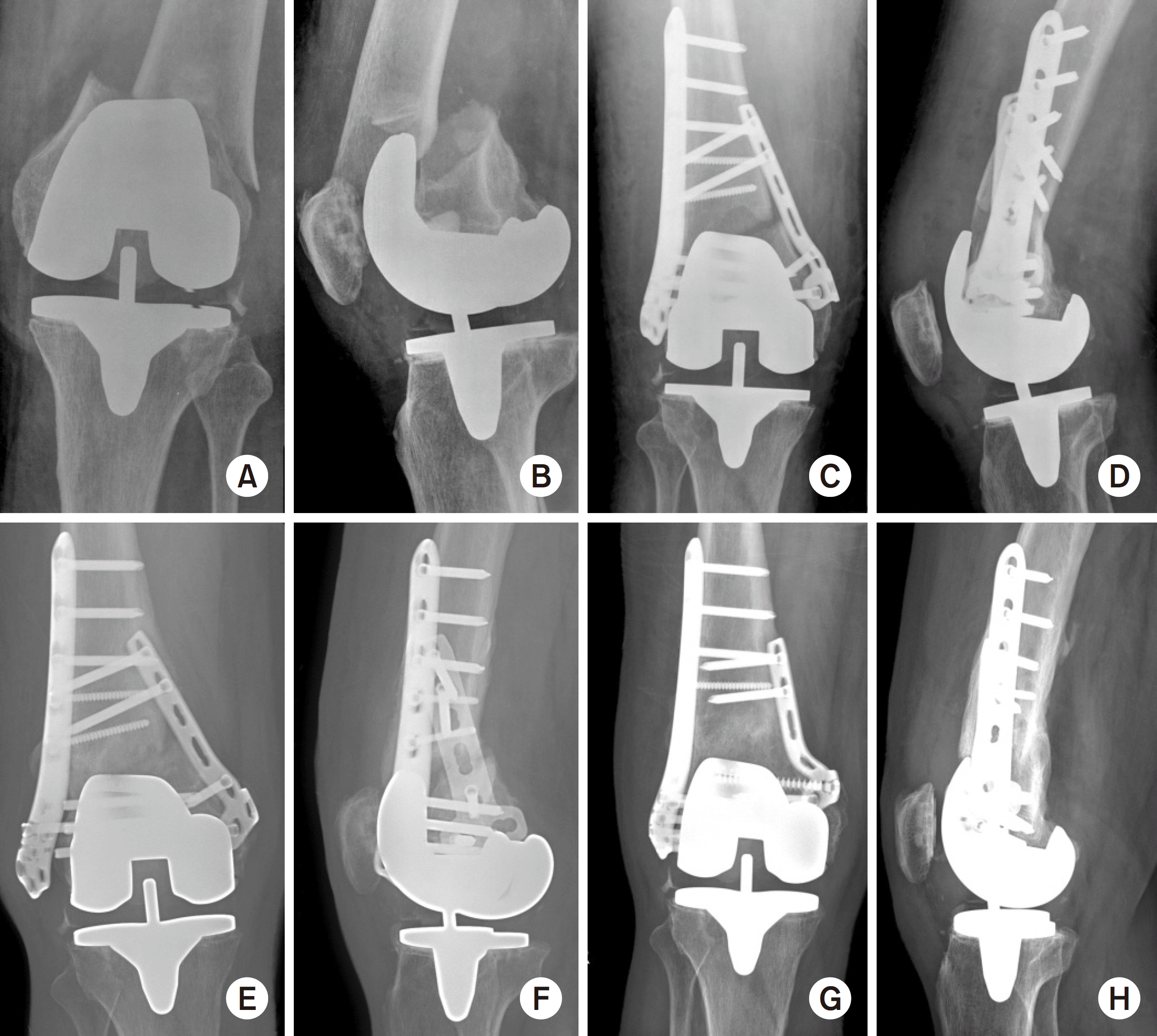
 | Fig. 2.A 76-year-old woman slipped down and developed a right peripros-thtic distal femur fracture (A, B). We performed open reduction and internal affixation with dual plates (C, D), but after 16 months there was no sign of bone union (E, F). Although there were some fracture gaps and slightly shorter plate lengths, oligotrophic nonunion occurred and not hypertrophic nonunion. From here, we performed reoperation with changing plates with using autogenous iliac bone grafting, and union was then achieved (G, H). |
Table 1.
Baseline Characteristics and Demographics
Table 2.
Selected Comorbidities, Postoperative Outcomes, Mortality Rates and Complications
Table 3.
Comparison between the Union and Nonunion Groups
Values are presented as number only, mean±standard deviation, or number (%). *Acceptable surgical factor means sufficient plate length, screw length, a great enough number of screws, and unacceptable surgical factor is a term used when any one of the above factors is not met. Fx.: fracture, BMD: bone mineral density, HTN: hypertension, DM: diabetes mellitus.
Table 4.
Multiple Logistic Regression Analysis of Nonunion


 PDF
PDF ePub
ePub Citation
 Print
Print


 XML Download