Abstract
Purpose
Radial head fractures, which account for 33% of all fractures, are treated depending on the Mason classification. In comminuted type 3 fractures, open reduction internal fixation (ORIF), and radial head arthroplasty are the treatment options. This study examined the clinical outcome of modified Mason type 3 radial head fractures using ORIF with a plate.
Materials and Methods
The medical records and image of 33 patients, who underwent ORIF for modified Mason type 3 radial head fractures, were reviewed retrospectively. The preoperative plain radiographs and computed tomography images were used to examine the location of the fracture of the radial head, the number of fragments, union, joint alignment, and traumatic arthritis at the final followup. The range of motion (ROM) of the elbow at the last follow-up, pain score (visual analogue scale), modified Mayo elbow score (MMES), and complications were analyzed for the clinical outcome.
Results
Of the 33 cases, 14 were men and 19 were women. The mean age was 41.8 years and the average follow-up period was 19 months. The functional ROM was divided into three groups according to the number of bone fragments: 141.2°±9.3° of 3 (n=20), 123.8°±18.5° of 4 (n=7), 100.7°±24.4° of more than 4 (n=6). Furthermore, the MMES were 88.2±2.9, 83.7±4.3, and 77.3±8.4, respectively (p=0.027). Depending on the radial head fracture location, the ROM and MMES were 130.7°±7.5° and 82.1±4.7, respectively, with poor outcomes on the ulnar aspect compared to 143.1°±3.8° and 89.9±3.2 on the radial aspect.
Conclusion
Various factors, such as the degree of crushing and location involved in the clinical outcome. In particular, the result was poor in the case of more than four comminuted fragments or chief position located in the ulnar aspect. In this case, radial head arthroplasty may be considered in the early stages.
Go to : 
References
1. Mason ML. Some observations on fractures of the head of the radius with a review of one hundred cases. Br J Surg. 42:123–132. 1954.

2. Ring D. Elbow fractures and dislocations. Rockwood CA, Green DP, Court-Brown CM, Heckman JD, McQueen MM, editors. Rockwood and Green's fractures in adults. Philadelphia, Baltimore, New York: Lippincott Williams & Wilkins;p. 905–944. 2015.
3. Johnston GW. A follow-up of one hundred cases of fracture of the head of the radius with a review of the literature. Ulster Med J. 31:51–56. 1962.
4. King GJW. Fractures of the radial head. Cohen MS, Green DP, Hotchkiss RN, Kozin SH, Pederson WC, Wolfe SW, editors. Green's operative hand surgery. Philadelphia: Elsevier;p. 734–770. 2017.
5. Burkhart KJ, Wegmann K, Müller LP, Gohlke FE. Fractures of the radial head. Hand Clin. 31:533–546. 2015.

6. Johansson O. Capsular and ligament injuries of the elbow joint. A clinical and arthrographic study. Acta Chir Scand Suppl, Suppl. 287:1–159. 1962.
7. Itamura J, Roidis N, Mirzayan R, Vaishnav S, Learch T, Shean C. Radial head fractures: MRI evaluation of associated injuries. J Shoulder Elbow Surg. 14:421–424. 2005.

8. Fowler JR, Goitz RJ. Radial head fractures: indications and outcomes for radial head arthroplasty. Orthop Clin North Am. 44:425–431. x,. 2013.
9. Lindenhovius AL, Felsch Q, Doornberg JN, Ring D, Kloen P. Open reduction and internal fixation compared with excision for unstable displaced fractures of the radial head. J Hand Surg Am. 32:630–636. 2007.

10. Crönlein M, Zyskowski M, Beirer M, et al. Using an anatomically preshaped low-profile locking plate system leads to reliable results in comminuted radial head fractures. Arch Orthop Trauma Surg. 137:789–795. 2017.

11. Hotchkiss RN. Displaced fractures of the radial head: internal fixation or excision? J Am Acad Orthop Surg. 5:1–10. 1997.

12. Wu PH, Shen L, Chee YH. Screw fixation versus arthroplasty versus plate fixation for 3-part radial head fractures. J Orthop Surg (Hong Kong). 24:57–61. 2016.

13. Tarallo L, Mugnai R, Rocchi M, Capra F, Catani F. Mason type III radial head fractures treated by anatomic radial head arthroplasty: is this a safe treatment option? Orthop Traumatol Surg Res, 103:. 2:183–189. 2017.

14. Ruan HJ, Fan CY, Liu JJ, Zeng BF. A comparative study of internal fixation and prosthesis replacement for radial head fractures of Mason type III. Int Orthop. 33:249–253. 2009.

15. Sun H, Duan J, Li F. Comparison between radial head arthroplasty and open reduction and internal fixation in patients with radial head fractures (modified Mason type III and IV): a metaanalysis. Eur J Orthop Surg Traumatol. 26:283–291. 2016.

16. Geel CW, Palmer AK, Ruedi T, Leutenegger AF. Internal fixation of proximal radial head fractures. J Orthop Trauma. 4:270–274. 1990.

Go to : 
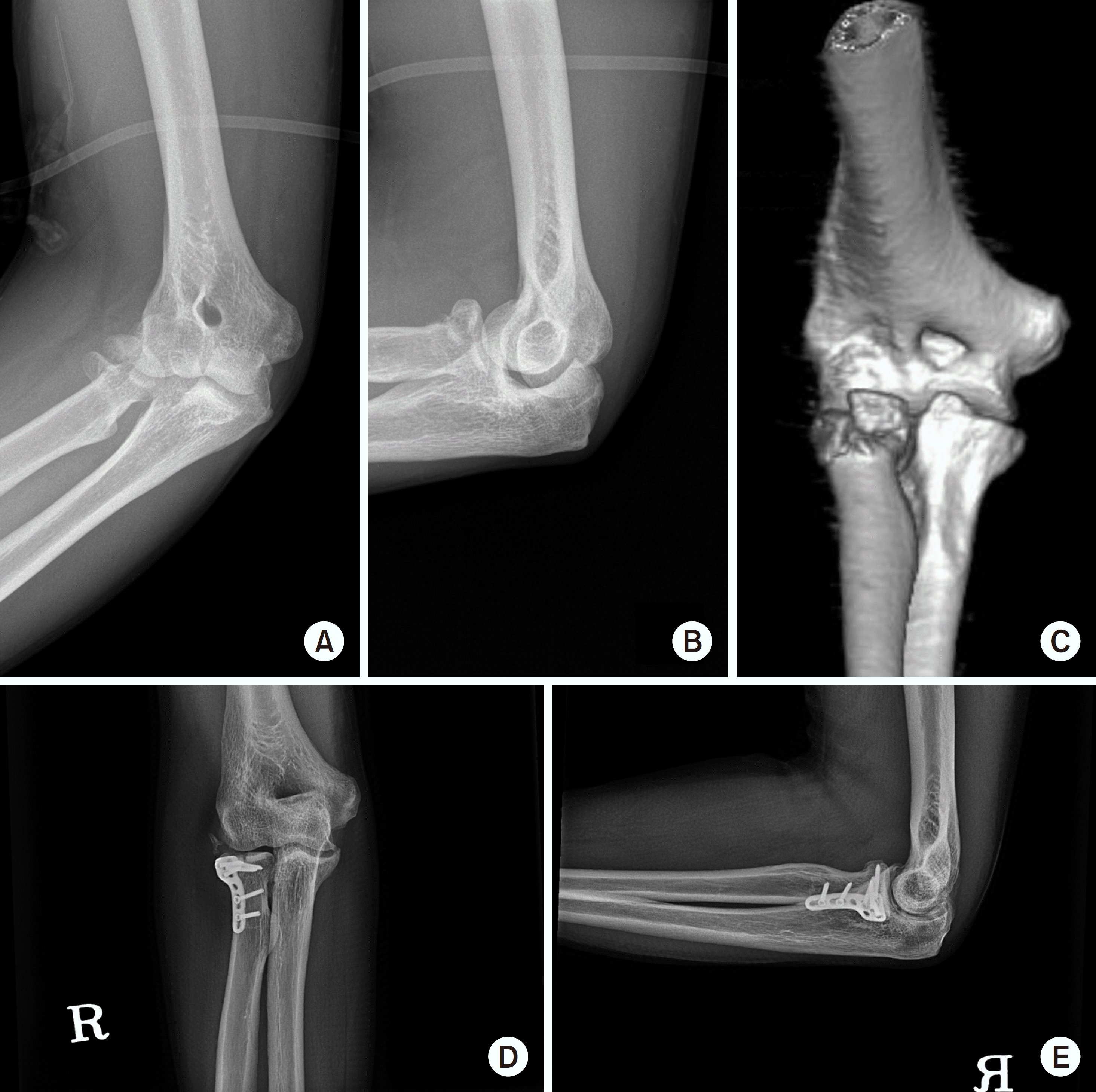
 | Fig. 2.A 57-year-old man sustained a comminuted radial head fracture of the right elbow after a fall from a 1 m height. (A-C) The plain X-ray and three-dimensional computed tomographic scans detected anterior aspect radial head fracture with three fragmentation. (D, E) Open reduction and plate fixation were carried out and solid union was achieved. |
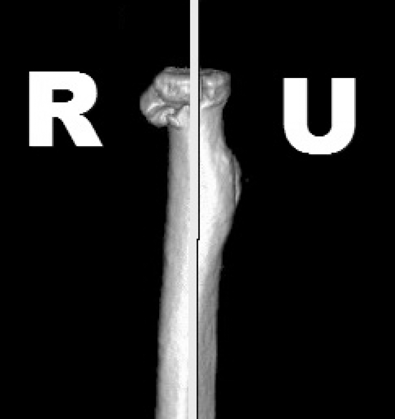
 | Fig. 3.Despite the small heterotrophic ossification at postoperative one year, the functional result was favorable with a range of motion (extension-flexion: 0°-130°, pronation-supination: 80°-80°) and 95 of the modified Mayo elbow score. |
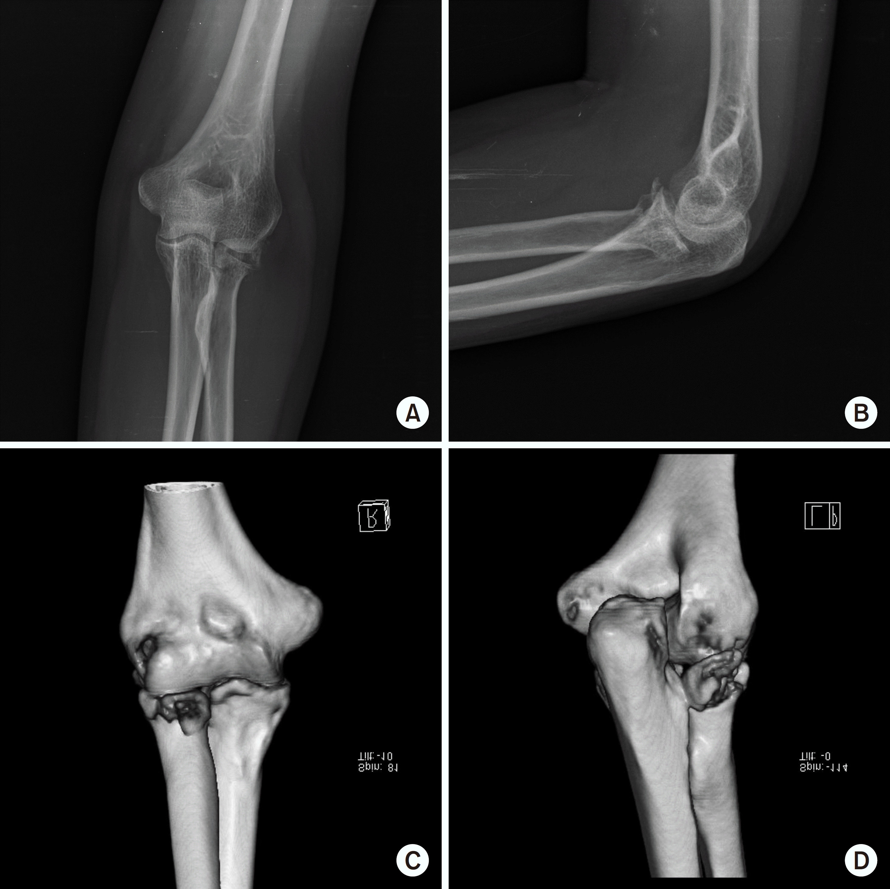
 | Fig. 4.(A, B) A 55-year-old woman fell on the ground with an outstretched hand and was diagnosed with a radial head fracture on the left elbow at the initial X-ray. (C, D) The three-dimensional computed tomographic scans revealed multi-fragmentary, five fragments, a radial head fracture with displacement in the overall radial head. |
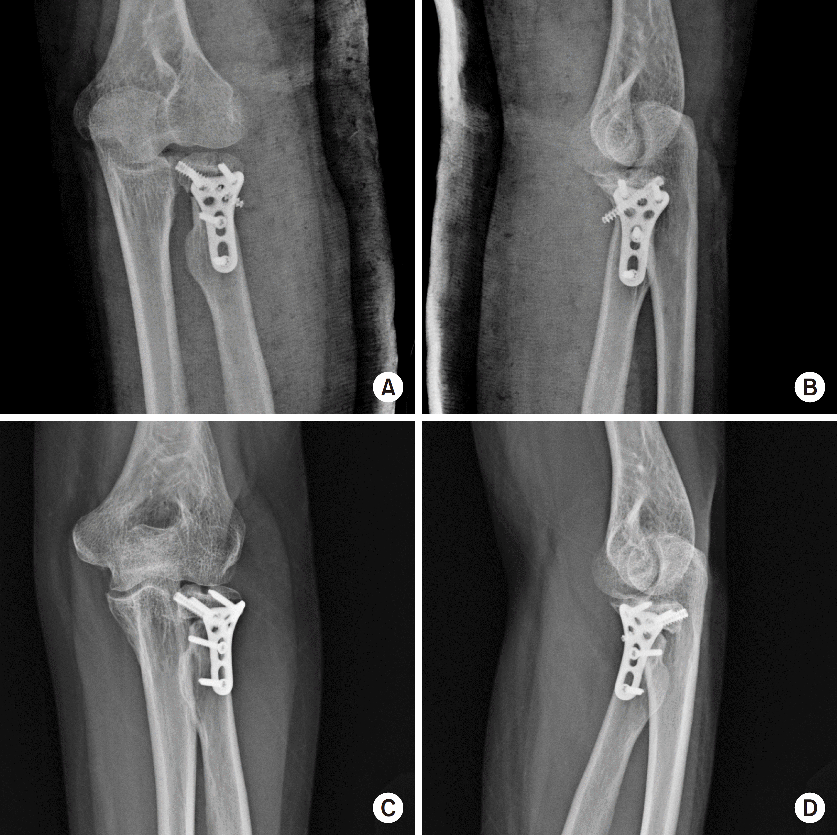
 | Fig. 5.Open reduction and internal fixation were conducted (A, B), but a postoperative 1 year, the patient presented with partial fragment nonunion (C, D). The range of motion was 10° to 120° flexion and the modified Mayo elbow score was 80. |
Table 1.
Baseline Data of the Patients
Table 2.
Overall Functional Outcome
| Variable | Value |
|---|---|
| Mean flexion (°) | 133.5 (90–150) |
| Mean extension (°) | 6.2 (0–40) |
| Mean pain VAS | 1.37 (0–4) |
| Modified Mayo elbow score | 87.1 (70–95) |
Table 3.
Functional Outcomes Depending on the Number of Fracture Fragments


 PDF
PDF ePub
ePub Citation
 Print
Print


 XML Download