

 PDF
PDF ePub
ePub Citation
 Print
Print


Introduction
Neonatal skull depressions are extremely rare, with an incidence of about 1/10,000 in western countries.1 A depressed fracture forms an inward buckling of the bones forming a ‘cup shape’ termed a ‘ping-pong (ball) fracture’.2 During a difficult delivery, trauma to the mother's abdomen or traumatic delivery with obstetric maneuvers may cause depressed skull fractures in neonates.2 However it may also occur in utero. Fetal head pressure against the maternal bony structures such as sacral promontory and exostosis of the lumbar vertebra, symphysis pubis, or ischial spine can result in ping-pong fractures.3 Also, fetal head pressure against the maternal pelvic mass such as uterine fibroids or tumor can result in fractures.4 Other mechanisms related to the fetus itself include skull compression by a twin or pressure exerted by the digits and fists of the newborn on his skull.5
The management of depressed skull fractures is controversial. Most ping-pong fractures are not severe enough to undergo surgery and the parents are usually afraid to observe without treatment for cosmetic and neurological concerns. Both surgical and non-surgical treatments are available and should be guided by the severity of the fracture and any underlying intracerebral injury. Being a less invasive procedure, we tried an obstetrical vacuum extractor immediately after birth. We hereby report with a brief review the cases of ping-pong fracture successfully elevated by means of obstetrical vacuum extractor.
Case
A term male infant was delivered in good condition of Apgar scores 9 and 10 at one and five minutes, respectively. Vaginal delivery was uneventful with no extraction instruments utilized at local obstetrical clinic. Birth weight was 2,830 g (25–50th percentile) and head circumference 33.0 cm (25–50th percentile). At birth, the infant was noted to have a depression in the right parietal region. The infant was transferred to the pediatric department in Sanggye Paik Hospital.
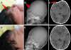
The pediatrician clinically examined and found the skull depression in the right frontal region, approximately 3 cm by 3 cm with 1 cm depth (Fig. 1A). There was a hard base suggesting the presence of bone. Otherwise, his skull was normally shaped and proportioned with patent fontanelles and regular sutures. There was no softening of the skull and skeletal survey was normal except cephalohematoma in right parietal area. There were no dysmorphic features and the eyes, neurology, respiratory, abdominal and skin examination were normal. The skull X-ray (Fig. 1B) and computed tomography (Fig. 1C) of the head revealed a ping-pong fracture in the right frontal bone.
The pediatrician consulted as to possibility of the depressed skull fracture treatment with the vacuum extractor to obstetrician. The infant underwent an elevation of the depression with an obstetrical vacuum extractor. We chose the diameter of the soft cup according to the diameter of the depression. We gradually increased the extraction pressure applied from 0.05 kg/cm2 to 0.3 kg/cm2 for 4 minutes, until we found the clinical restitution of the normal skull convexity. The depressed scalp was puffed back to normal position with mild scalp swelling (Fig. 1D). The skull X-rays (Fig. 1E) and computed tomography (Fig. 1F) were performed immediately following the procedure. After release of the pressure and removal of the cup, a circular patch of edema was observed. However, the scalp swelling disappeared within a day, revealing a normal head contour. The neurological examination remained normal and the magnetic resonance imaging did not show any cerebral lesion. The infant was observed in the hospital for a week, and reviewed by a pediatrician after 1 and 3 months. He developed normally and his parents had no concerns over his growth or behavior.
Discussion
In the past, the classic recommendation for a simple depressed fracture was to surgically elevate it on the basis of concerns regarding cosmetic effect, possible underlying pathological features, prevention of seizures, and improvement of focal neurological signs.6 Since 1960s, several studies have shown that for children without evidence of neurological or radiographic intracranial lesions, there was no difference between surgically treated and non-surgically treated patients in terms of future neurological sequels.7 Operative interventions are recommended if the fracture is combined with bone fragments in the cerebral tissue or if epidural or subdural hematomas exist.8 The least invasive treatment is a watchful waiting. In neonates and infants, the membranous sutures, the fontanelles, and the low level of calcium content in the fetal skull facilitate a great deal of plasticity.3 Because of these plasticity and relative thin skull bone, frequent crying with the resulting increase in intracranial pressure or cerebral edema had been presumed to result in the spontaneous elevation of depressed fractures.39 In addition, the Raynor-Parsa maneuver applying thumb pressure on opposite margins of the depression can be applied.10 Then a breast pump or an obstetrical vacuum extractor can be utilized for correction of the depression.
This is the first paper reported by pediatricians who can evaluate neonatal condition specially and obstetricians who can predict a fracture and use vacuum extractor skillfully. We summarize technic of obstetrical vacuum extractor below. The following brief review will be helpful to physicians who are involved in the perinatal period.
1. Indication
If the fracture status is not the indication that surgery should be considered first, it is an indication for vacuum extraction therapy. Surgical treatment is required in the cases where the fragments are depressed to the depth of at least one thickness of the skull, and in those with intracranial hematoma, cerebrospinal fluid leak, cosmetically deforming defects, gross wound contamination, and established wound infection.7 In theory, a 5 mm depression of the neonatal skull could impinge on the cerebral cortex and cause a focal area of decreased blood flow leading to tissue hypo-perfusion and cerebral edema.1 For infants with a minor depression (<5 mm in depth), conservative management only was given, after which patients were followed up with close observation. For a larger depression (>5 mm in depth and usually >2 cm in length), obstetrical vacuum extraction was used. There is no limit on the size of the fracture to be treated, if it is inside the cup. And the location of the fracture is not limited. The application of vacuum extraction seems to have age limitations. Most reports of vacuum reduction for simple skull depression featured newborns and infants, but the oldest age was 17 years old girl.6
2. Cup
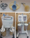
The cup is placed over the depressed region. The metal or soft cup should be able to cover the entire depression and to attach tightly to the infant's head without the likelihood of air leakage between the infant's skin and the periphery of the suctioning device (Fig. 2A). However, soft cup is preferred because extraction time is controlled under direct visualization.11 If a soft cup is not available, a transparent plastic cup (for example, a breast pump shield) can be substituted.12 The cup of breast pump is placed over the depressed region after gel is applied to the margin of the depression for obtaining the airtight seal between the scalp and the cup of breast pump. No traction is needed during the suction procedure. In our cases, a cup of twice the diameter of depression was large enough to seal the air.
3. Vacuum extractor
The obstetric vacuum extractor is the preferred instrument because it provides patent tubing unlikely to collapse during the application of suction. If not available, a suction generator which has been primarily utilized to suck all kinds of fluids at the operating room, intensive care unit, and emergency department can be substituted.12 We utilized two types of vacuum extractors (Fig. 2B, 2C). We prefer a button-type extractor, because it maintains the suction power more steadily (Fig. 2B).
4. Suction pressure and duration
The suction pressure has been reported to be 0.2 kg/cm2 for a 1 kg premature baby and 0.4–0.6 kg/cm2 for a full-term newborn, although it varied widely among reports.113 For older patients, a negative pressure of 0.8 kg/cm2 was sufficient.614 During the procedure, the elevation of the depression can be ascertained by direct visualization, an audible “click” sound or a “give” sensation accompanied by an instantaneous pressure release.11 Unfortunately, the pattern of pressure elevation was never described in the previous reports so far and we had to rely only on intuition. In our institute, we elevated pressure swiftly every 10–15 seconds (0–0.2 kg/cm2, yellow button on Fig. 2B), and elevated pressure slowly in every 30 seconds later on (0.3–0.6 kg/cm2, green button on Fig. 2B). The duration of negative pressure was determined on the clinician's discretion. From the literature, the duration of the application ranged from 10 seconds to 15 minutes.1112 Where necessary, a second application can be performed while observing the scalp swelling and skull contour.
5. Complication
Generally, the adverse effects of vacuum extractor are local edema, caput succedaneum and cephalohematoma.15 Artificial caput succedaneum is present when rigid vacuum cups are used, but is less common with soft cups.16 It usually disappears within hours to several days after procedure without sequelae. Cephalohematoma is an accumulation of blood beneath the periosteum of a cranial bone. No specific treatment of cephalohematoma is usually needed.15 They almost always resolve spontaneously in a matter of days, but occasionally may take several weeks or even months to completely regress. A close observation in intensive care unit for 24 hours is recommended as well because events such as apnea or bradycardia was temporarily observed during the first day.
In summary, the obstetrical vacuum extraction for ping-pong fracture of neonate is technically easy and less invasive. It should be considered first for the management of uncomplicated depressed skull fracture in the newborn. The vacuum extraction is especially warranted for the obstetricians, because the ping-pong fractures are mainly found immediately after birth.
 XML Download