

 PDF
PDF ePub
ePub Citation
 Print
Print


Penile amputation is an extremely rare traumatic event. The majority of cases involve adult self-mutilation due to psychological problems or surgical mishaps during circumcision of infants12. Prior to the advent of microsurgery, survival of the replanted penis depended on the adequacy of sinusoidal blood flow to re-approximated penile tissues1. Consequently, tissue necrosis, urethral stricture, loss of sensation, erectile dysfunction was often occurred.
Microsurgical replantation has the potential to reduce such complications, but most of the reported successes (approximately 30 cases) have focused on survival, rather than meticulous technique, and were not free of postoperative complications. To restore baseline penile function, the intrinsic anatomy of the penis must be maintained. We have achieved complete anatomic restoration of a severed adult penis using a microsurgical technique that eliminates complications. The details are described here in two case reports. Normal function and aesthetics resulted in each case.
CASE REPORT
1. Surgical technique
Under general anesthesia, blood loss from the penile stump was controlled by wrapping the base of the penis circumferentially with a small penrose drain or rubber nelaton catheter, secured by a hemostat; and percutaneous suprapubic cystostomy was performed for urinary diversion. Initially, the amputated segment and corresponding edge of penile remnant were carefully examined at high-power under a surgical microscope, tagging all arteries, veins, and nerves identified with 9-0 or 10-0 nylon suture. A 16 Fr Foley catheter inserted into urethra (severed and residual) and passed into the bladder functioned as a rigid support during surgery. The study was performed in accordance with the principles of the Declaration of Helsinki. The patient provided written informed consent for the publication and the use of her images.
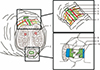
A 3-layer anastomosis of urethra was performed first. Following two z-plasty maneuvers with opposing 3-mm flaps (3 and 9 o'clock), vertical mattress suturing (6-0 polydioxanone [PDS™II]) of the mucosa was done. The muscular layer was sutured, avoiding damage to the sinusoidal spaces of spongiosum (Fig. 1, 2)3.
Next, the lower tunica albuginea (of corpora cavernosa) and the septum penis were repaired (4-0 PDS™II), anastomosis (10-0 nylon) of two cavernosal arteries (0.5 mm) which were situated in the center of corpus cavernosum took place under high-power microscopy, and repair of tunica albuginea and septum penis resumed for full restoration (Fig. 1). Any errors at this juncture could be a source of postoperative hematoma.
Two dorsal penile arteries (1 mm) and a deep dorsal vein (1.5 mm) were also repaired (10-0 nylon), as well as two proximal and two distal circumferential veins. Dorsal nerve bundles in the vicinity of deep dorsal and circumferential veins were identified for repair via epineural suture (10-0 nylon) (Fig. 2)3. Buck's fascia (deep penile fascia) was restored (5-0 PDS™II), as were other veins (10-0 nylon), including a superficial dorsal vein (3 mm) and superficial veins beneath dartos fascia (superficial penile fascia). Dartos fascia and skin were repaired (5-0 polyglactin 910 [Vicryl™]).
Prostaglandin E1 (5 mcg/mL/day), low molecular weight dextran (500 mL/day) and prophylactic antibiotics were administered intravenously for 5 days.
2. Patient 1
A 38-year-old male suffering from schizophrenia self-amputated his penis in a suicide attempt and was hospitalized 7 hours later. At presentation, the distal shaft (three-quarters) was completely amputated (Fig. 3). Warm ischemic time was approximately 7 hours, and cold ischemic time was 2 hours (total ischemic time, 9 hours).
Microsurgical replantation was performed as described above. Four arteries (2 dorsal penile and 2 cavernosal arteries), 6 veins (1 deep dorsal, 1 circumferential dorsal, 1 superficial dorsal, and 3 superficial veins beneath dartos fascia), and 5 dorsal penile nerves were repaired (Fig. 2)3.
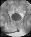
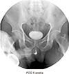
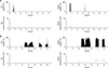
Mild edema developed during the first 2 postoperative days but resolved within five days, and no other complications occurred (Fig. 3). Following Foley catheter removal 2 weeks later, natural urinary volume (21 mL/sec) was within normal range by urine flowmetry, and a retrograde urethrogram (postoperative day 28) showed no evidence of stricture or fistula (Fig. 4). Nocturnal penile tumescence (NPT) tested at 3 weeks indicated three episodes of poorly sustained but rigid nocturnal erection (mean duration, 9 minutes). Repeat testing at 3 months revealed two episodes of well-sustained, completely rigid nocturnal erection (mean duration, 45 minutes) (Fig. 5). Patient's urethral patency was adequate for comfortable voiding.
3. Patient 2
A 43-year-old male with chronic alcoholism severed his penis with knife in a state of alcoholic delirium during intended detoxification. He has hospitalized 3 hours later. At presentation, the distal three-quarters of penile shaft was severed obliquely (Fig. 6). Vital signs were unavailable. Warm ischemic time was approximately 3 hours, and cold ischemic time was 1 hours (total ischemic time, 4 hours).
Microsurgical replantation was performed as described above. Four arteries (2 dorsal penile and two cavernosal arteries), 6 veins (1 deep dorsal, 1 superficial dorsal, and 4 superficial veins beneath dartos fascia), and 6 dorsal penile nerves were repaired (Fig. 2)3.
Mild subcutaneous edema developed during the first 3 postoperative days, resolving after five days. Following Foley catheter removal 2 weeks later, natural urinary volume (18 mL/sec) was within normal range by urine flowmetry. Retrograde urethrogram (postoperative day 30) showed no evidence of stricture (Fig. 7). NPT tested at 3 weeks indicated two episodes of poorly sustained but rigid nocturnal erection (mean duration, 12 minutes). Repeat testing at 5 months revealed five episodes of well-sustained, completely rigid nocturnal erection (mean duration, 101 minutes) (Fig. 8). Patient's urethral patency was adequate for comfortable voiding. Three months later, the patient was capable of normal sexual relations with his wife.
DISCUSSION
The objective of penile replantation is preservation of penile length, erectile function, and upright voiding capacity. A severed penis should be immediately and expeditiously repaired to prevent ischemia of the amputated segment. Amputations of the glans and distal penis may be viewed as composite grafts, treated by anastomosis of urethra and corpora, and subsequent suturing of skin. Such maneuvers rely on adequate sinusoidal blood flow and do not require a microvascular approach for success2. However, an array of complications, including skin necrosis, venous congestion, urethral fistula or stricture, poor sensation, and incomplete erection or impotence, have occurred postoperatively4. The first successful microsurgical replantation of the penis was performed by Cohen5 in 1976. It has become increasingly clear that outcomes are significantly improved by microsurgery.
The maximum ischemic time for return of testicular endocrine function is reported to be approximately 6 hours but for viability of a severed penis, the window of time is longer6. In addition, prolonged ischemia of a penile stump (18 hours) in a 4-year-old child has been reported and was not prohibitive, although the severed penis was cooled while ischemic to the extent that treatment for frostbite was needed7. In patient 1, duration of ischemia overall (including intraoperative period) was 10 hours 30 minutes, and no complications ensued. It is very likely that the actual ischemic insults incurred have been longer than published reports reflect. Nevertheless, ischemic time is the primary determinant of subsequent complications.
When initiating penile replantation, urethral repair should be performed first. An accepted method is one-layer suturing of urethra and corpus spongiosum, but this can easily lead to urethral stricture or fistula. Alternatively, two layer repair for improving viability, a spatulated technique, and an oval-end termino-terminal anastomosis have been devised to prevent urethral complications8. In the patients treated here, a three-layer repair with double opposing Z-plasty was used. Following double opposing z-plasty and suturing of mucosa, the muscular layer of urethra and fibrous capsule of corpus spongiosum were repaired in sequence, avoiding damage to the sinusoidal spaces of spongiosum. Above all, the integrity of blood supply must be assured to prevent ischemia or venous congestion and secondary necrosis.
There is currently no consensus on the number of arteries and veins it is necessary to repair during penile replantation for adequate perfusion or viability of the penis; but according to some sources, at least one dorsal penile artery and one deep dorsal vein may suffice for penile viability7. Anatomic restoration is paramount to regain baseline function, so as many vessels as feasible should be repaired. The patency of the cavernosal artery is the most important for erectile function. In instances where the cavernosal artery was incompletely repaired, results were poor, possibly owing to technical difficulties9. Cavernosal arteries of both patients here were completely repaired, restoring normal erectile function.
Rigorous repair of venous channels is also recommended to reduce postoperative edema and necrosis of the glans and foreskin10. In the present circumstances, six veins, including superficial and deep dorsal branches, were repaired, resulting in mild edema only. Anastomosis of the superficial vein beneath dartos fascia is especially important to prevent venous congestion of subcutis and skin necrosis.
Dorsal penile nerves distributed about the penile shaft within Buck's fascia innervate distal shaft, prepuce, and glans penis, are largely responsible for tactile and erogenous penile sensation. Dorsal neurorrhaphy is therefore essential for restoration of erectile/sexual capacity. In these patients, 5–6 dorsal penile nerve fascicles were repaired, with sensory impulses first to recover.
In conclusion, the goal of penile replantation is both functional and cosmetic, assuring penile length sufficient for normal erection and urethral patency adequate for comfortable voiding. We have presented two instances of successful microsurgical penile replantation after complete amputation. In this setting, a return to baseline status (as opposed to viability) is the most critical issue. Microsurgical replantation is thus the treatment of choice, and is aimed at restoring normal anatomy and function.


 XML Download