

 PDF
PDF ePub
ePub Citation
 Print
Print


Mallet finger is a very common finger disease. It occurs mainly on the ring finger, middle finger, and little finger12. In rare cases, there are two mallet fingers in one hand3. However, until now, to our knowledge, there have been no reported cases of three mallet fingers occurring simultaneously on one hand. Herein, we report a case of mallet fingers occurring on three ulnar fingers of the same hand and their successful treatment by surgical fixation.
CASE REPORT
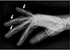
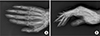
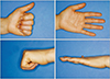
A 46-year-old female fell on the ground during a running race. She was injured on the middle, ring, and little fingers. Radiographically, bone fragments were observed on the dorsal side of the distal phalanx base on the middle, ring, and little fingers with more than 30% of intra-articular involvement (Fig. 1). Five days after the injury, all three fingers were treated surgically by the extension block method (EBM) (Fig. 2). Single mallet fingers are usually treated with finger splints, but this case involved three mallet fingers; thus, we applied a short arm splint. At this time, the index finger was movable and the splint was kept on for 10 days. Because of concerns about joint stiffness, we started passive range of motion (ROM) excercise starting from the 2nd week after surgery. K-wires on dorsal sides wire removed 3 weeks after surgery, and longitudinally fixated K-wires were removed 6 weeks after surgery. Finally, bone union was confirmed after 7 months (Fig. 3). During the outpatient visit 6 months after surgery, the ROM was good but extension lag was observed within 10° in all fingers (Fig. 4). According to Crawford's criteria, result as “Good” is defined as “0 to 10 degrees of extension loss, normal flexion, no pain”4. In this case, the condition is met.
Informed consent was obtained for the use of photographs from the patient in this study. The consent has been formally documented in the medical record.
DISCUSSION
Mallet finger can be divided into bony mallets and tendinous mallets. Conservative treatment with finger splints can be used for tendinous mallets. However, the appropriate treatment for bony mallet is still under discussion. The current literature describes several surgical and nonsurgical methods for managing mallet finger injuries such as pull-out suture, extension block with K-wire or splinting without surgery. If the bone fragment is bigger than one-third the joint surface or subluxation is severe, the bone fragment may remain unstable or may malfunction. In these cases, surgery is usually performed. Without instability or severe joint involvement, non-surgical treatment can be done. Gurnani et al.5 claimed that a bony mallet finger can also be treated using finger splints and conservative treatment56. There is no significant difference in deformity or pain when comparing splinting and conservative treatment with surgical treatments. Although there are no significant differences in complications resulting from splinting and surgery, we preferred surgical treatment in our case. The splint should be maintained for six to eight weeks if surgical intervention is not performed. If an extension lag remains, surgical treatment should be done. Therefore, the treatment period will be longer. Patients also tend to feel uncomfortable with the splint; thus, they often unwind the splint. But if the splint is not well-maintained, unfavorable results could happen. For this reason, we believe that surgical treatment shortens the duration of treatment and is safer for patients.
Among surgical methods, closed reduction is preferred to open reduction in recent years. Among the closed reduction surgical methods, the EBM and direct pining method (DPM) are used. EBM, developed by Ishiguro et al.7, is a good surgical method for treating mallet fingers8. However, it has been reported that complications, such as infection, joint failure, implant failure, and residual pain, can be increased when the K-wire is being fixed through the joint space. Recently, Han et al.9 also demonstrated that DPM is superior in that it offers better improvements in extensor lag and motion range. However, direct pinning to bone fragments can cause bone fragments to break. In our opinion, it is also possible to reduce the extension lag if early removal of the percutaneously inserted K-wire through the distal extensor tendon is performed. For this reason, EBM is preferred in our case, although it will depend on the individual tendency of the surgeon.
To our knowledge, mallet fingers occurring in simultaneously in three fingers have not been reported. In our case, hyperextension seems to have occurred in the bony mallet fingers10. The mallet fingers occurred simultaneously on three fingers, but they were treated as if they occurred on one finger. Surgical treatment with EBM was performed, and satisfactory results were obtained without any complication. However, because the size of the bone fragment varies among fingers, there were different results in terms of extension lags. There was also more extension lag in middle finger due to lack of bone fragment reduction. In conclusion, this case provides data on the treatment of multiple mallet fingers on the same hand.

 XML Download