

 PDF
PDF ePub
ePub Citation
 Print
Print


INTRODUCTION
Clostridium difficile (C. difficile), which is one of the important etiologic agents of pseudomembranous colitis in older children and adults, causes an asymptomatic infection in most neonates and young infants [1-3]. Likewise, in most cases, colonization of hospitalized preterm infants in the neonatal intensive care unit (NICU) is not apparently harmful; however, the association of C. difficile colonization (CDC) with necrotizing enterocolitis or nonspecific gastrointestinal symptoms such as diarrhea has been infrequently reported [4-7]. In addition, recent case reports suggested that C. difficile could be transmitted from infants with asymptomatic colonization to their nursing mothers, who subsequently develop recurrent pseudomembranous colitis [8,9]. C. difficile strains identical to those in adults were identified in asymptomatic infants even in the community [10]. Therefore, asymptomatic C. difficile infection in preterm infants may be medically important in the aspect of the potential of infected neonates to serve as reservoirs for C. difficile transmission in older children and adults, leading to symptomatic infection.
The establishment of healthy intestinal microbiota during the neonatal period is considered important in preventing certain diseases in the future lifetime based on a large amount of recent research on the intestinal microbiota [11,12]. According to the results of previous research, the candidate factors influencing the development of the intestinal flora in early life include the mode of delivery, feeding type (breast-milk or formula feeding), gestational age, history of hospitalization, and antibiotic use [13,14]. Consequently, preterm infants are unlikely to have a healthy intestinal microbiome because they are delivered via cesarean section, frequently supplemented with premature formula, mostly hospitalized for lengthy periods in the special medical environment of the NICU, and treated with antibiotics in many cases. However, the results of these previous studies are inconsistent, in contrast with those of studies of healthy term infants [15-17]. Moreover, CDC and its related factors in preterm infants have not been fully investigated because many studies did not differentiate between different species of the genus Clostridium [6,18,19]. In Korea, to our knowledge, only one study investigated CDC in preterm infants [20].
The aims of the present study were to monitor the prevalence of CDC and its toxin positivity and to examine the influence of a variety of potential determinants of CDC in the preterm infants by detecting C. difficile and its toxin in serially collected fecal samples from the infants in two NICUs over the first several weeks of life and collecting clinical information from their medical records.
MATERIALS AND METHODS
Subjects
From April 2007 until March 2008, preterm infants were recruited from the NICUs of University Hospitals A and B. Fecal samples from spontaneous defecations were serially collected within 72 h after birth and at 1, 2, and 4-6 weeks of age, although some of the 72-h fecal samples were obtained by normal saline enema because there was no spontaneous defecation in some of the infants. The preterm infants who had chromosomal abnormalities, congenital heart diseases, sepsis, and severe birth asphyxia were excluded. This study was conducted with informed consent from the infants' parents, and the protocols for this study were approved by the Institutional Review Board of Seoul Metropolitan Government Seoul National University Boramae Medical Center.
Methods
1. Stool collection and DNA isolation
Fecal samples were collected in sterile tubes and immediately frozen at -20℃. They were transferred to the laboratory on dry ice within 72 h of collection for further processing. Total bacterial DNA was extracted from each fecal sample using a commercial DNA isolation kit (QIAamp® DNA Stool Mini kit; QIAGEN, Germantown, MD, USA) with some modifications as described previously [21]. After quality assessment and quantitation, the extracted bacterial DNA was amplified using the GenomiPhi V2 DNA Amplification kit (GE Healthcare, Piscataway, UK) for polymerase chain reaction (PCR) assays. The bacterial DNA for C. difficile (ATCC 9689) was also isolated at the stationary growth phase as a positive control for PCR assays.
2. PCR assays
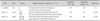
A total of 187 DNA samples were obtained from each fecal sample serially collected within 72 h after birth and at 1, 2, and 4-6 weeks of age for 49 infants (Hospital A: 26; Hospital B: 23). PCR amplification of the 16S rRNA gene of C. difficile was performed, and for DNA samples displaying positive results in this first PCR assay, two PCR assays were performed with primers directed at the toxin A gene (tcdA) and toxin B gene (tcdB) (Table 1) [22-25]. All PCR amplifications were performed using TaKaRa Ex Taq (Takara Bio Inc, Shiga, Japan). Reaction mixtures consisted of approximately 200 ng of template DNA, 10 pmol each primer, 1.25 U of Taq polymerase, 4 µl dNTP mixture (each 2.5 mM), 5 µl 10× Ex Taq Buffer, and water to a total volume of 50 µl. The reaction mixtures were subjected to amplification in a DNA thermal cycler (Mastercycler gradient®; Eppendorf AG, Hamburg, Germany) with the following cycling conditions: initial denaturation at 95℃ for 5 min followed by 35 cycles of denaturation at 95℃ for 20 s, annealing at the specified annealing temperature for 30 s (C. difficile), 2 min (toxin A), or 60 s (toxin B), and extension at 72℃ for 45 (C. difficile) or 40 s (toxin B). A final extension at 72 (C. difficile) or 74℃ (toxins A and B) for 5 min was performed. Two independent PCR reactions were performed for each sample. The amplification products were examined by electrophoresis in a 1.5% agarose gel and documented with the Bio-Rad Gel Doc 1000 Documentation System (BioRad, Hercules, CA, USA).
3. Neonatal characteristics influencing CDC
The neonatal characteristics examined were gestational age (weeks), birth weight, mode of delivery, the age of first feeding, the age of the first full feeding (the age of the preterm infant at which the amount of enteral feeding reached 100 ml/kg/day), presence/absence of nil per os (NPO) for more than 72 h after birth, exclusive breast-milk feeding (EBMF) for more than the first 7 days, EBMF after reaching full feeding, the use of antibiotics in the first week or after the first week, and the hospital (A or B).
4. Statistical analyses
We examined the data for normality by using the Kolmogorov-Smirnov test of normality. Categorical variables were summarized as the percentage of infants with evaluable data; continuous variables were summarized using the mean and standard deviation and the median, minimum, and maximum values for non-normally distributed continuous variables. Comparisons of categorical data were evaluated using Pearson's χ2 test. Comparisons of continuous data were evaluated using the independent-samples t-test for normally distributed data and the Mann-Whitney test for non-normally distributed data. To determine the factors associated with CDC, data were analyzed using multiple logistic regression with C. difficile positivity as the dependent variable and the mode of delivery, NPO beyond 72 hours after birth, EBMF for more than the first 7 days after birth, EBMF after reaching full feeding, initial antibiotics use, use of antibiotics after the first week, and the hospital (A vs. B) as the explanatory variables. All analyses were conducted using SPSS version 16.0 (SPSS, Chicago, IL, USA). All hypotheses were tested at an α of 0.05.
RESULTS
Prevalence of CDC and its toxin positivity
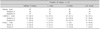
Paired PCR results for C. difficile were available for three different ages (within 72 h of birth, 1 or 2 weeks, 4-6 weeks) for 49 patients (hospital A: 26, hospital B: 23), whereas the results were available for all four postnatal ages for 40 infants.
The rates of C. difficile PCR positivity at 72 h and 1, 2, and 4-6 weeks of age were 34.7 (17/49), 37.2 (16/43), 41.3 (19/46), and 53.1% (26/49), respectively, indicating that the rate of positivity increased over the course of hospitalization, and the rates of positivity were similar between the two hospitals (Table 2). The C. difficile PCR assay results were persistently negative and positive in 12 (24.5%) and 12 (24.5%) preterm infants, respectively, whereas the results were transiently positive in one or two postnatal periods for 25 (51%) infants (Table 3). Moreover, 14 (28.6) infants exhibited C. difficile PCR positivity for only one postnatal age, consisting of 2, 2, 2, and 8 infants with positivity within 72 h after birth and at 1, 2, and 4-6 weeks of age, respectively.
Among C. difficile-colonized infants, the toxin positivity rate slightly increased from 23.5% (4/17) within 72 h after birth to 30.8% (8/26) at 4-6 weeks of age, although there were some fluctuations over time. Eight (66.7%) preterm infants with persistent CDC exhibited toxin positivity for at least two postnatal ages, compared with 6 of 25 (24.0%) preterm infants with transient CDC (p=0.001).
Potential determinants of CDC in the neonatal period
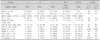
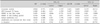
We compared the gestational age, birth weight, mode of delivery, feeding progression pattern, feeding method, use of antibiotics, time of antibiotic administration, and hospital among three groups of preterm infants: those displaying persistent PCR positivity comprised group A (n=12), those displaying persistent PCR negativity comprised group B (n=12), and those displaying transient PCR positivity comprised group C (n=25; Table 3). Among the various neonatal factors, only the feeding method during the first week after birth and the hospital were significantly different among the three groups (p=0.021, p=0.047). When we analyzed these relationships between the infants with persistent C. difficile PCR positivity (group A) and those with transient C. difficile PCR positivity or persistent C. difficile PCR negativity (groups B+C), only the proportion of exclusively breast-fed infants during the first week after birth was significantly different between the two groups (p=0.018). In multiple logistic regression analyses of the potential determinants of CDC in the two groups (A vs. B+C), only EBMF during the first week after birth significantly decreased the risk of persistent CDC compared to formula or mixed feeding (adjusted odds ratio (OR)=0.133, 95% confidence interval (CI)=0.02-0.898, p=0.038; Table 4). No other factor including the hospital exhibited a significant association with CDC patterns. When the relationship between those neonatal factors and C. difficile PCR positivity was analyzed for each postnatal age, the type of feeding during the first week after birth was also significantly associated with CDC at 4-6 weeks of age; EBMF significantly decreased the risk of CDC in this hospitalization period (adjusted OR=0.186, 95% CI=0.04-0.861, p=0.031; Table 5).
DISCUSSION
The present study monitored the CDC pattern and its toxin status in preterm infants in the NICU, which has been infrequently investigated in the literature, including only a single study from Korea [6,19,20,26,27]. In addition, to the best of our knowledge, this is one of the first reports revealing that EBMF during the first week of life may have protective effects against persistent CDC in preterm infants. Several studies have investigated the beneficial role of breast-milk feeding on CDC in term infants; however, its effects have been rarely investigated in hospitalized preterm infants, excluding one study that revealed no significant association between breast-milk feeding and CDC [19,28,29].
In our study, the C. difficile prevalence rate was 35-53%, which is comparable with the rates reported in previous studies of 33-90% [6,18-20,26,27]. The rates of CDC increased gradually over the course of hospitalization, which has been consistently reported among researchers and suggest that the hospital-acquired mode of C. difficile transmission is similar to that of later times of life. This hospital-acquired infectivity might explain why the prevalence of CDC in preterm infants, who usually are hospitalized for long periods, is higher than that of term infants and reflects the importance of the environmental source in CDC. Other potential explanations may be related to the immaturity of both gut and systemic immunity and the low colonization resistance of the intestinal microbiota of preterm infants, which have been considered important factors for CDC or infection in the human intestine [30,31]. Previous studies indicated that delayed colonization with a lower diversity and low prevalence and proportion of bifidobacteria is characteristic of the intestinal microbiota of preterm infants compared to that of healthy term infants [32]. In a recent large epidemiologic study including 11 preterm infants aged 1 month old (approximately 1% of total participants), the OR of premature birth for CDC was approximately 4.5-fold higher than that of term birth even after adjustment of history of hospitalization [13].
It is noteworthy that the results of our study suggest both the importance of the environment and host factors in CDC in preterm infants. In our study, the persistence of CDC was only associated with feeding type in the multiple regression analyses. Although still controversial, EBMF has been reported to favor the development of the so-called "healthy" microbiota even in preterm infants [15,17,33]. It is now generally accepted that Bifidobacterium species are the dominant beneficial microflora in the intestine of breastfed infants, and the presence and degree of their colonization were reported to be negatively associated with the colonization of pathogens including C. difficile [34,35]. In addition, the diversity of Bifidobacterium species has been reported to enhance the maturation of the mucosal immune response [36]. Because we did not examine the composition of the microbiota including Bifidobacterium species, it is not clear whether the protective effect of EBMF is mediated by bifidobacteria. Further large-scale studies are needed to elucidate the interactions among breast-milk feeding, bifidobacteria, and CDC in the intestinal microbiota of hospitalized preterm infants. It is also interesting that the type of feeding during the first week after birth exhibited a prolonged effect on subsequent CDC (i.e., 4-6 weeks after birth) in this study. This finding is consistent with the hypothesis that "the very first few days after birth" are critical for the development of intestinal microbiota in the neonatal period [37].
In our study, the use of antibiotics was not significantly different between the infants with CDC and those without colonization. Considering the relatively small number of participants in this study, this finding should be confirmed by additional large-scale studies; however, this finding was comparable with those of previous studies in which no significant association was observed between antibiotic usage and CDC in both term and preterm infants [2,13,17]. In our study, we also compared two different NICUs to examine the influence of environmental factors. Although the rates of CDC were not significantly different between the two NICUs over the entire observation period, the pattern of CDC during the course of hospitalization (persistently positive vs. transiently positive vs. persistently negative) was different between the two NICUs. In a recent study, colonization of hospitalized preterm infants by Clostridium species was only associated with the NICU in multiple regression analysis. Although C. difficile was not separately analyzed and the type of feeding was not investigated in that study, the association of the NICU itself with Clostridium species colonization may support the importance of environmental factors in the colonization process of C. difficile in preterm infants [2,16].
Meanwhile, the proportion of toxin positivity as assessed by PCR assays using total bacterial genomic DNA as a template was 25-37.5%, which corresponded to the lowest value reported in previous studies (33-100%) using both culture and direct fecal cytotoxin detection in cultured cells [10,19,26]. It has been reported that many asymptomatic infants under 2 years of age are frequently colonized with both nontoxigenic and toxigenic strains [10,38]. The reported proportion of toxigenic strains is 21-37.5%. Further studies are needed to answer whether the proportion of toxigenic strains in preterm infants is higher than that of term infants. In our study, toxin positivity was also positively associated with the persistence of colonization, and most cases of transient C. difficile PCR positivity were accompanied by toxin PCR negativity. However, a recent study reported no significant difference in composition of intestinal microbiota according to the toxin status of C. difficile in infants with CDC less than 2 years of age [34]. Therefore, the significance of our findings must be validated by further studies.
One of the limitations of our study is that traditional PCR assays were used to detect C. difficile instead of a real-time PCR method. Therefore, it is possible that the actual colonization rate is higher than the assessed rate because of the lower sensitivity of traditional PCR compared to that of real-time PCR. In addition, the quantitation of CDC in individual infants was limited even though a semiquantitative assessment was possible. We observed that the intensity and broadness of bands on electrophoretic gels tended to increase as the duration of hospitalization of the preterm infants increased. Further large-scale studies using real-time PCR assays could more precisely reveal the potential determinants of CDC in preterm infants.
In conclusion, the CDC rate increased as the duration of hospitalization increased, and toxin-positive C. difficile more frequently tended to be a persistent colonizer. Because EBMF during the first week of life appeared to prevent persistent CDC, it should be actively recommended for hospitalized preterm infants.

 XML Download