Journal List > Korean J Pediatr Gastroenterol Nutr > v.13(2) > 1138539

Park, Lim, Seo, Ko, Chang, Yang, Lim, and Kim: Small Bowel Intussusception in Children: Spontaneous Resolution vs. Surgical Intervention

Abstract

Purpose

Intussusception is one of the most common causes of an acute abdomen in infancy. The majority of pediatric cases of intussusception are of the ileocolic type and usually idiopathic. Small bowel intussusception is rarely diagnosed in children, and few cases have been reported. The purpose of this study was to determine the clinical features and causes of small bowel intussusception in children.

Methods

We retrospectively reviewed the clinical and radiologic findings of 21 children with small bowel intussusception who were admitted to Seoul National University Children's Hospital between March 2005 and January 2010.

Results

The clinical presentation of small bowel intussusception included abdominal pain or irritability (85%), vomiting (23%), fever (14%), bloody stools (14%), and abdominal masses (4%). Six patients required surgical management. Ultrasonography showed that the mean diameter of the lesions and mean thickness of the outer rims were 1.6±0.7 and 1.7±1.8 mm, respectively. Eleven lesions were located in the left abdominal or paraumbilical regions. Children who underwent surgical management were older than children with transient small bowel intussusception (mean age, 51 vs. 109 months). The mean diameter of the lesions and mean thickness of the outer rims were greater in the surgically-managed group. The location of intussusception was not significantly different between the two groups.

Conclusion

Small bowel intussusception was spontaneously reduced in a large number of pediatric patients. However, sonographic demonstration of larger size, older age, and pathologic lead point warrant surgical intervention.

REFERENCES

1. DiFiore JW. Intussusception. Semin Pediatr Surg. 1999; 8:214–220.
crossref
2. Lehnert T, Sorge I, Till H, Rolle U. Intussusception in children-clinical presentation, diagnosis and management. Int J Colorectal Dis. 2009; 24:1187–1192.
crossref
3. Ko SF, Tiao MM, Hsieh CS, Huang FC, Huang CC, Ng SH, et al. Pediatric small bowel intussusception disease: feasibility of screening for surgery with early computed tomographic evaluation. Surgery. 2010; 147:521–528.
crossref
4. Saxena AK, Seebacher U, Bernhardt C, Hollwarth ME. Small bowel intussusceptions: issues and controversies related to pneumatic reduction and surgical approach. Acta Paediatr. 2007; 96:1651–1654.
crossref
5. Hur NJ, Ryu MH, Lee DJ, Kwon JH. A clinical observation on children with transient small bowel intussusception. Korean J Pediatr Gastroenterol Nutr. 2000; 3:160–168.
crossref
6. Kornecki A, Daneman A, Navarro O, Connolly B, Manson D, Alton DJ. Spontaneous reduction of intussusception: clinical spectrum, management and outcome. Pediatr Radiol. 2000; 30:58–63.
crossref
7. Parikh M, Samujh R, Kanojia R, Sodhi KS. Does all small bowel intussuseption need exploration? Afr J Paediatr Surg. 2010; 7:30–32.
8. Ko SF, Lee TY, Ng SH, Wan YL, Chen MC, Tiao MM, et al. Small bowel intussusception in symptomatic pediatric patients: experiences with 19 surgically proven cases. World J Surg. 2002; 26:438–443.
9. Strouse PJ, DiPietro MA, Saez F. Transient small-bowel intussusception in children on CT. Pediatr Radiol. 2003; 33:316–320.
crossref
10. Tiao MM, Wan YL, Ng SH, Ko SF, Lee TY, Chen MC, et al. Sonographic features of small-bowel intussusception in pediatric patients. Acad Emerg Med. 2001; 8:368–373.
crossref
11. del-Pozo G, Albillos JC, Tejedor D, Calero R, Rasero M, de-la-Calle U, et al. Intussusception in children: current concepts in diagnosis and enema reduction. Radiographics. 1999; 19:299–319.
crossref
12. Bisset GS 3rd, Kirks DR. Intussusception in infants and children: diagnosis and therapy. Radiology. 1988; 168:141–145.
crossref
13. Lim HK, Bae SH, Lee KH, Seo GS, Yoon GS. Assessment of reducibility of ileocolic intussusception in children: usefulness of color Doppler sonography. Radiology. 1994; 191:781–785.
crossref
14. Lee HC, Yeh HJ, Leu YJ. Intussusception: the sonographic diagnosis and its clinical value. J Pediatr Gastroenterol Nutr. 1989; 8:343–347.
15. Verschelden P, Filiatrault D, Garel L, Grignon A, Perreault G, Boisvert J, et al. Intussusception in children: reliability of US in diagnosis--a prospective study. Radiology. 1992; 184:741–744.
crossref
16. Pracros JP, Tran-Minh VA, Morin de Finfe CH, Deffrenne-Pracros P, Louis D, Basset T. Acute intestinal intussusception in children. Contribution of ultrasonography (145 cases). Ann Radiol (Paris). 1987; 30:525–530.
17. Doi O, Aoyama K, Hutson JM. Twenty-one cases of small bowel intussusception: the pathophysiology of idiopathic intussusception and the concept of benign small bowel intussusception. Pediatr Surg Int. 2004; 20:140–143.
crossref
18. Kim JH. US features of transient small bowel intussusception in pediatric patients. Korean J Radiol. 2004; 5:178–184.
crossref
19. Lee HS, Chung JY, Koo JW, Kim SW, Kim SH. Clinical characteristics of intussusception in children: comparison between small bowel and large bowel type. Korean J Gastroenterol. 2006; 47:37–43.
20. Lim HT, Park JH, Choi HJ, Kim JS, Shin HK, Gu CH. Clinical approach of ultrasonography in the diagnosis of intussusception in infant and children. J Korean Pediatr Soc. 1994; 37:649–654.

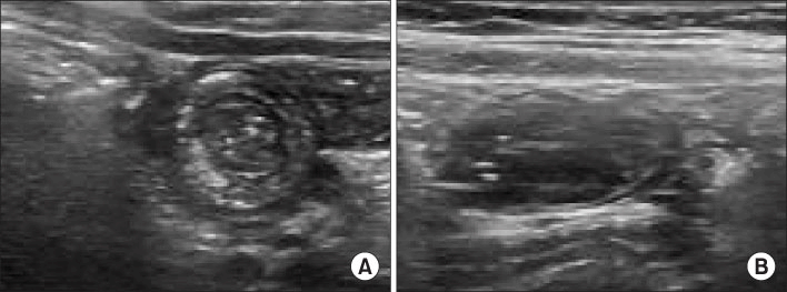
Fig. 1.
Typical benign small bowel intussusceptions (jeju-nojejunal) in a 4 year old boy with abdominal pain. Transeverse (A) and longitudinal (B) US scan showed doughnut (A) and sandwich signs (B).
kjpgn-2010-13-2-128f1.tif
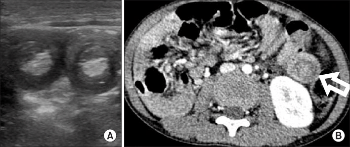
Fig. 2.
US (A) and CT scan (B) of a 5-year-old boy with recurrent abdominal pain and melena who underwent surgical reduction of small bowel intussusceptions at multiple sites (arrow). Based on biopsy, he was diagnosed with Burkitt lymphoma.
kjpgn-2010-13-2-128f2.tif
Table 1.
Clinical Characteristics of Small Bowel Intussusception
Case Age (months) Sex Irritability/Pain Vomiting Bloody stool Abdominal mass Underlying disorder Management
1 6 M - SR*
2 17 M - SR
3 17 M - SR
4 22 F - SR
5 27 M - SR
6 38 M - SR
7 39 M - SR
8 51 M - SR
9 56 M - SR
10 57 M - SR
11 57 M Duplication cyst SR
12 149 M T-cell lymphoma SR
13 173 F Peutz-Jeghers SR
14 34 M Hydronephrosis SR
15 23 M - SR
16 179 M DLBL OP (resection)
17 67 M Burkitt lymphoma OP (manual)
18 119 M - OP (manual)
19 19 M - OP (manual)
20 164 M Peutz-jeghers OP (resection)
21 107 F Peutz-jeghers OP (resection)

* SR: spontaneous reduction,

DLBL: diffuse large B cell lymphoma,

OP: operation.

Table 2.
Transient Small Bowel Intussusception and Surgically Managed Small Bowel Intussusception
Transient SBI (n=15) Surgically managed SBI* (n=6) p value
Clinical features
Male:Female 13:2 5:1 0.658
Mean age (mo) (range) 51±20 (6 mo∼14 yrs) 109±17 (19 mo∼14 yrs) 0.045
Symptoms
Pain/irritability 12 (85%) 5 (83%) 0.658
Bloody stool 1 (6%) 2 (33%) 0.184
Mass 1 (6%) 0 (0%) 0.714
Bowel ischemia/necrosis 0 (0%) 2 (33%) 0.071
Recurrence 3 (18%) 1 (16%) 0.684
Sonographic findings
Location (Right; Left; Middle) 7; 6; 2 2; 2; 1 (1: multifocal) 0.956
Transverse diameter (cm) 1.6±0.4 (0.9∼2.0) 3.2±0.5 (2.3∼3.5) 0.004
Thickness of outer rim (mm) 1.6±0.8 (1.0∼2.5) 5.8±1.4 (4.4∼7.8) 0.004
Lead point 1 (6%) 4 (67%) 0.031
Ascites 1 (6%) 4 (67%) 0.017
Bowel distention 8 (53%) 5 (83%) 0.221

* SBI: small bowel intussusception,

Fisher’s exact test,

Mann-Whitney U-test.

TOOLS
Similar articles