

 PDF
PDF ePub
ePub Citation
 Print
Print


INTRODUCTION
Intraductal papillary mucinous neoplasm (IPMN) is a cystic precursor of pancreatic cancer characterized by dilated main and/or branch ducts with papillary projections composed of dysplastic mucinous epithelium.1234 IPMN has a wide histological spectrum ranging from low-(LGD), intermediate-(IGD), high-grade dysplasia (HGD) to invasive carcinoma (IC), and the management of IPMN continues to evolve.1234
IPMNs have unique features, and multiple sites of occurrence in the same pancreas are often observed. IPMN probably represents a pancreatic “field defect”, which means all pancreatic ductal epithelial cells are at risk of dysplastic change, and this can be apparent in patients with multifocal BD-IPMNs.24 Also, it is recognized that a proportion of resected patients for IPMN evolve over time and can become metachronous IPMN that patients are at an increased risk of developing conventional pancreatic ductal adenocarcinoma (PDAC) elsewhere in the residual gland. Because of this, all patients with IPMN, including even those with non-invasive IPMN with negative surgical margin, should undergo surveillance after resection to detect the development of a new IPMN requiring surgery or concomitant PDAC.4567 However, the risk factors and characteristics of metachronous occurrence of IPMNs in the remnant pancreas have been unclear. As a result, there has been still debated the details for follow-up, such as, indication, methods, interval, and timing of follow-up.234
The cell lineage of the “papillary component” of IPMNs, such as, gastric, intestinal, pancreatobiliary, and oncocytic forms has been known clinicopathologic significance.24891011 The prognostic significance of these subtype showed heterogeneous results for recurrences and metachronous IPMN development in remnant pancreas after initial partial pancreatectomy.691011 Thus, the aim of this study was to identify the factors including these subtypes that predict recurrences and metachronous occurrence high-risk lesions (HRL) in the remnant pancreas after partial pancreatectomy for IPMN, and to make comprehensive follow-up strategy after resection of IPMN.
PATIENTS AND METHODS
After our institutional review board approval (No. 2017-07-016-005), clinicopathologic and surveillance data of consecutive 346 patients who underwent pancreatectomy for IPMN at the Samsung Medical Center from January 2005 to December 2016 were reviewed retrospectively from prospectively maintained electronic database system (MDBⓒ, Seoul, Korea).
Surgical indication
Before 2014, surgical indications followed International Association of Pancreatology (IAP) Sendai guidelines in 2006,1 including main duct type IPMNs, branch duct type IPMNs with cysts larger than 30 mm, main pancreatic duct dilatation exceeding 5 mm, the appearance of new mural nodules, or the presence of any symptoms. Surgery was also performed when cysts showed significant growth or when there was increased suspicion of malignancy. From 2014 to 2016, surgical indication was modified according to IAP Fukuoka guidelines in 2012,2 and were categorized as ‘high risk stigmata’, that are ‘obstructive jaundice in a patient with cystic lesion of the head of the pancreas’, ‘enhancing solid component within cyst’, and ‘main pancreatic duct >10 mm in size’.
Surgical procedure
Surgical procedures, such as, pancreaticoduodenectomy, central pancreatectomy or distal pancreatectomy, were determined according to the location and extent of the IPMN. If no invasive lesions were detected preoperatively, then laparoscopic procedures or limited resection were undertaken. In case of multifocal lesions, lesions without high-risk indications were not resected, in accordance with the 2006 IAP Sendai guidelines and Fukuoka guidelines in 2012.12 Most of pancreatic resection margins were accessed intraoperatively using frozen sections. Additional pancreatic resection or completion total pancreatectomy was done if HGD or IC was recognized.
Postoperative surveillance
Postoperative follow up based on radiologic examination and blood test including tumor markers was performed every 3 to 6 months. Radiologic examination, such as Computed Tomography (CT) or Magnetic Resonance Imaging (MRI) was carried out every 3 months during the initial 1 to 2 years after surgery for invasive IPMN and/or concomitant PDAC, and every 6 months thereafter. Additional endoscopic ultrasonography and/or endoscopic retrograde pancreatography were conducted in case of abnormal findings such as elevation of tumor markers, worsening diabetes mellitus, presence of new lesions, dilation of the main pancreatic duct, or morphological changes in residual IPMNs. After 2014, the 2012 IAP Fukuoka surveillance guidelines have been followed.2
Histologic classifications of IPMN
Pancreas specimens were serially sectioned at 5 mm intervals. The degree of dysplasia was classified to four distinct categories, such as, LGD, IGD, HGD, and IC. Also, all of IPMNs were subclassified as gastric, intestinal, pancreatobiliary, and oncocytic type.8910 All of surgical specimen were reviewed by single pancreas-specialized pathologist with over 20 years of experiences to determine subtypes of IPMN.
Definition and recurrence or metachronous high risk lesion
Tumor recurrence detected with CT or MRI was confirmed by biopsy if possible. New tumors including remnant pancreas, resection margin, peri-pancreatic area, or systemic metastasis were included recurrences. However, recurrences resulting from malignancies in other organs were not included in the analysis. Remnant pancreas were thoroughly examined, and the occurrence of new IPMNs was monitored. Metachronous HRL was defined by all newly occurring IPMNs in the remnant pancreas except resection margin, which was examined histologically as HGD or IC. In non-resected cases, HRLs were diagnosed by cytological examination or radiological findings, which strongly suggested malignancy according to IAP guidelines.124
Statistical analysis
The Kaplan-Meier method was used to estimate survival. Variables that were significant in univariate analysis were analyzed by multiple regression analysis using the Cox proportional hazards regression model to determine independent predictive factors. Two-sided p-value less than 0.05 was considered statistically significant. All statistical analyses were conducted using SPSS 23.
RESULTS
Clinicopathologic characteristics
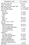
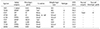
Demographic findings of the study are shown in Table 1. Mean age of patients was 63.2 and 70% of patients were male. All of IPMN were classified as main duct (n=64, 18.5%), branch duct (n=171, 49.4%), and mixed type (n=111, 32.1%). Forty-eight patients (13.9%) experienced recurrence during follow-up. Among these, 9 patients (3.0%) were identified to metachronous development of HRL in the remnant pancreas (Table 1).
Risk factors analysis for recurrences after initial surgery
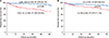
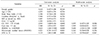
Patients with HGD/IC had significantly lower 5-year disease-free survival (5Y DFS) than among those with LGD/ IGD (5Y DFS 69.5% vs. 90.1%, p=0.008; Fig. 1A). Also, the patients with intestinal or pancreatobiliary subtype IPMNs had significantly lower DFS than those with gastric subtype IPMNs (5Y DFS 71.1% vs. 89.4%, p=0.001; Fig. 1B). After univariate analysis, high serum CA 19–9 level (HR 2.941, 95% CI 1.028– 8.416, p=0.048), cyst size larger than 3 cm (HR 2.568, 95% CI 1.035– 4.953, p=0.042), HGD/IC compared to LGD/IGD (HR 4.624, 95% CI 1.265– 14.002, p=0.008), and intestinal/ pancreatobiliary subtype compared to gastric subtype (HR 3.135, 95% CI 1.681– 5.848, p=0.001) were identified risk factor for recurrence. However, HGD/IC was only independent risk factor for recurrence (HR 3.688, 95% CI 2.124– 12.524, p=0.009) after multivariate analysis (Table 2).
Risk factors analysis for metachronous development of high risk lesion
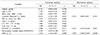
After uni- and multivariate analysis, the independent risk factors for metachronous development of HRL were HGD/IC (HR 8.414, 95% CI 4.310– 16.426, p=0.001), and intestinal/pancreatobiliary subtype compared to gastric subtype (HR 7.874, 95% CI 3.650– 27.027, p=0.010) (Table 3). All of patients details who developed metachronous HRL were described in Table 4.
DISCUSSION
It is now widely accepted that patients with IPMN who undergo partial pancreatectomy reportedly remain at risk of developing metachronous IPMN or concomitant PDAC.571112 Moreover, some patients have secondary IPMNs with HGD/IC, which is an indication for resection, although the initial surgical margins were free from neoplastic lesions.1113 However, the rate of new recurrence of metachronous IPMN following resection is difficult to determine from the literature,2 because MPD dilation in the distal pancreas following resection may be secondary to anastomotic stenosis or caused by true IPMN recurrence, and better imaging in the postoperative setting may reveal a previously undetected IPMN. Because of these limitations, the recurrence rates in the remnant gland were reported to be 0–20%,25111314 but the characteristics of patients harboring such lesions have not been well assessed.
In this study, intestinal or pancreatobiliary subtype is one of the independent risk factors for metachronous development compared to gastric subtype (Table 3). It is well known that intestinal subtype IPMNs can have invasive carcinoma with relatively indolent behavior.28 Also, pancreatobiliary type is regarded by some as an aggressive and high-grade version of the gastric type.2489 There was a similar report that partial pancreatectomy for pancreatobiliary subtype of IPMN was a predictor for the metachronous development of concomitant PDAC in the pancreatic remnant.611 However, Ideno et al.10 showed that IPMN having concomitant PDAC were frequently of gastric subtype even regarded that gastric type is typically low grade, with only a small percentage developing into carcinoma.249 As a result, to determine the relationship for risk of metachronous development and subtype of IPMN, the larger population study such as nationwide-based cohort study will be needed.
From the point of view that a metachronous lesion is a kind of multifocality, there is no convincing evidence that the risk of invasive IPMN multiplies according to the number of lesions. In one study, patients with unifocal BD-IPMN carried a higher risk than those with multifocal BD-IPMNs,15 whereas another reported a higher rate of IC or HGD in multifocal BD-IPMNs.16 Also, Miyasaka et al.11 reported that initial HGD/IC was an independent predictive factor for metachronous HGD/IC in the remnant pancreas. In this study, HGD/IC is one of the independent risk factors for metachronous development of HRL (Table 3). As a result, we think HGD/IC indicate that close attention should also be paid to the remnant pancreas for the possible development of metachronous HRL.
In this study, type of IPMN, such as, main duct or mixed type, was not a risk factor for metachronous development (Table 3). However, a recent molecular analysis demonstrated the possibility of monoclonal skip implantation in main duct type IPMN could cause potential metachronous lesions.17 Also, Kang et al.5 reported that the rate of recurrent IPMN in the remnant pancreas requiring surgical treatment was higher in main duct type than in branch duct type IPMNs. To find out this discrepancy, we have a plan to perform nationwide multicenter study to identify relationship between the type of IPMN and metachronous development.
Although invasive IPMN may be associated with a poor prognosis, the long-term outcome after resection of most cases of IPMN is globally better than that of conventional PDAC. In this study, Patients with HGD to IC had a significantly higher rate of recurrence (Table 2) and a lower DFS rate (Fig. 1A) than patients with LGD to IGD in agreement with previous results.518 There were some studies that the subtype of IPMN appears to be an independent predictor of patient prognosis that intestinal or pancreatobiliary type IPMN showed poor survival than gastric type IPMN.48 However, in this study, the subtype of IPMN was even correlated with the DFS rate (Fig. 1B), but multivariate analysis revealed that there was no correlation between the subtype of IPMN and recurrence rate (Table 2).
The decision to follow an IPMN is a matter of clinical judgment based on the patient age, family history, symptoms, comorbidities, perceived pancreatic cancer risk, and patient preference. There is still controversy in the literature to guide the frequency and type of surveillance for IPMNs.1234 American Gastroenterological Association guidelines3 in 2015 restricted indications for surgery more stringently and recommended physicians to stop surveillance if no significant change had occurred in a pancreatic cyst after five years of surveillance, or if a patient underwent resection and a nonmalignant IPMN was found. However, there are little good long-term data to indicate whether surveillance can be safely discontinued after long-term stability.2 On the contrary, most of studies suggested that long-term surveillance in patients with IPMN should be necessary and important because of the potential for secondary or recurrent development of HRL, such as concomitant PDAC.245711 These metachronous lesions often develop more than 5–10 years after initial operation and the cumulative 5-year incidence of the development of concomitant HRL ranges from 2.2 to 8.8%.45671112192021 In this study, one patients experienced metachronous HRL after 96.8 months from initial distal pancreatectomy (Table 4). As a result, we think surveillance should continue as long as the patient remains fit for surgery, especially for patient with HGD/IC, intestinal/pancreatobiliary subtype of IPMN.
This study has some limitation. At first, we identified 9 metachronous HRL after initial surgery. Only one papatient received secondary completion total pancreatectomy who revealed with IC. The other 8 patients were strongly suspicious for HRL, such as, main pancreatic duct dilatation or enhancing mural nodule in CT or MRI but not conformed histologically because of unfit or refusal for secondary surgery. At second, we excluded oncocytic subtype for analysis as risk factor for metachronous development because of very small population (n=5, Table 1) and no patient with oncocytic subtype developed metachronous lesion (Table 4). A larger study population is necessary to clarify the relationship between oncocytic subtype and metachronous development.
In conclusion, the risk of recurrence and metachronous development of high risk lesion does not diminish over time following resection, and surveillance should continue indefinitely as long as the patient remains fit for surgery. Also, special attention should be paid to the remnant pancreas in case of initial pathology with high grade dysplasia/ invasive carcinoma, and intestinal/ pancreatobiliary subtype.
 XML Download