

 PDF
PDF ePub
ePub Citation
 Print
Print


INTRODUCTION
Various gallbladder (GB) diseases are characterized by generalized or localized wall thickening of the GB on computed tomography (CT) or ultrasound (US), including gallbladder adenomyomatosis (GBA), chronic cholecystitis, GB polyp, and early-stage, wall-thickening-type gallbladder cancer (GBC).12 Among them, the differentiation GBA from GBC is still required because of the similarity in appearance, despite some reports being published concerning their imaging findings using US, CT, and magnetic resonance imaging (MRI) since 1981.23 In this regard, Ching et al.2 reported that the differential diagnostic performance of contrast-enhanced CT for GBA and GBC showed 30% sensitivity and 93% specificity. MRI imaging is known to be useful because it can sensitively depict the pearl necklace sign, which is pathognomonic of adenomyomatosis and directly indicates the presence of Rokitansky-Aschoff sinuses in the thickened wall.45 Recently, Joo et al.6 reported that high-resolution ultrasound (HRUS) and MRI with MR cholangiopancreatography have comparable sensitivity and accuracy.
Although there have been significant advances in diagnostic imaging technology that can be helpful for distinguishing GBA from early-stage GBC,789 some issues remain unsolved. As mentioned above, MRI, one of the most useful diagnostic tools, is expensive and requires patients to hold still for long periods of time. HRUS, while offering the ability to overcome the drawbacks of conventional US, is not yet widely available except in large general hospitals.
In order to accurately distinguish between the two conditions, it is necessary to analyze the perioperative demographic and imaging data for diagnostic purposes. Therefore, this study aimed to compare perioperative and clinical outcomes in patients undergoing laparoscopic cholecystectomy for GBA or early-stage GBC to evaluate the diagnostic performance of differences in preoperative demographics and imaging findings between the two conditions.
MATERIALS AND METHODS
Patients
This retrospective study was approved by our institutional review board and the requirement for informed consent was waived. Between January 2011 and December 2017, a total of 2389 patients underwent laparoscopic cholecystectomy at a hospital, including 194 diagnosed with GBA (GBA group) and 30 diagnosed with GBC (GBC group). No patients had both adenomyomatosis and cancer and patients were assigned to separate groups according to the pathological result. We performed a retrospective analysis of the perioperative and clinical outcomes of 224 consecutive patients.
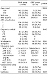
The GBA group (n=194) consisted of 110 males and 84 females with a mean age of 50.5±14.0 years (range 17–86 years). The mean body mass index (BMI) was 23.9±3.4 kg/m2 (range 16.2–38.6 kg/m2). Laparoscopic cholecystectomy was performed because of gallstone-related symptoms in 122 patients and gallbladder wall thickening in 72 patients.
The GBC group (n=30) consisted of 18 male and 12 female patients with a mean age of 66.6±10.2 years (range 45–86 years). The mean BMI was 24.0±3.9 kg/m2 (range 15.8–32.8 kg/m2). In terms of TNM staging by the American Joint Committee on Cancer (8th edition), all tumors of the GBC group were classified as T1a or T1b. The tumor was located in the fundus in 9 patients, in the body in 12 patients, in the neck in 3 patients, and in the entire gallbladder in 5 patients.
Statistical analysis
Results are presented as mean and standard error of the mean. Patient demographics and clinical characteristics were compared using the χ2 test or Fisher exact test for categorical variables and t-test or Mann-Whitney test for continuous variables, as appropriate. In assessing risk factors associated with GBC, only variables statistically significant by univariate analysis were included in the multivariate analysis, which was performed using logistic regression. All statistical analyses were performed using SPSS, version 21.0 (IBM, Armonk, NJ), with p-values < 0.05 considered statistically significant.
RESULTS
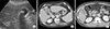
In most patients, preoperative diagnostic examination made it difficult to differentiate gallbladder diseases with wall thickening and the preoperative diagnostic images were inaccurate when compared to postoperative pathology reports (Fig. 1). The demographic and perioperative findings of both groups are listed in Table 1. There were no significant differences in sex (1.0:0.8 vs. 1.0:0.7, male: female, p=0.734), BMI (23.9±3.4 vs. 24.0±3.8 kg/m2, p=0.916), or preoperative liver function tests between the GBA and GBC groups. The GBC group was significantly older (50.5±14.1 vs. 65.9±10.6 years, p<0.001) and had a higher ASA grade (40.3 vs. 63.4, grade II or III (%), p=0.043) than the GBA group. The operation time of the GBC group was significantly longer than that of the GBA group (p=0.036) and there was a significant difference in the rate of cases with gallstones between 2 groups (68.6% vs. 40.0%, p=0.004). Although there was no significant difference in preoperative diagnostic methods (p=0.442), the GBC group showed a significantly higher rate of disparity between preoperative imaging and postoperative histopathological findings (30.9% vs. 53.3%, p=0.011).
DISCUSSION
According to the Korea Central Cancer Registry's annual report of 2016, as published by Korean Ministry of Health and Welfare, GBC and other biliary tract cancers account for 2.9% of all cancers in Korea.10 GBC is silent during the early-stage and remains asymptomatic until it gets to an advanced and unresectable stage. Therefore, early diagnosis and treatment of GBC is very important.
Inflammatory or obstructive GB changes may induce GBA at 2–5% of prevalence in any cholecystectomy specimen.1112 In surgery for pain aggravation or other symptoms, accurate differential diagnosis between GBA and GBC is a major factor for choosing the adequate treatment. Although GBA has not been considered to have malignant potential, several reports have suggested a relationship between GBA and GBC.21314 Kai et al.11 reported GBC was associated with GBA in 25% of cases. Additionally, patients with GBC and GBA presented with a more advanced TNM stage than those without GBA. Given the differences in prognosis according to the TNM stage of GBC, preoperative differential diagnosis between GBA and GBC is indispensable to avoid nefarious consequences.
Despite the technical advances in imaging modalities (HRUS, multi-detector CT, and MRI), it is still difficult to distinguish between GBA and GBC before surgical resection. According to Ching et al. at 2007, the differential diagnostic performance of CT for GBA and GBC was 30% sensitivity and 93% specificity.2 However, Bang et al. found improved values of 50% sensitivity and 98.2% specificity.15 The improvement in diagnostic performance of US is most likely the result of the technological advances which have been utilized since 2000, such as harmonics, compounding techniques, and speckle reduction. In a previously published study, the diagnostic performance of HRUS was equivalent to that of MRI for differentiating GBA from GBC.15 The presence of either intramural echogenic foci or cystic spaces, which indicate cholesterol crystals/stones or bile within the pathognomonic Rokitansky-Aschoff sinuses, respectively, had a sensitivity of 80.0%, specificity of 85.7% and accuracy of 82.2% for the diagnosis of GBA on HRUS.616 MRI may be superior to HRUS for the depiction of intramural cystic spaces. As shown above, multiple imaging modalities would be helpful for evaluating and choosing treatment strategies since each modality has different advantages.
As with most other epithelial cancers, there is a strong relationship between age and gallbladder cancer.17 In this study, 76.7% of patients in the GBC group were over 60 years old; patients in this group were also significantly older than those in the GBA group. The absence of cholelithiasis was an independent risk factor for GBC. The association between GBA and gallstones ranges from 36 to 95%, and gallstones have been also found to be associated with GBC in varying frequency.18 In this study, the GBA group showed a significantly higher rate of presence of gallstones compared to the GBC group (68.6 vs. 40.0%, p=0.004). If gallstones are absent in patients with an unclear distinction between GBA and GBC on preoperative imaging, the presence of GBC may be considered.
The study has some limitations. First, this was a retrospective study. Thus, it was difficult to determine the exact diagnosis of patients and surgical plan. However, this study included only patients who underwent laparoscopic cholecystectomy for early stage GBC; thus, we consider that the selection bias associated with retrospective studies was minimized. Second, patients enrolled in this study did not undergo a variety of diagnostic tests. More specifically, no patients underwent preoperative EUS in the GBC group, no matter how few patients in that group (n=30). Therefore, in many cases, the preoperative diagnosis was different from that after surgery.
In conclusion, this study suggests that the possibility of early-stage GBC should be considered in older patients hospitalized for biliary colic without gallstones but with a thickened gallbladder wall with inflammation on preoperative examination.
 XML Download