

 PDF
PDF ePub
ePub Citation
 Print
Print


Abstract
Purpose
This study is to evaluate orbital rim uptake on bone scan and to discuss their clinical significance.
Materials and Methods
From January 2011 to August 2013, 3149 cases of bone scans were analyzed to check for existence of uptake abnormalities in the orbital rim with relative size and position. The bone scans were compared with either positron emission tomography-computed tomography (PET/CT) or computed tomography (CT). For cases without other imaging examinations, comparisons were made with other bone scans.
Results
In total, 13 cases of the orbital rib uptake were ultimately evaluated. In 6 cases, the intake abnormalities of the orbital rim appeared in superior lateral aspect of the orbital rim to occupy the highest frequency (46.2%). Distinctively, bone scans showed no abnormal uptake in medial and inferior aspect of orbital rim. The 10 cases are compared with PET/CT or CT and as a result, there are no abnormalities that correspond to the orbital lesions of bone scans. The 3 cases were compared with other bone scans and no changes in the orbital lesions were confirmed between the bone scans.
Go to : 
References
1. Moon DH, Ryu JS. Recent advances in musculoskeletal nuclear medicine.J Korean Med Assoc. 2003; 46:196–203.
2. Sohn MH. Normal variants and artifacts in bone scan: potential for errors in interpretation.Korean J Nucl Med. 2004; 38:1–20.
3. Thang SP, Tan AE, Goh AS. Bone scan “Hot Spot” at the superior lateral orbital margin fronto-zygomatic suture uptake characterized with Tc-99m MDP SPECT/CT.World J Nucl Med. 2011; 10:139–140.
4. Swie˛taszczyk C, Pilecki SE. Enhanced accumulation of bone seekers at superior lateral orbital margin: potential origin.World J Nucl Med. 2014; 13:3–5.
5. Senda K, Itoh S. Evaluation of diffusely high uptake by the calvaria in bone scintigraphy.Ann Nucl Med. 1987; 1:23–26.
6. Suematsu T, Yoshida S, Motohara T, Fujiwara H, Nishii H, Komiyama T, et al. [Diffusely increased uptake in the skull in normal bone scans]. Kaku Igaku. 1992; 29:599–605.
7. Jacobsson H, Haverling M. Hyperostosis cranii. Radiography and scintigraphy compared.Acta Radiol. 1988; 29:223–226.

8. Ceylan E, Caner B. Evolution of hyperostosis frontalis interna butterfly sign demonstrated by bone scan. Clin Nucl Med. 2003; 28:78–79.
9. Novetsky GJ, Berlin L. Bone scintigraphy in hyperostosis frontalis interna. Clin Nucl Med. 1982; 7:265–266.
10. Harbert J, Desai R. Small calvarial bone scan foci–normal variations. J Nucl Med. 1985; 26:1144–1148.
Go to :
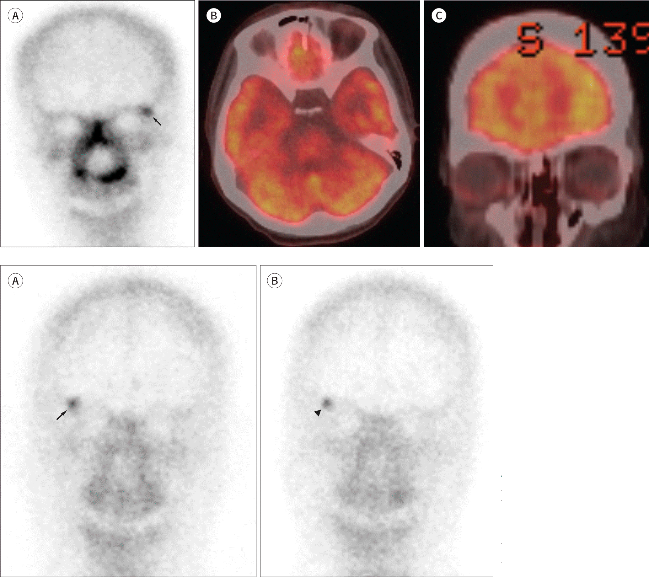 | Fig. 1.A 48-year-old female with nasal septal epithelial-myoepithelial carcinoma. A. Anterior view of the bone scan shows discrete focal activity in the left superior lateral margin of the orbital rim (arrow). B, C. Axial and coronal fused positron emission tomography-computed tomography images demonstrate no corresponding abnormal uptake in the left superior lateral margin of the orbital rim. |
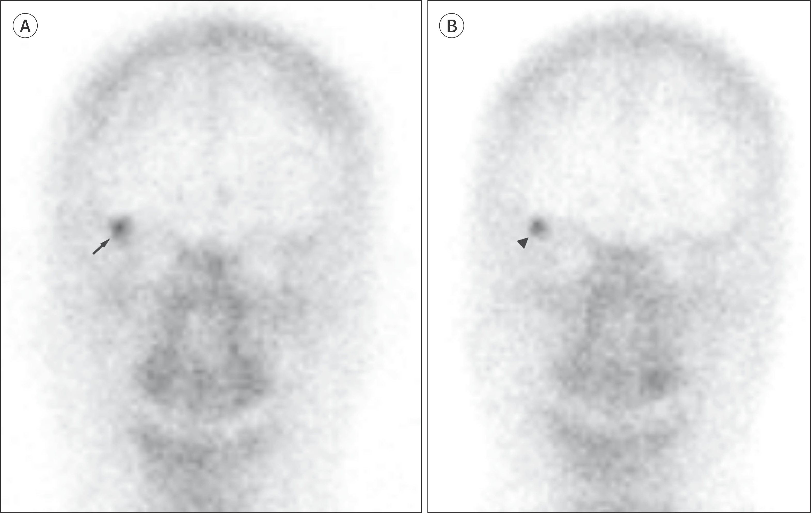 | Fig. 2.A 32-year-old male with urinary bladder cancer. A. Anterior view of the bone scan shows discrete focal activity in the superior lateral margin of the orbital rim (arrow). B. The bone scan performed at the 2-year follow up demonstrates no remarkable change over time (arrowhead). |
Table 1.
Characteristics of Patients and Patterns of Orbital Rim Uptake in Bone Scans
 XML Download