Abstract
Gastrointestinal tract involvement in undifferentiated pleomorphic sarcoma (UPS) is extremely rare. To the authors' knowledge, only 21 cases of primary small bowel UPS have been reported in the literature available in English. Reported CT findings in primary small bowel UPS have been nonspecific, and MRI findings have been reported in only one case. The present article describes a case involving a 72-year-old male with histologically confirmed primary UPS arising from the ileum, focusing on both CT and magnetic resonance enterography findings. On CT, primary small bowel UPS was noted as a heterogeneously enhanced small bowel mass without obstruction. Magnetic resonance enterography revealed heterogeneous intermediate T1 and T2 signal intensity, with hemorrhagic or necrotic foci within the mass and heterogeneous enhancement. The differential diagnosis included malignant gastrointestinal tumor; however, the prognosis of UPS is worse, with higher incidences of extra-abdominal metastasis.
Go to : 
References
1. Karki B, Xu YK, Wu YK, Zhang WW. Primary malignant fibrous histiocytoma of the abdominal cavity: CT findings and pathological correlation. World J Radiol. 2012; 4:151–158.

2. Kim YR, Lee YH, Yoon KH, Yun KJ. CT findings of primary undifferentiated pleomorphic sarcoma in the small bowel: a case report. J Korean Soc Radiol. 2015; 73:323–327.

3. Giuliani J. Primary malignant fibrous histiocytoma of Vater's Papilla: first reported case.J Gastrointest Cancer. 2013; 44:366–367.
4. Kobayashi K, Narita H, Morimoto K, Hato M, Ito A, Sugiyama K. Primary malignant fibrous histiocytoma of the ileum: report of a case. Surg Today. 2001; 31:727–731.

5. Meyers SP.MRI of bone and soft tissue tumors and tumorlike lesions: differential diagnosis and atlas. Stuttgart and. New York: Thieme;2008.
6. Amzallag-Bellenger E, Oudjit A, Ruiz A, Cadiot G, Soyer PA, Hoeffel CC. Effectiveness of MR enterography for the assessment of small-bowel diseases beyond Crohn disease.Radiograph/iics. 2012; 32:1423–1444.
7. Kroep JR, Bovée JV, van der Molen AJ, Hogendoorn PC, Gelderblom H. Extra-abdominal subcutaneous metastasis of a gastrointestinal stromal tumor: report of a case and a review of the literature.J Cutan Pathol. 2009; 36:565–569.
8. Tran T, Davila JA, El-Serag HB. The epidemiology of malignant gastrointestinal stromal tumors: an analysis of 1458 cases from 1992 to 2000. Am J Gastroenterol. 2005; 100:162–168.

9. Wang ZS, Xiong CL, Zhan N, Xiong GS, Li H, Hu H. Primary malignant fibrous histiocytoma of the small bowel: a report of an additional case in duodenum. Int J Gastrointest Cancer. 2005; 36:105–112.

10. Katsourakis A, Noussios G, Hadjis I, Evangelou N, Chatzitheoklitos E. Primary malignant fibrous histiocytoma: a rare case.Case Rep Med. 2011; 2011:134801.
Go to : 
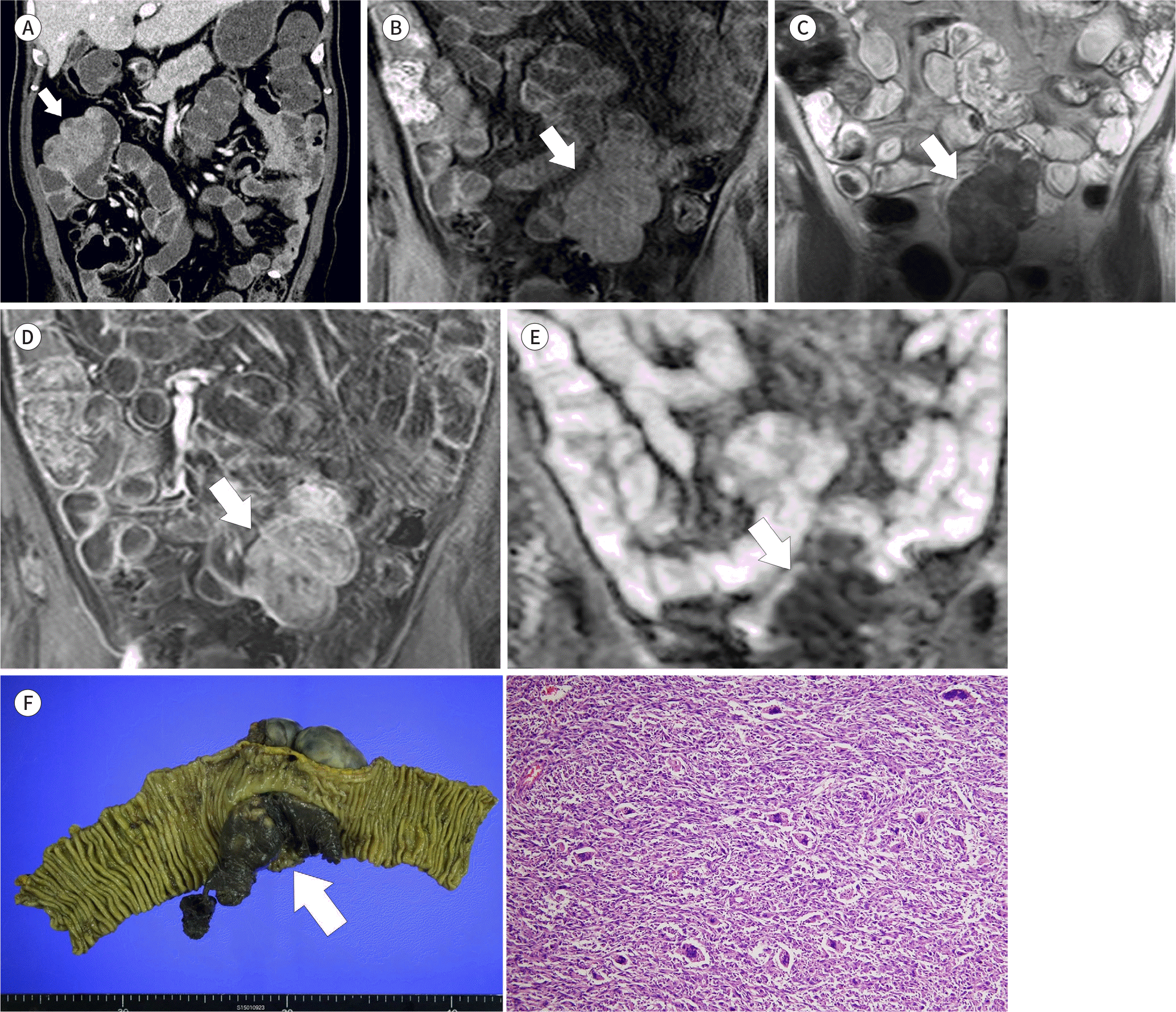
 | Fig. 1.Primary undifferentiated pleomorphic sarcoma of the small bowel in a 72-year-old male patient. A. CT enterography revealing a mass 6.8 cm in size at the ileum, exhibiting endoluminal and exoluminal growth. On the portal phase, the mass exhibits heterogeneous enhancement (arrow). B. On T1WI in MR enterography, the mass exhibited heterogeneous intermediate signal intensity (arrow). C. On T2WI, the mass exhibited heterogeneous intermediate signal intensity with T2 hyperintense foci (arrow). D. On T1WI (fat suppressed), 70 s after intravenous contrast administration, the mass exhibited heterogeneous enhancement with non-enhancing foci (arrow). E. On the ADC map, the mass exhibited diffuse restriction with a low ADC value (arrow). F. On gross examination, a 12.4 × 7.9 × 3.8 cm exophytic soft mass (arrow) was identified in the small intestinal wall. Microscopic examination (hematoxylin and eosin, × 100) revealed solid sheets of malignant tumor cells, which are mixture of spindle cells with vague fascicular pattern, pleomorphic cells, and a considerable number of bizarre multinucleated tumor giant cells. ADC = apparent diffusion coefficient, WI = weighted imaging |
Table 1.
Previously Reported CT Findings of Small Bowel Undifferentiated Pleomorphic Sarcoma in the English Literature


 PDF
PDF ePub
ePub Citation
 Print
Print


 XML Download