

 PDF
PDF ePub
ePub Citation
 Print
Print


INTRODUCTION
Despite its decreasing incidence, gastric cancer is the fifth most common cancer and the third leading cause of cancer-related death worldwide.1 In particular, its incidence in East Asia, including Korea and Japan, is the highest in the world.2 According to the Korea Central Cancer Registry, 30,504 gastric cancer cases were diagnosed in 2016, making gastric cancer the most common cancer (age-adjusted incidence rate: 34.0 per 100,000). In men, it was the most common cancer (age-adjusted incidence rate: 49.6 per 100,000), and the incidence was high in those between the ages of 35 to 64 (age-adjusted incidence rate: 85.6 per 100,000).3 The prognosis of gastric cancer depends on many factors, including the depth of invasion, nodal involvement, and distant metastasis.4 Among them, the stage of gastric cancer is the most important prognostic factor. Early gastric cancer (EGC) usually has a good prognosis with a 5-year survival rate of more than 90%. Therefore, it is very important to detect gastric cancer in the early stages to allow for earlier treatment and to cure this disease.5
Population-based screening for gastric cancer has been implemented in several countries with a high incidence of gastric cancer. On the other hand, the recommended screening methods, age, and intervals vary.6789 Based on the fact that gastric cancer is rare before the age of 40 years, the Korean National Gastric Cancer Screening Program recommends that upper endoscopy be performed biennially for people aged ≥40 years.9 On the other hand, the incidence of gastric cancer in patients younger than 40 years of age has been increasing in western countries over the past two decades. In particular, this tendency is prominent in patients 30 to 39 years of age.10 Several reports have suggested that younger patients are frequently diagnosed with advanced-stage tumors and have poorer prognoses than older patients.11 Genetic factors and late diagnosis have been proposed as the main causes of the poorer prognoses.12131415 Therefore, proper screening and early management may be important for this age group. This study examined whether the early diagnosis of gastric cancer by screening gastroduodenoscopy in individuals <40 years of age is helpful by comparing the characteristics of gastric cancer in young patients whose disease was detected at a health checkup with those whose disease was detected by gastroduodenoscopy, which was undertaken because of symptoms.
SUBJECTS AND METHODS
1. Patients
For this retrospective study, this study enrolled 84 patients diagnosed with gastric cancer before 40 years of age at three tertiary centers (Dongguk University Ilsan Hospital, Seoul National University Boramae Hospital, and Seoul National University Bundang hospital) from May 2007 to February 2017. The medical records were analyzed retrospectively to evaluate the patient characteristics, including smoking status, family history of gastric cancer (at least a first-degree relative with gastric cancer), Helicobacter pylori (H. pylori) infection status, laboratory findings, stage at the time of diagnosis, cancer location, pathologic findings, and clinical outcomes. The patients were classified into two groups according to the purpose of the upper endoscopy, which is a health checkup regardless of symptoms (screening group) or diagnosis of symptoms, including abdominal pain, dyspepsia, or upper gastrointestinal bleeding (diagnostic group). The admission notes were reviewed to confirm whether the purpose of endoscopy was screening or diagnosis. Patients were classified as the screening group if they were diagnosed with gastric cancer at the outpatient department with simple dyspepsia and the purpose of endoscopy was screening. Although they were diagnosed with gastric cancer in screening endoscopy, patients who had symptoms, including dyspepsia at that time were classified as the diagnostic group. In addition, an older patient group was formed by one- to two-stage matching to compare the characteristics of young and older patients (aged 40–50 years) diagnosed with gastric cancer by screening endoscopy in the same year. The study protocol was approved by the Institutional Review Board of each hospital (IRB no. DUIH 2014-119).
2. Classification and staging of gastric cancer
All gastric cancers were categorized pathologically as EGC or advanced gastric cancer (AGC). EGC is, by definition, confined to the mucosa and submucosa regardless of the tumor size or regional lymph node metastasis. In AGC, the tumor invades beyond the submucosa. The morphological classification of EGC was categorized according to the Japanese classification of gastric carcinoma. AGC was classified according to the Borrmann classification.16 The location of the cancer and the depth of invasion were also described.16 Histopathologically, gastric cancers were classified as well, moderately, and poorly differentiated tubular or papillary adenocarcinomas and signet ring cell carcinomas according to the Japanese classification of gastric carcinoma.16 In addition, the histological subtypes of gastric cancers were classified as intestinal and diffuse types according to Lauren's criteria.17 Cancer was staged according to the TNM classification based on the final pathology, abdominal CT, or endoscopic ultrasound. The 8th American Joint Committee on Cancer TNM staging classification for carcinoma of the stomach was used to determine the cancer staging.18 A H. pylori infection was defined as a positivity of at least one test among the rapid urease test, urea breath test, and histology for H. pylori.
3. Statistical analysis
The sequential data are expressed as the median and interquartile range. The continuous variables were compared using a Mann-Whitney U test or Student's t-test as appropriate. The categorical variables were compared using a Pearson's χ2 test or Fisher's exact test. The survival curves were calculated using the Kaplan-Meier method. Statistical analyses were conducted using IBM SPSS Statistics software for Windows v.22.0 (IBM Corp., Armonk, NY, USA). All tests were two-sided and statistical significance was considered when the p-value was <0.05.
RESULTS
1. Baseline characteristics of the study population
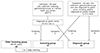
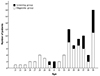
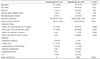
In this study, 84 patients were enrolled. Twenty-three patients <40 years of age with gastric cancer were classified into the screening group. Fourteen patients were diagnosed with gastric cancer in three tertiary health checkup centers. The estimated prevalence of gastric cancer in patients younger than 40 years in screening endoscopy was 7.2 per 100,000 (14/195,711). One patient was transferred to another hospital by the patient's direction. In outpatient clinics, 12 patients diagnosed with gastric cancer during screening endoscopy. Among them, two patients were transferred to other hospitals. As a result, 23 were included in the screening group. An additional 61 patients who had visited the hospital because of symptoms were also diagnosed with gastric cancer (Fig. 1). The male to female sex ratio was 0.83, and the median age of the study population was 35 years (range, 21 to 39 years) (Fig. 2). In total, 48 (57.1%) gastric cancer patients had ages ranging from 35 to 39, and 22 (26.2%) patients were 30 to 34 years of age. Fourteen (16.7%) patients were younger than 30 years of age; all of these patients belonged to the diagnostic group. The study population was comprised of 36 cases of EGC and 48 cases of AGC. The median follow-up period was 16.1 months (interquartile range 6.5-38.4). Pathologically, there were 40 cases of adenocarcinoma and 42 cases of signet ring cell carcinoma (well differentiated carcinoma, 5; moderately differentiated carcinoma, 13; poorly differentiated adenocarcinoma, 22; signet ring cell carcinoma, 42). The most common stage was stage 1A (31 cases, 36.9%), followed by stage 4 (24 cases, 28.6%) (Table 1).
2. Comparison of the clinical characteristics of the screening and diagnostic groups
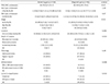
Abdominal pain was the most common symptom in the diagnostic group (34 cases, 40.5%). No patients had underlying diseases except for two patients who had type 2 diabetes. Three patients had a familial history of gastric cancer (one case in the screening group, and two cases in the diagnostic group). The median age of the screening group was significantly higher than that of the diagnostic group (37 vs. 35 years, p=0.03). The ratios of patients with normal hemoglobin and hematocrit were significantly lower in the diagnostic group (47.5% vs. 73.9%, p=0.048, 42.6% vs. 73.9%, p=0.010). The ratio of patients with a high CEA level was higher in the diagnostic group (17.9% vs. 0%, p=0.040). No significant differences were found in the other laboratory findings. The proportion of EGC in the screening group was significantly higher than that of the diagnostic group (78.3% vs. 29.5%, p<0.01). The number of patients who received chemotherapy was significantly higher in the diagnostic group (13.0% vs. 41.0%, p<0.01). The proportion of patients who underwent a curative resection was significantly higher in the screening group (95.7% vs. 52.5%, p<0.01). During the follow-up period, 21 deaths were observed, which all occurred in the diagnostic group (Table 2). All 21 deaths were due to gastric cancer. The 5-year estimated mortality rate was significantly lower in the screening group than in the diagnostic group (0% vs. 48.9%, p<0.01).
3. Comparison of the pathologic characteristics of the screening and diagnostic groups
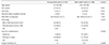
Among the 36 cases of EGC, the depressed type (IIc) was the most common type, regardless of the presence of symptoms. Among the 48 cases of AGC, the ulceroinfiltrative type (Borrmann type 3) was most common. The stage at the time of diagnosis was significantly earlier in the screening group than in the diagnostic group (p<0.01), and the proportion of EGC was significantly higher (78.3% vs. 29.5%, p<0.01). The depth of invasion of primary cancer in the diagnostic group was significantly deeper than that in the screening group (p=0.010), and poorly differentiated carcinoma was more common (56.5% vs. 83.6%, p=0.006). The location and pathological type were similar in the two groups (Table 3).
4. Comparison of the characteristics of young and older patients diagnosed with gastric cancer with a screening endoscopy
A one to two stage-matched older patient group (aged 40–49 years) was formed to compare the characteristics between young and older patients diagnosed with gastric cancer with a screening endoscopy. The young and older groups showed a similar smoking status, family history of gastric cancer, and survival. The ratio of patients with poorly differentiated adenocarcinoma or signet ring cell carcinoma was higher in the young patients (56.5% vs. 32.6%, p=0.072). Diffuse type cancer was more prevalent in the young screening group (69.6% vs. 23.9%, p=0.002) (Table 4).
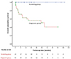
5. Comparison of the overall survival in the screening and diagnostic groups
Fig. 3 presents a schematic diagram of the overall survival of patients according to the purpose of their endoscopy. The overall 1-, 2-, 3-, and 5-year survival rates for the screening group were higher than those of the diagnostic group (100% vs. 77.1%, 100% vs. 72.1%, 100% vs. 67.2%, and 100% vs. 51.1% respectively, p<0.01).
DISCUSSION
This retrospective study evaluated the role of screening gastroduodenoscopies in young patients indirectly by comparing the screening and diagnostic groups, which differed in the purpose of endoscopy. To the best of the authors' knowledge, this is the first report of gastric cancer in young patients detected during a health checkup. These results show that young patients aged <40 years, who were diagnosed with gastric cancer after a screening gastroduodenoscopy, tended to be diagnosed in the earlier stages than those who underwent upper endoscopy because of symptoms. In addition, this group showed a significantly longer survival than the symptomatic group.
Previous studies have investigated the features of gastric cancer in young patients with gastric cancer. The definition of young onset gastric cancer is controversial, but it is usually defined as gastric cancer diagnosed in patients <40–45 years of age.19202122 A Mexican study that evaluated gastric cancer in patients under 30 years of age from 1985 to 2006 reported a tendency towards a late diagnosis of gastric cancer in young patients.12 Almost all cancers were already at an advanced stage at the time of diagnosis, and 25 patients (83%) were diagnosed with stage 4 disease.12 On the other hand, this study enrolled only symptomatic patients. Therefore, it is difficult to apply this result to asymptomatic young patients. In the present study, the survival rate of asymptomatic patients was significantly higher than that of symptomatic patients. A European study comparing the clinicopathologic features of young gastric cancer patients with older patients >45 years old, showed that young-onset gastric cancer tends to be more advanced, but the prognosis is similar to that of older patients.23 In the younger age group, diffuse type carcinoma (73%), lymph node metastasis (59%), and stage IV disease (49%), and non-curative treatment (36%) are more prevalent than in older patients. These results are similar to the results of a diagnostic group in the present study.
In a retrospective Korean study, the prognosis of young patients (<40 years old) with early gastric carcinoma was similar to that of older patients with EGC. They insisted that the belief that the prognosis of young gastric cancer patients is poorer than that of aged patients should be abandoned and that early detection is critical to achieve a good prognosis.24 Another study reported that young gastric cancer patients (<40 years old) present with poorly differentiated cancer and with more metastatic and advanced disease.25 In addition, they concluded that the patient's health status, tumor localization, smoking status did not confer a significant difference in the disease state, which coincides with the present data showing a poorly differentiated histology in 52% of the study population. Another Korean study revealed the predominance of female patients and histologically undifferentiated carcinoma.20 In the present study, there was no difference in sex and location of the primary cancer between the screening and diagnostic groups, and adenocarcinoma and signet ring cell carcinoma showed a similar incidence. On the other hand, poorly differentiated adenocarcinoma was more common in the diagnostic group than the screening group (p<0.01).
In terms of screening an upper endoscopy for gastric cancer, a Korean study previously reported the characteristics of gastric cancer diagnosed at a health screening.26 EGC represented 81 out of 111 cases (73%) in this study. The mean overall survival was 103 months and the cumulative probability of survival at 5 and 10 years was 82.7% and 67%, respectively. Five patients (4.5%) under the age of 40 years were diagnosed with gastric cancer. Another study showed that annual endoscopic screening in a region with a high incidence of gastric cancer improved the detection of early-stage and endoscopically treatable gastric cancer.27 A recent large-scale Korean study, using nationwide screening program results, reported that gastric cancer screening with upper endoscopy reduced gastric cancer mortality.28 A recent meta-analysis of observational studies of gastric cancer mortality reduction after endoscopic screening suggested that patients might achieve a 40% reduction in gastric cancer mortality after endoscopic screening but screening did not reduce incidence in Asian countries, including South Korea.29 These results support the role of screening endoscopy for gastric cancer in individuals aged ≥40 years.
A comparison of the characteristics of a young screening group in this study with stage-matched gastric cancer patients aged 40 to 49 years diagnosed by screening endoscopy revealed similar characteristics, including the proportion of EGC and survival, except for the Lauren's classification. Previous studies reported that gastric cancer at a young age is more frequently associated with diffuse and undifferentiated histology types.30313233 In the present study, diffuse type cancer was more prevalent in the young screening group. The ratio of patients with poorly differentiated adenocarcinoma or signet ring cell carcinoma tended to be higher in the young patients, which was not statistically different. The lack of significant differences might be due to the relatively small number of patients in the study population.
A higher ratio of patients with lower serum hemoglobin or hematocrit levels in the diagnostic group may be related to their advanced disease. When this study was designed, it is believed that the serum CEA and CA 19-9 levels in the screening group would be different from those of the diagnostic group. Patients with abnormal CEA levels were more prevalent in the diagnostic group and the CA 19-9 levels tended to be higher in the diagnostic group.
This study had several limitations. First, this study was conducted as an observational, retrospective study. Therefore, biases, such as length-time bias and lead-time bias, could not be controlled. Slow progressive cancers are more likely to be detected in screening endoscopy. Therefore, these are likely to be classified as the screening group, and show a better prognosis. In contrast, rapidly progressive cancer tends to cause symptoms rapidly and is unlikely to be found during screening endoscopy; these were classified as a diagnostic group. In the present study, the ratios of patients with poorly differentiated carcinoma or signet ring cell carcinoma and diffuse type cancer, which are characterized by rapid progression and a poorer prognosis,34 were significantly lower in the screening group than in the diagnostic group. Regarding the lead-time bias, patients diagnosed at the early stage during screening endoscopy may show increased survival without affecting the course of the disease. Therefore, a well-designed, randomized controlled trial is needed to prove the effectiveness of screening endoscopy for survival gain in young patients. Second, this was a retrospective study using medical records. Therefore, data, such as the underlying disease, symptoms at the time of diagnosis, and familial history could be inaccurate. In addition, the patients were classified into two groups according to the purpose of endoscopy based on the medical records. Hence, misclassification bias could occur. Third, only a small number of patients were included in the screening group. In addition, this study included patients who were referred to the three territory centers from other clinics with abnormal endoscopy finding suggesting gastric cancer. Therefore, it was impossible to calculate the exact incidence of gastric cancer. Future large-scale, prospective studies should resolve these limitations.
In conclusion, asymptomatic patients aged <40 diagnosed with gastric cancer by screening endoscopy may allow a downward migration of the stage, and in some populations, screening endoscopy could result in a better prognosis compared to those by diagnostic endoscopy for symptoms. A well designed, multi-center, large-scale study for the effects of screening endoscopy in individuals aged <40 will be needed.
 XML Download