

 PDF
PDF ePub
ePub Citation
 Print
Print


INTRODUCTION
Dentoalveolar protrusion is commonly treated by premolar extraction followed by anterior retraction to achieve a harmonious facial profile and an adequate incisor relationship. The applied biomechanical system should ensure adequate retraction of the anterior teeth for proper function, esthetics, and stability. In severe protrusion cases, maximum anchorage is often required. For anterior retraction, miniscrew anchorage was reported to be more effective than conventional methods for anchorage reinforcement.1 En masse anterior retraction is often performed using sliding mechanics with the retraction force applied from a hook crimped or soldered to the archwire and interdental miniscrews. In the presence of a rigid continuous archwire, the line of action of the retraction force relative to the center of resistance of the entire arch will dictate the displacement pattern of the dentition.2 The retraction force at the archwire level will result in rotation of the entire arch around the center of resistance, producing a tendency toward posterior open bite, anterior deep overbite with lingual inclination of the anterior segment, and steepening of the occlusal plane.3
To avoid this problem, the “biocreative therapy technique” was introduced by Chung et al.4 In this technique, brackets are bonded only on the six anterior teeth, and temporary anchorage devices (TADs) are used as the only source of anchorage. To provide bodily tooth movement during retraction, anterior torque moment is generated on the anterior teeth by using gable bends in labial biocreative therapy type I,4 and an overlay intrusion arch in type II.5 No change in the posterior occlusion is expected, since the forces generated during anterior retraction are not against the posterior teeth, but rather applied against the mini-implants. This approach can also minimize iatrogenic effects on the periodontium, since the posterior segments are not engaged during anterior retraction. After en masse retraction, short-term fixed appliances or clear aligners can be used as the finishing stage.
However, according to Mo et al.,6 this protocol requires adequate stability of the mini-implant against the moment applied to secure torque and vertical control over the anterior segment during retraction. They recommended using the C-implant (sandblasted, large-grit, acid-etched mini-implant; C-implant, Seoul, Korea), which can resist rotational moments.
In biocreative therapy type I, gable bends on 0.016 × 0.022-inch (in) stainless steel utility archwires are used for anterior torque control during retraction.4 The archwire is placed into the hole of the C-implant. Changes in the length of the retraction hook and the degree of the gable bend will affect anterior torque and vertical control during retraction. The biocreative therapy type II technique can be considered as an improved method of applying Burstone's segmented intrusion arch technique.7 An intrusion overlay archwire is inserted posteriorly into the hole of the mini-implant and ligated anteriorly to the archwire, between the two central incisors. This produces forces that control both the torque and the vertical position of the incisor segment.
On the other hand, lingual biocreative therapy was introduced8 to overcome many of the disadvantages of conventional lingual orthodontics, such as excessive chair time, patient discomfort, and expensive lab procedures. Lingual biocreative therapy uses a bonded lingual retractor and a palatal plate for en masse anterior retraction. A soldered hook on the lingual retractor carries the point of application of the retraction force close to the center of resistance of the anterior segment to provide torque control during retraction. Biocreative therapy can thus offer significant advantages, including controlled tooth movement, no need for complex appliances, and skeletal anchorage with minimal reliance on patient compliance.
Several case reports were published showing successful anterior retraction using labial,459 and lingual biocreative therapy.810 However, only a few studies have analyzed the results of anterior retraction using these techniques. In 2009, Kim et al.11 conducted two retrospective studies; one to evaluate labial biocreative therapy in 200911 and the other to evaluate lingual biocreative therapy in 2011.12 They found that significant anterior retraction was achieved with maximum anchorage using TADs as the only source of anchorage.
Using finite element analysis, factors that affect effective torque control during en masse anterior retraction by using labial biocreative therapy type I and type II techniques613 and the lingual biocreative technique were evaluated by Mo et al.14 in 2013. Torque control was found to vary depending on the height of the anterior retraction hook in both techniques as well as the amount of intrusion force used in labial biocreative technique type II. Jee et al.15 compared the effects of a preformed C-wire with those of a conventional C-wire for en masse retraction, with TADs as the only source of anchorage. Full anterior retraction with controlled tipping was found without alteration of the posterior occlusion in both groups. However, they reported that a preformed C-wire can allow for simultaneous leveling and space closure, and thus ensure faster treatment.
The existing body of research on the biocreative technique mainly consists of case reports,45910 three-dimensional finite element analyses,6131416 and retrospective studies.111217 No randomized controlled trial has been published comparing the treatment effects using labial versus lingual biocreative techniques. Accordingly, this study was conducted to compare the type of tooth movement during en masse anterior retraction using labial versus lingual biocreative therapy.
MATERIALS AND METHODS
This study was a two-arm randomized clinical trial with a 1 : 1 allocation ratio. The trial was performed in the outpatient clinic at the Department of Orthodontic, Ain Shams University. The trial was registered at ClinicalTrials.gov with the identifier NCT03239275. The protocol of this study was approved by the Ethical Committee of the Faculty of Dentistry, Ain Shams University (approval number: FDASU-RECID091408). Before treatment, all participants signed a detailed written consent form. Participants for the study were adult female patients showing maxillary dentoalveolar protrusion in need for extraction of the maxillary first premolars and anterior retraction with maximum anchorage. Participants were judged to have maxillary dentoalveolar protrusion when they had a convex profile, upper incisor to A-Pog linear measurement more than 5 mm, and procumbent lips. Subjects were excluded if they showed severe crowding in the maxillary anterior segment, previous orthodontic treatment, obvious periodontal disease, and signs of bone loss or systemic diseases (such as bleeding disorders, bisphosphonate therapy, chemotherapy, and radiotherapy).
A computerized random sequence table was generated, and the randomization was made in blocks to ensure a 1 : 1 allocation ratio. Patients were randomly assigned to either group A or B and allocated into the two groups using sequentially numbered, opaque, sealed envelopes. A person not involved in the trial was responsible for implementing the randomization and opening of the envelopes. Patients in group A (14 patients, 20.5 ± 2.1 years) were treated with labial biocreative therapy type II, while those in group B (14 patients, 21.1 ± 2.5 years) were treated with the lingual biocreative therapy technique.
Blinding of the patients and the operator to the type of intervention was impossible. Blinding the outcome assessor was not possible during analysis of cone beam computed tomography (CBCT) scans because of the presence of the brackets in the labial group. Blinding regarding the time point of the scans was also not possible because of the presence of the extraction space in the pre-treatment scans.
Interventions
In group A (labial biocreative therapy group), preadjusted straight wire brackets with a 0.018 × 0.025-in slot were bonded to the maxillary six anterior teeth. Leveling and alignment were then performed till a 0.017 × 0.025-in stainless steel wire was obtained. To ensure that the wire was passive, it was left in place for 4 weeks before starting retraction. During that time, extraction of the right and left first premolars was performed and en masse retraction of the six anterior teeth was started. Two AbsoAnchor (Dentos Inc., Daegu, Korea) self-drilling bracket head mini-implants (right and left-handed screws, 1.6 mm diameter and 8 mm length) were placed buccally in the mucogingival junction between the maxillary second premolar and first molar. Next, 10-mm-long hooks were crimped distal to the lateral incisors. An overlay reverse curve of 0.016 × 0.022-in nickel-titanium (NiTi) wire was inserted posteriorly into the hole of the mini-implant (hole diameter, 0.75-mm) and ligated anteriorly (one-point contact) onto the wire at the midline between the two central incisors. Closed NiTi coil springs (G4™ NiTi closed coil springs; G&H Wire Company, Franklin, IN, USA) were used to provide a consistent force of 200 g per side for en masse retraction of the anterior teeth (Figure 1).
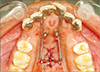
In group B (lingual biocreative therapy group), the lingual retractor was fabricated from a chrome cobalt alloy and sandblasted to provide adequate bond strength. It was bonded to the lingual surface of the six anterior teeth so that they were rigidly splinted together and had 10-mm retraction hooks that were located between the central and lateral incisors, as recommended by Kim et al.12 and Mo et al.14 A cross-type miniplate (C palatal plate; Gebrüder Martin GmbH, Tuttlingen, Germany) was fixed mid-palatally to provide skeletal anchorage for en masse retraction of the anterior segment. Its main arm had three holes for inserting the microscrews and two horizontal arms with holes for the NiTi closing coil springs. A retraction force of 200 g per side was used (Figure 2).
In both groups, participants underwent follow-up examinations every 6 weeks. Retraction was stopped when a Class I canine relationship was achieved and an adequate incisor relationship was obtained (Figures 3 and 4).
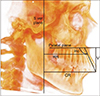
The main outcome of the study was the type of tooth movement during anterior retraction. Secondary outcomes were molar anchorage loss, vertical movement of the crown of the six maxillary anterior teeth, inclination of the maxillary occlusal plane, and time required to complete the retraction phase. CBCT scans were obtained for every subject before and after retraction by using the iCAT scanner (Model 17/19 series; Imaging Sciences International, Hatfield, PA, USA). The CBCT scanner parameters were set to 120 kVp at 5 mA for a total scan time of 8.9 seconds, and the field of view was 17- × 23-mm. The Digital Imaging and Communications in Medicine (DICOM) files were processed into volumetric images using InVivo 5 ver. 5.2 software (Anatomage, San Jose, CA, USA).
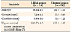
CBCT analysis involved orienting the CBCT unit, defining the desired landmarks, creating the reference lines and reference planes, and then obtaining linear and angular measurements. The landmarks, reference lines, and planes are shown in Tables 1 and 2. Table 3 shows the linear and angular measurements used (Figure 5).
Sample size calculation
Sample size calculation was based on the studies by Kim et al.11 and Kim et al.12 G*power software (Universität Düsseldorf, Düsseldorf, Germany) showed that group sample sizes of 12 each would achieve 80.09% power to reject the null hypothesis of equal means with a significance level (alpha) of 0.05 using a two-sided two-sample equal variance t-test. To compensate for possible dropouts, a sample size of 28 patients was selected and 14 patients were included in each group.
Statistical analysis
The statistical analysis was performed using IBM SPSS ver. 20 (IBM Corp., Armonk, NY, USA). The statistician was blinded and data from the patients were presented with no indication of which treatment the patients received. All variables were measured for both sides, and averages were then taken for the right and left sides. Shapiro–Wilk test of normality was used to test the normality hypothesis of all quantitative variables. Paired t-test was performed to compare the pre- and post-treatment CBCT measurements within the labial and lingual groups. An independent-sample t-test was performed for comparing the mean treatment changes between the two groups. The significance level was set at p < 0.05.
Intra-examiner and inter-examiner error analysis for the CBCT measurements were performed using concordance correlation coefficients (CCC). The closer the CCC was to 1.0, higher was the reliability of the measurement.
RESULTS
Twenty-eight patients were enrolled in the trial. Four patients were lost to follow-up. The details are provided in the CONSORT flow diagram (Figure 6). The baseline demographic characteristics are presented in Table 4. Participants' recruitment and follow-up was carried out over 20 months from January 2016 till September 2017. In the error analysis for the CBCT measurements, the CCC values ranged between 0.796 and 0.997, indicating good to excellent agreement. Shapiro–Wilk test of normality showed that variables were normally distributed; therefore, parametric tests were used for analyzing the data.
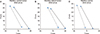
Retraction durations for both groups are shown in Table 5. No statistically significant differences were found between the two groups. Table 6 shows descriptive statistics for the intragroup and intergroup comparisons for all variables measured. The mean change in the maxillary central incisor inclination was −4.41 ± 2.33° in the labial group and −10.26 ± 4.70° in the lingual group. When comparing both groups, significant differences were found. The inclination of the central incisor was reduced more in the lingual group (i.e., more lingual tipping; mean difference, 5.85 ± 1.85°). The canine showed significant distal tipping in the lingual group (mean difference, 6.98 ± 1.25°). Figures 7 and 8 show tooth axis graphs for the maxillary central and lateral incisors and canine before and after retraction. No significant differences were found between the two groups in relation to the amount of anterior retraction, except for coronal retraction of the maxillary central incisor (1.35 mm more retraction in the lingual group) and root retraction of the canine (1.8 mm more root retraction in the labial group).
The maxillary central incisor showed a mean intrusion of 1.46 ± 0.54 mm in the labial group and 1.45 ± 1.02 mm in the lingual group, with no significant difference between the two groups. However, significant differences in intrusion of the lateral incisor and canine were found between the two groups. The canine was significantly more intruded (mean difference, 1.67 ± 0.49 mm) in the lingual group.
Good anchorage control was achieved in both groups. The mean molar mesial movement was 0.63 ± 0.51 mm in the labial group and 0.47 ± 0.26 mm in the lingual group. Significant improvement in the facial profile was achieved in both groups.
Adverse effects
No serious adverse effects were observed other than gingivitis associated with plaque accumulation. One miniscrew failed in the labial group due to poor oral hygiene. Retraction was stopped, oral hygiene measures were given to the patient, and the miniscrew was reinserted after 6 weeks. No other problems occurred.
DISCUSSION
The aim of this study was to compare the treatment effects observed when using labial versus lingual biocreative therapy for en masse retraction of the maxillary anterior teeth. Regarding the mean retraction period, no statistically significant differences in the duration of the retraction phase were found between the two groups. Jee et al.15 reported the duration to be 13.44 ± 4.30 months in the conventional C-wire group, and Kim et al.11 reported an average duration of 13.94 ± 5.61 months, which is close to our results.
In labial biocreative therapy type II, various patterns of tooth movement could be obtained through the combination of intrusion force, retraction force, and length of the retraction hook. In the labial group, 10-mm-long crimpable hooks were used to achieve bodily retraction of the anterior segment.618 An overlay reverse-curved 0.016 × 0.022-in NiTi wire would produce an intrusion force of 70 g6 and a counterclockwise moment that was used for anterior vertical and torque control during retraction. Two AbsoAnchor bracket head (right and left) mini-implants were used instead of the partially osseointegrated C-implants recommended by Chung et al.5 They were self-drilling and could be loaded immediately. The right bracket head mini-implant was inserted into the right side and turned clockwise during insertion. The left bracket head mini-implant was inserted into the left side and turned counterclockwise during insertion. The tip-back moment created by the overlay reverse curve wire on the mini-implant would serve to stabilize rather than loosen the screw, tightening it even further.
The primary outcome of our study was to assess the type of tooth movement during retraction. Kim et al.11 reported a mean change of 15.33 ± 6.85° for the central incisor and Jee et al.15 observed uprighting of the maxillary anterior teeth with a 13.77° change in central incisor inclination. Accordingly, in comparison with other studies, our results show better torque control for the maxillary incisors with only a mean change of 4.41 ± 2.33°.
Sung et al.19 suggested that en masse bodily retraction of the anterior teeth seems to be difficult with conventional sliding mechanics by using orthodontic miniimplants. We completely agree with this. Even though the line of action of force was applied close to the center of resistance for the six anterior teeth and a counterclockwise moment was added via the reverse curve, exactly equal amount of crown and root retraction for the central and lateral incisors was not achieved. Despite the many advantages of conducting a finite element study, the accuracy of such studies might be affected by major factors such that the results can only represent a theoretical scenario that cannot be achieved clinically; a clinical study is essential for confirmation. Remarkable tipping of the anterior segment during retraction was frequently reported by clinicians despite using forces and moments that were adjusted to achieve pure retraction of the anterior teeth. Furthermore, it was previously reported that the desired bodily movement of the anterior segment requires further correction of the force system, even though the biomechanical design employed should theoretically provide bodily movement of the segment.20 This is quite close to our experience in this study. Our results are based on a clinical study rather than a theoretical model and can be well applied in clinical practice, resulting in anterior retraction with good torque control.
As for the lingual biocreative therapy group, the “vertical bowing effect” occurred during anterior retraction, with lingual tipping of the incisors and distal tipping and intrusion of the canines. The maxillary central and lateral incisors showed controlled tipping movement. The mean change in central incisor inclination was similar to that reported by Seo et al.,17 where a 10.65 ± 5.17° change in the labiolingual inclination of the central incisor occurred. Kim et al.12 found a decrease of 7.8° in the SN-U1 angle in the group where 10-mm long retraction hooks were used, while the group with shorter retraction hooks showed a decrease of 12.1°. Therefore, it can be recommended that for cases with flared incisors that will need controlled lingual tipping, a retraction hook length of 10 mm should be used. On the other hand, a longer hook should be used if bodily tooth movement is desired. One important consideration is the morphology of the palatal vault. Furthermore, the exact location of the center of resistance of the anterior segment is affected by the morphology of alveolar bone and roots.21 In some challenging cases, we might need to resort to additional TADs or other complex biomechanics. This may warrant further study.
As for anterior vertical control, similar to our results, Kim et al.11 reported a 0.63-mm intrusion of the maxillary central incisor using the labial technique and Seo et al.17 reported a 1.18 ± 2.04 mm intrusion for the central incisor using the lingual biocreative technique. The often-reported adverse effect of increasing the incisor display and creating a gummy smile, which is associated with anterior retraction in extraction, did not occur with these mechanics. Significant differences in the intrusion of the lateral incisor and canine were found between the two groups. The canine was significantly more intruded in the lingual group than in the labial group. Different measures could be applied for proper vertical control of the canine during retraction using the lingual biocreative technique. The use of anterior triangular elastics from buttons bonded to the maxillary canine and the lower arch was recommended.14 Jang et al.16 recommended using torqueing springs for canine vertical control. However, they reported that the length of the retraction hook and the resultant line of action of force were more important factors than the incorporation of the torqueing springs. Mo et al.14 recommended sectioning the canine from the anterior segment to allow individual control of the canine. At the end of the retraction phase, full fixed appliances had to be bonded to the maxillary and mandibual arches to continue the levelling and alignment phases, followed by finishing and settling of the occlusion.
Similar to previous studies,1112 good anchorage control was achieved in both groups. This is of paramount importance to maximize anterior retraction in patients with dentoalveolar protrusion and protrusive lips. Furthermore, both groups showed significant soft tissue changes following anterior retraction, with a significant increase in nasolabial angle and reduction of the interlabial gap and the linear distance from Labrale Superius and Labrale Inferius to E-Line. Kim et al.11 reported significant soft tissue changes using the labial technique, where the upper and lower lips to the E-line moved posteriorly 1.87 ± 0.91 mm and 2.75 ± 1.80 mm, respectively.
Limitations
Blinding of the patients and the operator to the type of intervention was impossible. Blinding of the outcome assessor was not possible during analysis of the CBCT scans because of the presence of the brackets in the labial group. Two patients were lost to follow-up in each group. However, the final sample size was equal to the sample size calculated and the number of patients analyzed was the same in both groups. The vertical positions of the miniscrews are different in the two techniques, and this might have had an impact on the final inclination of the incisors. This should be considered while interpreting the results.
CONCLUSION
Significant differences were found between the two groups regarding the type of tooth movement. The inclination of the incisors was reduced more in the lingual group (i.e., more lingual tipping). The canine showed significant distal tipping and intrusion in the lingual group.
Using the labial biocreative therapy with a 10-mm anterior retraction hook and a reverse curve overlay, anterior retraction with good torque control, vertical control, and anchorage control were achieved.
Using the lingual biocreative therapy with a 10-mm anterior retraction hook, anterior retraction occurred with controlled tipping movement. Clockwise rotation of the anterior segment occurred with significant distal tipping and intrusion of the canine.
No statistically significant differences in the duration of the retraction phase were found between the two groups.


 XML Download