

 PDF
PDF ePub
ePub Citation
 Print
Print


INTRODUCTION
Whereas stabilized atherosclerotic lesions progress slowly, vulnerable plaques suddenly rupture and cause thrombosis, resulting in acute coronary syndrome (ACS). Vulnerable plaques can be clinically identified by intracoronary imaging modalities such as intravascular ultrasonography (IVUS) or optical coherence tomography (OCT) and are known to have the imaging features of thin-cap fibroatheromas (TCFAs). However, in recent clinical studies, the identification of vulnerable plaques by IVUS failed to improve the predictability of cardiovascular risk when compared to existing models.1)2) In some interesting studies, only a small number of vulnerable plaques actually ruptured, and most vulnerable plaques showed a silent clinical course even if they were ruptured.3) Therefore, the concept of vulnerable plaques has recently been challenged and requires further perspectives to be identified.4)5) Some new factors are proposed to distinguish the “real vulnerable plaque” that consequently develops into clinical events and to define “vulnerable patients”.6) Morphological and physiological factors, such as microcalcification, cholesterol crystals, the apoptosis of macrophages and endothelial shear stress (ESS), seem to play an important role in causing the instability of plaques and the inflammation of local atherosclerosis.7)8)9)10) Here, we summarize the characteristics of vulnerable plaques and review the latest pathological and physiological mechanisms of plaque formation and rupture.
EVOLUTION OF THE CONCEPT OF VULNERABLE PLAQUES
Historically, since the first report by autopsy data in 1844, the main cause of acute myocardial infarction (MI) has been known as plaque rupture accompanied by superimposed coronary artery thrombosis.11) Afterwards, thrombosis caused by fissures and erosions in the intimal surface of coronary arteries was reported.12)13) The authors introduced the term “intramural atheromatous abscess” and reported the existence of a necrotic material accompanied by a thrombus. Davies14) demonstrated the role of the inflammatory mechanism associated with the progression of plaque instability and the pattern of plaque disruption. In 1989, the nomenclature of the vulnerable plaque was adopted by James E. Muller et al., and the importance of identifying high-risk lesions amenable to treatment was raised in 2003.15)16) The concept of a TCFA, a major precursor of ACS, was presented as a rupture-prone plaque with a thin fibrous cap (<65 µm thick) accompanied by an infiltration of many inflammatory cells and a few smooth muscle cells, a large necrotic core, spotty calcification and positive outward remodeling.17)18)
NEWLY DISCOVERED MECHANISM OF PLAQUE RUPTURE: THE HIDDEN PATHOLOGICAL CONCEPT OF VULNERABLE PLAQUES
Microcalcification in the fibrous cap
Microscopic calcification or calcified nodules are risk factors for thrombosis, whereas plaques with severe calcification show clinically stable outcomes.19)20) The apoptosis of smooth muscle cells and the release of matrix vesicles by macrophages are key mechanisms in developing intimal microcalcification.7) When these microcalcifications progressively aggregate and create a large mass, they form calcified sheets or plates. This process is more pronounced in healed plaques and fibroatheromas and is rarely observed in fibrous plaques. The calcium sheets later form calcified protrusions with cutting edges after breakage, called calcified nodules, which are evaluated as a potential substrate of acute thrombosis.20) Previous studies using IVUS and coronary computed tomography (CT) demonstrated that the lesions consisting of spotty calcification were associated with plaque rupture and the incidence of ACS.21)22) In addition, a recent 18F sodium fluoride positron emission tomography (PET) study, which can trace only the active calcification, better discriminated between culprit and nonculprit plaques in ACS.23) In recent computational fluid dynamics studies, the presence of microcalcification in the fibrous cap played a role in promoting cap disruption by exaggerating the mechanical force applied to the cap during the cardiac cycle.5)24) Subsequent studies using micro-CT also demonstrated that if multiple microcalcifications larger than 5 µm are very close, the stress can be increased exponentially to create “explosive voids” that can cause plaque rupture. However, micro-CT is limited in the clinical detection of microcalcifications smaller than 15–20 µm, even if high-resolution imaging devices such as OCT are used.25)
Cholesterol crystal-induced inflammasome activation: the basis for the design of the Canakinumab Anti-Inflammatory Thrombosis Outcomes Study trial
A theory has recently been proposed that cholesterol crystallization enhances thrombogenesis through plaque fissure and acute volumetric expansion.8) This theory is based on changes in local plaque temperature, pH, and hydration status during cholesterol crystallization. In another study, the authors demonstrated that the secretion of the mature human pro-inflammatory cytokine interleukin (IL)-1 was induced through inflammasome-mediated pathways during the phagocytosis of cholesterol crystals by human macrophages.25) Currently, micro-optical chemistry is being used to identify the link between cholesterol metabolism and plaque inflammation. In addition, based on the above phenomena and experimental evidence that inflammation contributes to the pathogenesis of atherosclerosis, the Canakinumab Anti-Inflammatory Thrombosis Outcomes Study was recently completed.26) High-risk patients with prior MI and residual inflammation with elevated high-sensitivity C-reactive protein were enrolled and randomized to canakinumab, a human monoclonal antibody targeting IL-1β, or placebo. Canakinumab significantly lowered the risk of major adverse cardiac and cerebral events.
Apoptosis of intraplaque macrophages
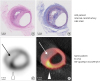
A key component of vulnerable atherosclerotic plaques is the large necrotic core. The necrotic core is formed primarily by the apoptosis of advanced lesional macrophages and the combination of defective efferocytosis.27) Factors such as delayed endoplasmic reticulum stress and oxidative stress or the activation of death receptors cause apoptosis in macrophage foam cells.28) These apoptotic cells are not effectively removed by macrophages due to the defective efferocytosis of advanced plaques, resulting in secondary cell necrosis.29) Therefore, apoptosis is considered a proper imaging target that evaluates the vulnerability of plaques.9) Currently, 99mTc-annexin A5 imaging is available for inflamed carotid plaques, but there are limits due to low resolution and specificity. Moreover, the small plaque sizes and motion artifacts due to heartbeat or the act of breathing act as hurdles. Recently, our group has succeeded in creating a novel PET probe to image plaque apoptosis: 18F-ApoPep1. In vivo PET imaging after 18F-ApoPep1 clearly imaged vulnerable plaques containing many apoptotic cells in apoprotein-E-deficient mice (unpublished data) (Figure 1).
Figure 1
In vivo H&E staining and ex vivo PET imaging of 18F-ApoPep1 to detect plaque apoptosis and vulnerability. (A) H&E staining of the left anterior descending artery in an autopsy coronary specimen who suddenly died of acute myocardial infarction (kindly provided by Dr. In-Beom Kim) showed the features of vulnerable plaques and (B) its tunnel stain demonstrated plenty of apoptotic cells in the plaque. (C, D) 18F-ApoPep1 PET and fusion imaging with micro CT clearly imaged apoptotic process occurring in the vulnerable plaque.
AMI = acute myocardial infarction; CT = computed tomography; H&E = hematoxylin and eosin; PET = positron emission tomography.
CURRENT STATUS OF THE MOLECULAR IMAGING OF VULNERABLE PLAQUES
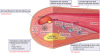
Although molecular imaging technology in small animals, including fluorescence imaging, bioluminescence imaging, ultrasound, micro-PET, micro-single photon emission CT, micro- CT, and high-field small-animal magnetic resonance imaging (MRI), has tremendously advanced, imaging molecular signals in human coronary plaques is still not easy. We can expect the development of an atherosclerotic molecular imaging field if human imaging platforms or probes make rapid progress.30)31) Many researchers have studied the applicability of molecular MRI to detect macrophage activity, apoptosis, and adhesion molecules in plaques using gadolinium chelates or iron oxide nanoparticles.32) Whereas MRI is suitable for fast-moving coronary arteries due to its high spatial and time resolution, it has lower imaging sensitivity, which requires a large amount of potentially toxic imaging agents for molecular imaging.33) Thus, an imaging platform using PET and its probe is highly anticipated for the introduction of the molecular imaging of vulnerable plaques in the clinics (Figure 2). The identification of an asymptomatic vulnerable plaque before it ruptures and its treatment with aggressive and/or advanced medical therapy with or without revascularization is an unmet clinical need in interventional cardiology.
Figure 2
Newly added pathophysiological concept of vulnerable plaques and the applicable imaging modalities to detect the process of plaque rupture.
CCTA = coronary computed tomography angiography; CFD = computational fluid dynamics; ESS = endothelial shear stress; IL = interleukin; IVUS = intravascular ultrasonography; LDL = low-density lipoprotein; MRI = magnetic resonance imaging; OCT = optical coherence tomography; PET = positron emission tomography.
CHALLENGES OF THE CONCEPT OF A THIN-CAP FIBROATHEROMA AS A VULNERABLE PLAQUE
The concept of a TCFA as a vulnerable plaque has been challenged lately because very few TCFAs cause ACS.34) The natural history of TCFAs varies and, in most cases, has an indolent course transforming into a more stable plaque.35) Moreover, asymptomatic plaque rupture may also occur in the condition of less severe stenosis or less thrombus formation. Abrupt vessel occlusion accompanying thrombus by rupture was more likely to occur in severe stenoses and in the condition of a “vulnerable patient”. The concept of a “vulnerable patient” requires altered coagulation or thrombosis, endothelial dysfunction, and hemodynamic factors.36) Recently, introduced intravascular imaging techniques, such as IVUS and OCT, help to see the characteristics of lesions in detail, but this imaging-guided approach failed to improve clinical outcome. Currently, fractional flow reserve (FFR) is the most powerful tool in assessing the potential ischemic risk of lesions and in reducing future clinical events in patients with symptoms.
NEWLY DISCOVERED MECHANISM OF PLAQUE RUPTURE: THE HIDDEN PHYSIOLOGICAL CONCEPT OF VULNERABLE PLAQUES
Recently, there have been efforts to explain the mechanism of plaque rupture by hemodynamic and physiological factors, such as shear stress and fluid dynamics. The ESS resulting from friction on the surface of the endothelium is closely related to the pathogenesis of atherosclerosis, plaque formation, and the progression of plaque vulnerability. It has been reported that low ESS is a powerful stimulus that precedes atherosclerosis by inducing lipid aggregation, neovascularization and expanding plaque volumes in previous pig and human data.37)38)39) In addition, the low ESS increases the activity of the major elastolytic matrix metallopeptidases (MMPs) and cathepsins. It also affects endogenous inhibitors such as tissue inhibitors of metalloproteinases and cystatin C, resulting in the fragmentation of the internal elastic lamina.37) Low ESS causes inflammatory cells to migrate to the media, resulting in the degradation of the matrix and vessel remodeling. Furthermore, the low ESS increases the activity of collagenolytic MMPs, resulting in the degradation of collagen and the thinning of the fibrous cap.38)39) Recent clinical studies with IVUS and OCT demonstrated that low ESS is an independent predictor of plaque progression and expansive remodeling with lumen narrowing.40)41) The ESS is associated with plaque rupture as well as the production of atherosclerotic plaques.42) Stenotic vulnerable plaques create a heterogeneous local ESS environment, such as low ESS in the upstream shoulder, high ESS in the neck, and low ESS or oscillatory stress in the downstream shoulder of plaques.43) The majority of ruptures occur on the upstream side of the plaques and result from low ESS and high local wall stress.44) Rupture can also occur when very fast blood flow and high ESS are accompanied by the maximal stenotic lesions. A very recent study investigated the prognostic value of the ESS measure in the upstream shoulder to predict MI in 441 patients who were deferred with FFR≤0.80.45) Higher ESS in the proximal segments of stenotic lesions was predictive of MI and had incremental prognostic value in addition to the FFR value.
CONCLUSIONS
Plaque vulnerability and plaque rupture develop from a complex interplay of anatomical and hemodynamical factors. The identification of TCFAs by imaging modality; the imaging detection of molecular signals in plaques including microcalcification, apoptosis, inflammation, and/or angiogenesis; and the hemodynamic assessment of ESS and local wall stress by computational fluid dynamics all contribute to the better identification of plaque vulnerability, resulting in a better clinical outcome by adopting state-of-the-art therapeutics and selective coronary interventions.
 XML Download