

 PDF
PDF Citation
 Print
Print


Fractures of the distal femur are relatively common in cats, accounting for 20 to 32% of all femoral fractures [12]. In immature animals, these fractures often occur at the physis, with Salter-Harris (SH) fractures types I and II being the most common [134].
Several choices are available for the treatment of distal femoral physeal fractures in cats, including single and multiple intramedullary pins [56], modified Rush pins [7], and cross-pins [8].
The present report describes the use of a stainless steel wire in a figure-of-eight configuration for the treatment of distal femoral SH fractures types I and II in 10 cats. To the best of the authors' knowledge, there are few reports on the use of steel wires for the treatment of distal femoral physeal fractures [9].
Table 1 lists the 10 cats treated with the figure-of-eight stainless steel wire technique. Anesthesia was not standardized, and was determined individually for each cat depending on their overall status. Perioperative first generation cephalosporin (Cefalotin Villerton, Villerton Invest SA, Luxembourg) was administered intravenously at 25 mg/kg prior to surgery.
The patient was positioned in dorsal recumbency with the affected limb suspended vertically for draping. The limb was prepared routinely for surgery. A standard lateral or medial parapatellar approach to the stifle and distal femur was used and the patella was reflected medially or laterally, exposing the fracture. A bilateral approach was used if additional exposure was needed. Following inspection, the fracture fragments were cleared carefully of blood clots and debris. The stifle was then flexed and with the tarsocrural joint held in extension the fracture was reduced by applying cranial traction to the proximal tibia.
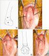
With the fracture held in reduction by a stifle extension and digital pressure, 2 parallel holes were drilled in the femur using either a 1.5 mm Kirschner wire and a hand chuck, or a power drill. The hole in the distal fragment was drilled from lateral to medial (if a lateral approach was used) or from medial to lateral (if a medial approach was used) cranioproximal to the extensor fossa. The hole in the proximal fragment was placed at an equal distance from the fracture in the cranial part of the femoral diaphysis (Fig. 1A). Ensuring that the holes were placed in the cranial part of the bone was important because a more caudal position would not adequately counteract the caudal pull of the gastrocnemius and caudal thigh muscles on the distal fragment, and could result in downward tilting and caudal displacement of the distal fragment.
A 0.8 mm stainless steel wire (Cerclage wire, Synthes, USA) was introduced through each of the predrilled holes (Fig. 1B). The wires were placed in a figure-of-eight configuration with twists on each side of the femur, just proximal to the cross but abaxial enough to ensure that the wires would not interfere with the patellar function. The wires were then tightened with a wire-tightening instrument alternately on the lateral and medial sides until the wires were seated tightly and the fracture was stable (Fig. 1C-E).
If maintaining reduction of the fracture during drilling was difficult, holes were drilled in the distal and proximal fragment before the fracture was reduced. The steel wires were preplaced and the fracture was then reduced gradually while the wires were carefully tightened alternately on the lateral and medial sides.
The wires were cut with a wire cutter leaving 3 twists on each side. The patella was returned to its normal position and the joint was lavaged. The arthrotomy and surgical site were closed routinely. The technique was simple to perform and there were no intraoperative complications.
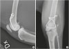
Postoperative radiographs were taken to evaluate the fracture stabilization (Fig. 2). Postoperative analgesia was adjusted individually. Postoperative management was routine, including restricted activities and revisits for clinical evaluations and follow up radiographs at 2- to 4-week intervals, depending on the patient's age.
Healing was uneventful in 8 out of 10 cats. Most cats began to use the affected limb 1 to 2 days after surgery and lameness had usually resolved completely by 4 weeks after surgery.
Early fixation failure occurred in 1 cat and required revisional surgery. This was suspected to be due to inadequate tightening of the wire. Once stability was achieved, the fracture healed uneventfully. In 1 cat, lameness recurred 4 weeks after the fracture had been considered healed. Breakage of the stainless steel wire was suspected to be the reason for the lameness and the wire was removed, after which the lameness resolved.
For the long-term follow-up, the owners were contacted for a telephone interview (Table 1). A long-term clinical evaluation and radiography were performed 9.8 years postoperatively in case 2, and 1.5 years postoperatively in case 10. None of the 2 cats showed lameness of the affected limb, but case 2 had mild disuse muscle atrophy that was indicative of intermittent lameness. None of the cats showed pain upon palpation or upon flexion or extension of the stifle joint. Standard radiographs of the femur and stifle showed no signs of complications related to the stainless steel wire and there were no signs of femoral shortening, recurvatum or medial or lateral deviation. Case 2 displayed stifle osteoarthritis with severe osteophytosis, which was considered the reason for the lameness in this cat. In case 10, there were signs of mild osteoarthritis.
The distal femoral physis in cats features 4 metaphyseal protuberances fitting in corresponding depressions in the epiphysis [13]. This conformation aids in the reduction of SH fractures types I and II, and provides stability against rotational and shear forces. The stability provided by the anatomical reduction allows for less rigid fixation. Commonly used techniques include cross-pins, Rush pins, and single or multiple intramedullary pins [3]. In 1974, Whittick [9] described the use of a stainless steel wire placed in a figure-of-eight configuration in the distal fragment, but in a horizontal mattress fashion at the fracture line. The technique described here is slightly different because the figure-of-eight configuration is placed at the fracture line. Because of the caudal muscular pull of the gastrocnemius, semimembranosus and semitendinosus muscles on the distal fragment, a stainless steel wire placed in the cranial part of the bone will act as a tension band wire. Placing the wire in a figure-of-eight manner ensures that the longest possible lever arm is maintained, thereby adding more compression and stability [101112].
In contrast to traditional techniques, the figure-of-eight stainless steel wire technique allows for a gradual reduction of the fracture. This can be beneficial, particularly in more chronic fractures, where maintaining reduction during fixation can be difficult.
Complications occurred in 2 of the 10 cases in this case series. This is similar to the complication rates reported using traditional techniques [67]. A possible concern with the figure-of-eight stainless steel wire technique is the proximity of the wire to the patellar articular surface, particularly with the twists being at risk of causing irritation of the patella. Care should be taken when placing the twists to place them close enough to the cross to make a mechanically optimal twist, but abaxial enough to not interfere with the patellar function. When adhering to these recommendations, signs of problems related to irritation of the patella were not observed in this case series.
At the long-term follow-up, 1 of the 6 cats showed a mild intermittent lameness, which was attributed to osteoarthritis of the stifle joint. The development of osteoarthritis after the repair of distal femoral physeal fractures is not surprising given the juxta-articular nature of these fractures, and stifle osteoarthritis has also been reported at long-term follow-ups following repair using traditional techniques [17].
Historically, the controversial point when using a stainless steel wire for the repair of physeal fractures has been that it places compression over a physis, causing premature closure of the physis. On the other hand, other factors, such as the original trauma to the physis at the time of injury, surgical trauma, and placement of pins through the physis, have also been shown to cause premature closure of the physis [1314]. Premature closure of the physis appears to occur in the majority of cases following the repair of distal femoral physeal fractures, regardless of the technique used, with the only significant predictor of a growth disturbance being the age at the time of injury [1101314]. Premature closure of the physis was considered unlikely to cause any problems in the cats in the present report with a median age of 14 months [15].
The main limitation of this report was the limited sample size. In addition, bias might have occurred because the long-term follow-up was based mainly on telephone interviews. Future prospective studies will be needed to evaluate the technique further, also in younger cats with major bone growth after fracture repair.
In conclusion, the figure-of-eight stainless steel wire technique appears to be a good alternative for the repair of distal femoral SH types I and II fractures in cats.

 XML Download