

 PDF
PDF Citation
 Print
Print


INTRODUCTION
Despite a continuous decline in smoking rates for decades, lung cancer remains the most commonly diagnosed cancer and the leading cause of cancer-related deaths worldwide.1 In 2018, lung cancer was newly diagnosed in 2.1 million people (11.6% of the total number of cancer cases), and 1.8 million people (18.4% of the total number of cancer cases) died of lung cancer worldwide.1 In Korea, lung cancer has been the most common cause of cancer death since 1999 and accounted for 22.6% (17,399/76,855) of all cancer deaths in 2015.2 Surgical resection is the only curative treatment that improves survival for early-stage lung cancer,3 and in Korea, 22% of newly diagnosed lung cancer cases are candidates for surgery.4
Over the past few decades, clinical information changed significantly in patients with surgically resected lung cancer. Patient screening was performed via low-dose computed tomography (LDCT) and an increased proportion of factors such as women gender, stage I, and adenocarcinoma have contributed to improved survival outcome of lung cancer surgery.56 As such, it is crucial to analyze the demographics, tumor-related background, and prognosis that can affect the incidence of disease and treatment modalities. In line with other countries,789 there have been two studies analyzing the trend on lung cancer surgery in Korea410 However, one study based on Korean national data does not have information on primary diagnosis, histologic type, and survival outcome.4 In addition, the other study included a relatively small number of patients and is out of date.10 Therefore, we sought to investigate recent trends and surgical outcomes of lung cancer surgery conducted in Korea on a large cohort collected by a single institute between 2002 and 2016.
METHODS
Patients
We reviewed the records of patients who underwent pulmonary resection for primary lung cancer between January 2002 and December 2016 from a prospectively collected database at Asan Medical Center, Seoul, Korea. Patients who underwent surgery for diagnostic intent were excluded from the study. Each cancer case of patients with more than one primary lung cancer was included. The patient population was pathologically staged according to the recommendations by the 8th edition American Joint Committee on Cancer (AJCC) in a retrospective manner.11 Tumor histology was categorized according to the World Health Organization classification.12 Lobectomy with systematic lymph node dissection was adopted as a standard procedure for primary lung cancer, but some patients with old age, borderline lung function, and early stage adenocarcinoma received sublobar resection. Patients with clinical stage I-IIIA, including N2 node metastasis, were indicated for surgical resection. All patients were followed either until death or the last follow-up date (March 1, 2019).
Follow-up information on all patients was obtained via notes taken during clinical follow-up, which was conducted every 3–6 months during the first 2 years after surgery and every 6–12 months thereafter. Chest CT scans were performed in sync with clinical visits or at any time when disease recurrence was suspected. Treatment modalities and chemotherapeutic regimens in relapsed cases were determined at the discretion of the attending physician.
Outcome measurements
Considering that the number of comorbid diseases per patient is superior to individual comorbidities (hypertension, diabetes mellitus, smoking, chronic bronchitis, coronary artery disease, cardiac arrhythmia, congestive heart failure, liver cirrhosis, cerebral vascular event, renal insufficiency, history of malignant disease, and prior thoracic surgery) in a predictive model for postoperative mortality,13 we considered it to be a categorical variable. Major complications were evaluated according to the joint definitions provided by the Society of Thoracic Surgeons and the European Society of Thoracic Surgeons.14 Postoperative mortality was defined as any deaths occurring within the first 30 days after operation and those occurring during the same hospitalization. Overall survival (OS) was defined as the time interval between the date of operation and the date of death, which was obtained by reviewing the records from the Korean National Security Death Index Database.
Statistical analysis
Continuous variables were presented as means and standard deviations, and the categorical variables were presented as a percentage. To effectively perform a trend analysis, the operation years were divided into three categories over 5-year intervals (2002–2006 vs. 2007–2011 vs. 2012–2016). For comparing among the three groups, we used parametric test (ANOVA) for continuous variables and χ2 test for categorical variables. Survival analysis was conducted using the Kaplan–Meier method and the results were assessed using the log-rank test. Cox proportional hazards model was used for univariable and multivariable analyses to identify prognostic factors for OS. The final multivariable model was selected by adopting the stepwise model selection approach (P ≤ 0.1 for entering the model and P ≤ 0.05 for staying in the model). Cox proportional hazard analysis was also utilized to adjust for baseline covariates and to find significant differences between adjacent operation year categories.
Annual percent changes (APC) in incidences, proportions, and their statistical significance were calculated using the Joinpoint software ver. 4.6.0.0 from the Surveillance Research Program of the United States National Cancer Institute.15 The other statistical calculations were performed using R version 3.4.2 (The R Foundation for Statistical Computing, Vanderbilt University, Nashville, TN, USA) by employing the Survival, ggplot2, GGally, survminer, and rms packages. P values of < 0.05 were considered to be statistically significant.
RESULTS
Patient characteristics
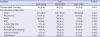
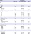
From January 2002 to December 2016, a total of 7,485 patients with primary lung cancer were included in this study. Characteristics of the enrolled patients are summarized in Table 1.
Table 1
Demographic and clinical characteristics of enrolled patients (n = 7,485) according to the operation period
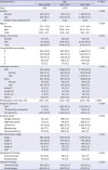
Continuous variables were presented as means ± standard deviations, and categorical variables as number (%).
ADC = adenocarcinoma, SqCC = squamous cell carcinoma, ADSqCC = adenosquamous cell carcinoma, VATS = video-assisted thoracic surgery.
![]()
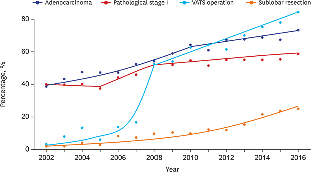
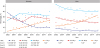
Over the study period, the number of lung cancer surgeries in both genders has continued to increase (Table 1 and Fig. 1). Especially, the proportion of women to the total number of surgeries has increased significantly from 24.0% to 41.9% (APC, 3.31; P < 0.05) (Fig. 1). The mean age of patients with lung cancer surgery in both genders increased gradually, and the number of comorbidities per patient also correspondingly increased (Table 1). The overall smoking rate among patients declined from 66.7% to 54.1% owing to the increased proportion of women with low smoking rates (Table 1). The proportion of patients with adenocarcinoma, sublobar resection (wedge resection or segmentectomy), and pathological stage I has significantly increased from 45.6% to 69.0% (P < 0.001), 4.3% to 19.9% (P < 0.001), and 40.6% to 56.0% (P < 0.001), respectively (Table 1 and Fig. 2). The proportion of patients with video-assisted thoracic surgery (VATS) has also significantly increased up to 84.4% in 2016; especially in 2008, a dramatic increase has been observed (APC 45.17; P < 0.05) (Fig. 2). As the proportion of patients with stage I patients increased, the rates of neoadjuvant and adjuvant therapy decreased (Table 1). Postoperative mortality decreased gradually from 2.8% to 0.3%. The incidences of detailed postoperative complications over time are described in Table 2.
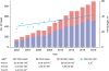 | Fig. 1Changes in the number of lung cancer surgeries for total, men, and women patients and the proportion of women to total patients according to the year. The numbers at the bottom of each variable in the legend indicate APC during the years shown in parentheses.APC = annual percent change.
*Significantly different from zero at (P < 0.05).
|
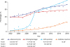 | Fig. 2Changes in the proportion of adenocarcinoma, pathological stage I, VATS operation, sublobar resection according to the year. The numbers at the bottom of each variable in the legend indicate APC during the years shown in parentheses.APC = annual percent change, VATS = video-assisted thoracic surgery.
*Significantly different from zero at (P < 0.05).
|
Table 2
Postoperative outcomes according to operation period
Values are presented as number (%).
ARDS = acute respiratory distress syndrome, BPF = bronchopleural fistula.
![]()
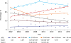
Time trend of age
The age distribution of lung cancer surgery showed a different trend depending on gender and time (Fig. 3). There was a significant increase in the proportion of patients who were aged ≥ 70 years in both genders (APC for men and women: 7.56 and 6.41, respectively; all P < 0.05). The proportion of men in other age groups declined significantly (APC for < 50 years, 50–59 years, and 60–69 years −3.70, −1.73, and −1.49; all P < 0.05), whereas only the proportion of women aged < 50 years showed a significant decrease (APC, −11.39; P < 0.05). Patients aged 60–69 years accounted for the largest percentage of lung cancer surgeries, regardless of gender, and women were younger than men (Table 1).
Time trend of tumor size
Tumor size was divided into six categories at 1-cm size intervals, and the distribution over time was plotted in Fig. 4. A significant increase was observed in tumors measuring 1–2 cm and 2–3 cm (APC for 1–2 cm and 2–3-cm sized tumor, 4.06 and 3.67; all P < 0.05). In particular, only the proportion of 1–2-cm sized tumors have significantly increased since 2009. On the contrary, the proportions of tumors measuring 3–4 cm, 4–5 cm, and > 5 cm in size have shown a significant decrease, with the largest reduction in the proportion of tumors measuring > 5 cm in size from 20% to 11% (APC, −4.13; P < 0.05). The proportion of tumors measuring < 1 cm in size was relatively consistent.
Time trend in OS
The median follow-up time was 50 months. At the end of follow-up, 2,495 of the 7,485 patients (33.3%) died, and the remaining 4,990 patients (66.7%) were alive or censored. The median survival time (MST) and 5-year survival rate for OS were 107 months (95% confidence interval [CI], 102–113) and 68.9%.
As shown in Fig. 5, a significant escalation in the survival curve was observed with each passing operation year period (all P < 0.001). The 5-year OS rate was 61.1%, 67.8%, and 72.1% in 2002–2006, 2007–2011, and 2012–2016, respectively. Multivariable Cox proportional hazard analyses were performed to identify independent prognostic factors for OS such as age, gender, operation year, smoking history, number of comorbidities, histologic type, surgical approach, surgical extent, pathological stage, neoadjuvant radiotherapy, adjuvant chemotherapy, and adjuvant radiotherapy (Table 3). After adjusting them as covariates, the survival outcome in the recent period (2012–2016) was superior to that in the previous periods (P < 0.001) (Fig. 5). We also performed a subgroup analysis of OS according to pathological stage (Fig. 6). Significant differences were found in patients with pathological stages I and II (P < 0.001 in stage I and 0.017 in stage II) (Fig. 6A and B). In contrast, there were no significant differences in patients with pathological stages III and IV (P < 0.273 in stage III and 0.857 in stage IV) (Fig. 6C and D).
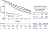 | Fig. 5Kaplan-Meier estimates of overall survival according to the operation period. The P values in the survival graph were calculated using the log-rank test, and the P values in the table were assessed by multivariate Cox analysis.HR = hazard ratio, MST = median survival time, n.a. = not available.
|
Table 3
Multivariable Cox proportional hazard analyses for overall survival
![]()
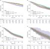 | Fig. 6Stage-specific Kaplan-Meier estimates of overall survival according to the operation period. Kaplan-Meier survival curve for (A) pathological stages I, (B) pathological stage II, (C) pathological stage III, and (D) pathological stage IV. The P values in the survival graph were calculated using the log-rank.
|
DISCUSSION
In this study, we evaluated recent trend analysis of demographics, surgery, and prognosis of lung cancer surgery for a large cohort from a single Korean institute over a 15-year period. Overall, despite the decrease in smoking rates, the number of lung cancer surgeries continued to increase. The mean age of patients with lung cancer surgery increased gradually, whereas tumor size declined. The proportions of patients with adenocarcinoma, VATS operation, sublobar resection, and pathological stage I was also increased significantly. These changes led to the improved 5-year OS rate from 61.1% to 72.1% in patients who received lung cancer surgery.
The main cause of the continued increase of lung cancer surgeries can be explained by the aging population in Korea. From 1999 to 2015, the crude incidence rate per 100,000 individuals (total number of cases divided by the mid-year population for the particular year) of lung cancer was increased from 42.1 to 66.8 in men and from 15.1 to 28.4 in women. During the period, the age-standardized lung cancer incidence rate (a weighted average of the rates by age, where the weights are the percentage of persons in the corresponding age group of the standard population)15 changed from 50.9 to 42.3 in men and from 12.4 to 14.3 in women, which continued to decline since 2002 for men and 1998 for women.216 This means that the Korean population is increasingly concentrated in the age group where lung cancer is common, that is, the elderly individuals. This phenomenon is directly related to the increasing age of patients who received lung cancer surgery in our study. While the proportions of other age groups have been similar or decreased, the proportion of elderly patients (≥ 70 years) in both genders rapidly increased (Fig. 2). Considering the improved perioperative care for elderly patients and the report from United Nations,17 the number of elderly individuals and the frequency of lung cancer surgery for elderly patients have been expected to increase over the years.
Contrary to squamous cell carcinoma, the proportion of adenocarcinoma continued to increase and become the most common histologic type in both men and women (Table 1). This trend is similar to that previously reported in the study conducted based on the data from the Korea Central Cancer Registry18 and those conducted in other countries.91920 The US Surgeon General noted that since the 1950s, the predominance of adenocarcinoma in histologic type can be attributed to changes in the design and consumption of cigarettes, such as lower tar, lower nicotine, and filtered cigarettes.21 However, this argument is challenged by the contradictory evidences that the trend was observed long before the changes in cigarettes and that changes in cigarette design has no relation with the increase in the incidence of adenocarcinoma in epidemiological studies.222324 Moreover, adenocarcinoma is more common in never-smokers, especially among Asian women.25 These findings suggest that other unidentified etiologic factors might be related to the increase in the incidence of adenocarcinoma.
Recently, lung cancer in never-smokers (LCINS) has gained attention owing to the increasing number of related cases and its distinctive nature. LCINS is regarded as a different disease entity from lung cancer among smokers not only because most affected patients are women having adenocarcinoma but also because of distinct underlying molecular mechanisms.26 Global statistics estimate that LCINS accounts for 25% of the total lung cancer cases worldwide.27 Its prevalence is especially higher in Eastern Asia, constituting 33% and 38% of lung cancer cases in Japan28 and Korea,29 respectively. During the study period, the number and the proportion of LCINS surgeries have increased owing to the increasing number of women patients with low smoking rates (Table 1). The overall LCINS incidence based on the Korea Central Cancer Registry also showed an increasing trend similar to that observed in our results.18
The widespread use of LDCT screening system for lung cancer has enabled the detection of early-stage lung cancer.5 Early detection of lung cancer patients has resulted in smaller tumor size (Fig. 4) and increased proportions of VATS operation, sublobar resection, and pathological stage I (Fig. 2). The increasing trend of pathological stage with a low malignant potential has also reduced the rate of bilobectomy, pneumonectomy, neoadjuvant therapy, and adjuvant therapy (Table 1). Along with the increase in the proportion of adenocarcinoma, the abovementioned factors have contributed to an improved survival rate of lung cancer surgery. The 5-year OS rate significantly improved from 61.1% to 72.1% (P < 0.001). Nonetheless, this trend was not just caused by demographics changes. Operation period alone remained a significant prognostic factor in multivariable Cox analysis (P < 0.001). Based on our data, relatively greater increase in survival rate was observed in patients with pathological stage I or II than that in the patients with advanced stage (Fig. 6). The reasons for these findings can be explained by several factors, such as improved surgical technique, standardized postoperative care, and development of adjuvant therapy. Especially in adjuvant therapy, platinum-based chemotherapy30 and target therapy (tyrosine kinase inhibitors in patients with activating mutations in epidermal growth factor receptor)31 might have contributed to improving the prognosis of recurrent lung cancer as well as primary lung cancer. However, because this study was targeted at patients who underwent lung cancer surgery, these findings could not be generalized to the entire population of lung cancer patients. Considering medical advances within the last decades, such as development of angiogenesis inhibitors, molecular therapy, and immunotherapy, survival rate of patients with advanced stage has improved and will continue to improve in the future.
Our current study has a few limitations stemming from its retrospective nature, as well as using observational data from a single institution. First, we acknowledge that our results cannot represent the trend for all the lung cancer surgeries performed in Korea, given that our research is not based on national data. However, being one of the largest tertiary referral hospitals having patients from all over of the country, our hospital encountered approximately 800 cases of primary lung cancer surgery in 2014, compared with 6,000 cases (including recurred cases) in Korea.4 Thus, we believe that our data, with relatively consistent quality and standardized protocols, fully reflect the trend for lung cancer surgery in Korea. In addition, because our results are based on patients undergoing surgical treatment, the survival curves might be different from those of general patients with lung cancer, especially those with stage III and IV.
In conclusion, this study demonstrated important temporal changes in the demographics, surgery, and prognosis of lung cancer surgery at Asan Medical Center from 2002 to 2016. The number of lung cancer surgeries and the proportion of patients who underwent VATS operation or sublobar resection, or those who were women or non-smokers or had adenocarcinoma or pathological stage I, increased significantly. These changes led to an improvement in the 5-year OS rate from 61.1% to 72.1% in patients who underwent lung cancer surgery.
 XML Download