

 PDF
PDF Citation
 Print
Print


INTRODUCTION
Dental adhesive systems are essential for restorations to be clinically successful. The gold standard for the marginal adaption of tooth substance and restorations is the three-step system of etching, priming, and bonding [1-3]. Resin-based dental luting agents have traditionally required a separate etching step for the adhesive to penetrate into the tooth structure [4]. However, conventional dentin-bonding agents have been reported to yield incomplete adhesive diffusion throughout the demineralized dentin [5]. It is conceivable that collagen exposure resulting from the discrepancy between the etching depth and adhesive penetration causes post-operative sensitivity [6]. In addition, the adhesive procedure used in three-step systems is complex and technically sensitive [7].
Self-adhesive resin cements were designed to adhere to the tooth structure without separate adhesive or etchant, to require a reduced application time, and to be less technique-sensitive [8,9]. Acidic and hydrophilic monomers in self-adhesive resin cements simultaneously demineralize and infiltrate enamel and dentin, resulting in a strong bond [10]. Therefore, pretreatments of the tooth substrate with conditioner or primer are not required. The hydrophilic acidic moieties of adhesive monomers are broadly classified into two categories: carboxylic acid groups and phosphoric acid groups [11]. Commercially utilized carboxylic acid–type adhesive monomers include 4-methacryloyloxyethoxycarbonylphthalic anhydride (4-META), 4-methacryloyloxyethoxycarbonylphthalic acid (4-MET), and 4-acryloyloxyethoxycarbonylphthalic anhydride (4-AETA), while phosphoric acid–type adhesive monomers include 2-methacryloyloxyethyl phenyl hydrogen phosphate (phenyl-P), 10-methacryloyloxydecyl dihydrogen phosphate (10-MDP), and 6-methacryloyloxyhexyl phosphonoacetate (6-MHPA). Carboxylic acid groups and phosphoric acid groups are expected to react with calcium in hydroxyapatite. A comparative study on the adhesive performance of functional monomers reported that 10-MDP was the most stable functional monomer for chemical bonding to calcium [12]. The purpose of this study was to evaluate the influence of the concentration of 10-MDP added as an adhesive monomer on the adhesion and physical properties of self-adhesive resin cements.
MATERIALS AND METHODS
Experimental resin cements
The experimental resin cements were formulated by Kuraray Noritake Dental (Tokyo, Japan). The chemical compositions of the experimental resin cements used in this study are shown in Table 1. The experimental resin cements were dual-curing resin cements composed of two pastes. The pastes included 3 different concentrations of 10-MDP monomer: 3.3 wt% (RC1), 6.6 wt% (RC2), and 9.9 wt% (RC3). The structure of a 10-MDP monomer is shown in Figure 1.
Table 1
Chemical composition of the experimental resin cements

![]()

Evaluation of bonding strength to dentin
Thirty extracted, caries-free human premolars were used in this study. The teeth were cut horizontally using a diamond disc at slow speed (Isomet, Buehler, Lake Bluff, IL, USA) to expose a flat area of superficial dentin. The dentin surfaces were ground on wet #180 and #600 SiC papers (60 seconds). The Research Ethics Committee of Okayama University reviewed and approved this study under protocol No. 189.
The dentin specimens were divided randomly into 3 experimental groups (n = 10) based on the concentration of 10-MDP. Estenia C&B (Kuraray Noritake Dental) was used to create composite resin blocks with dimensions of 10 mm × 10 mm × 5 mm. The following treatments were applied to the bonding surface of the Estenia C&B: alumina sand blasting for 5 seconds, ultrasonic cleaning for 2 minutes, phosphoric acid (K-Etchant Gel, Kuraray Noritake Dental) etching for 5 seconds, rinsing, air-drying, and silane treatment (Clearfil Porcelain Bond Activator, Kuraray Noritake Dental). Dentin specimens were bonded to Estenia C&B with experimental resin cement and light-cured for 40 seconds from each direction with a light output not less than 1,000 mW/cm2 (PenCure 2000, J. Morita Mfg., Kyoto, Japan). All bonding procedures were carried out by a single operator at room temperature.
After storage in distilled water at 37°C for 24 hours, the bonded specimens were sectioned into Estenia C&B–dentin sticks (1.0 mm × 1.0 mm) for micro-tensile bond strength (µTBS) testing. The sticks were attached to a jig with a cyanoacrylate glue (Model Repair II Blue, Dentsply-Sankin, Ohtawara, Japan) and tested to tensile failure in a universal testing machine (AGS-10KND, Shimadzu, Kyoto, Japan) at a crosshead speed of 1.0 mm/min. The cross-sectional area at the site of failure was measured with digital calipers (model CD-S10C, Mitutoyo, Tokyo, Japan), and this area was used to calculate the µTBS value, using the following formula:
The µTBS values obtained from the sticks of the same experimental resin cements were averaged.
Failure mode analysis
After µTBS testing, the fractured specimens were mounted on an aluminum stub, and then coated with gold. The fracture mode was determined using a scanning electron microscope (SEM; DS-720, TOPCON, Tokyo, Japan) at an accelerating voltage of 15 kV. The dentin side and the Esthenia C&B side of the fractured surface were observed at ×3,000 magnification. The failure modes were classified into the following four categories: cohesive failure in the dentin, cohesive failure in the resin cement, adhesive failure (less than 10% of the resin cement remaining), and mixed failure (co-existence of adhesive failure between the Estenia C&B and resin cement, cohesive failure in the resin cement, and/or adhesive failure between the dentin and resin cement).
Flexural strength testing
To compare the flexural strength of the experimental resin cements, 7 sticks (25 mm × 2 mm × 2 mm) were made for each cement. The experimental resin cements were filled into a mold, covered with a polyester film (KerrHawe, Bioggio, Switzerland), and light-cured from the center towards the edge in five overlapping sections with the PenCure 2000. Specimens were stored in distilled water for 24 hours at 37°C before testing.
A three-point bending test was performed using a universal testing machine with a crosshead speed of 1.0 mm/min. The dimensions of the specimens were measured with a digital caliper. The flexural strength (σ) in MPa was calculated using the formula:
where F is the load at fracture (N), L is the support span (20 mm), b is the width (mm) of the specimen, and d is the depth (mm) of the specimen.
Water sorption and solubility tests
Water sorption and solubility tests were carried out based on ISO 4049. Five specimen discs with a thickness of 1.0 ± 0.1 mm and a diameter of 15.0 ± 0.1 mm were made using a split-ring mold. The discs were finished with silicon carbide abrasive paper of #1,000 to #1,500 grit. All specimens were prepared at room temperature. The discs were immersed in distilled water at 37°C for 30 days. Water sorption (Wsp), in micrograms per cubic millimeter, was calculated for each of the 5 specimens using the following formula:
where m1 is the mass of the specimen in micrograms (µg) after immersion in water for 30 days, m2 is the initial dry constant mass (µg) before water immersion, and V is the volume of the specimen in cubic millimeters (mm3). After obtaining Wsp, the same specimens were used for determining water solubility (Wsl) using the following formula:
where m3 is the mass of the specimens after they were dried in a desiccator at 37°C for 24 hours.
Weight measurements were made using an analytical scale with a precision of 0.001 g (1712-MP8, Sartorius, Göttingen, Germany).
Statistical analysis
The data for µTBS, flexural strength, water sorption, and water solubility were statistically analyzed using one-way analysis of variance followed by the Tukey post hoc test at a significance level of 0.05. All statistical analyses were performed using IBM SPSS Statistics version 21 (IBM Corp., Armonk, NY, USA).
RESULTS
Micro-tensile bond strength testing
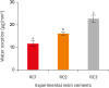
The mean values and standard deviations of the µTBS of each group are summarized in Figure 2. The bond strengths of RC2 (16.7 ± 3.5 MPa) and RC3 (17.1 ± 3.5 MPa) were significantly higher than that of RC1 (8.6 ± 1.6 MPa). However, there was no significant difference between the bond strengths of RC2 and RC3.
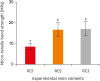 | Figure 2Micro-tensile bond strength of the experimental resin cements. RC1, RC2, and RC3 are the experimental resin cements containing 3.3 wt%, 6.6 wt%, and 9.9 wt% concentrations of 10-methacryloyloxydecyl dihydrogen phosphate (10-MDP) monomer, respectively.The same letters indicate the absence of a significant difference.
|
Fracture modes and SEM observations
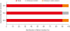
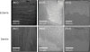
The results of the failure mode analysis are shown in Figure 3. The main fracture mode was mixed failure in all groups. Less cement remained on the dentin surface of RC1 than on the dentin surfaces of RC2 and RC3. No samples showed adhesive failure. Representative SEM images of fractured dentin and Estenia C&B surfaces after µTBS testing are shown in Figure 4.
Flexural strength testing
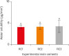
The flexural strength values of the experimental resin cements are shown in Figure 5. The flexural strength of RC1, RC2, and RC3 was 134.3 ± 9.9 MPa, 139.0 ± 8.8 MPa, and 139.6 ± 8.3 MPa, respectively. There was no significant difference among them.
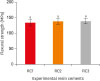 | Figure 5Flexural strength of the experimental resin cements. RC1, RC2, and RC3 are the experimental resin cements containing 3.3 wt%, 6.6 wt%, and 9.9 wt% concentrations of 10-methacryloyloxydecyl dihydrogen phosphate (10-MDP) monomer, respectively.The same letters indicate no significant difference.
|
Water sorption and solubility
Upon water immersion, all the experimental resin cement specimens increased in weight. The lowest water sorption (11.6 ± 1.25 μg/mm3) was found for RC1, followed by RC2 (16.0 ± 0.77 μg/mm3), and RC3 showed the highest value (22.6 ± 1.58 μg/mm3) (Figure 6). The water sorption of the experimental resin cements increased significantly as the 10-MDP concentration increased. The water solubility values of RC1, RC2, and RC3 were 4.04 ± 0.40 μg/mm3, 4.08 ± 0.66 μg/mm3, and 4.20 ± 1.46 μg/mm3, respectively (Figure 7). There was no significant difference among them.
 | Figure 6Water sorption of the experimental resin cements. RC1, RC2, and RC3 are the experimental resin cements containing 3.3 wt%, 6.6 wt%, and 9.9 wt% concentrations of 10-methacryloyloxydecyl dihydrogen phosphate (10-MDP) monomer, respectively.The same letters indicate no significant difference.
|
 | Figure 7Water solubility of the experimental resin cements. RC1, RC2, and RC3 are the experimental resin cements containing 3.3 wt%, 6.6 wt%, and 9.9 wt% concentrations of 10-methacryloyloxydecyl dihydrogen phosphate (10-MDP) monomer, respectively.The same letters indicate no significant difference.
|
DISCUSSION
Functional monomers chemically interact with hydroxyapatite that remains within the submicron-scale hybrid layers produced by mild self-etch adhesives. The typical molecular structure of an acidic adhesive monomer consists of three parts: a polymerizable functional group, a flexible alkylene spacer (connecting group), and a hydrophilic acidic moiety [11]. Bonding performance to dental hard tissues is predominantly influenced by the ability of the hydrophilic acidic moiety to interact with hydroxyapatite in dental hard tissues. In particular, 10-MDP readily adheres to hydroxyapatite, with a stable bonding potential [12]. This study aimed to investigate the most appropriate concentration of 10-MDP for incorporation into self-adhesive resin cements. When the 10-MDP concentration in the experimental resin cement was 6.6 wt% or more, the dentin bond strength improved compared with a concentration of 3.3 wt%. However, there was no significant difference between the µTBS of the cement with a 10-MDP concentration of 6.6 wt% and the µTBS of the cement with a 10-MDP concentration of 9.9 wt%. The µTBS results showed that a 10-MDP concentration of 9.9 wt% did not enhance bond strength.
There was no significant difference in flexural strength among all three experimental resin cements tested in this study. Dental resin composites encompass 3 main components: a resin matrix, an inorganic filler, and a coupling agent. The properties and performance of resin composites are dependent upon the 3 basic components of the material. Some physical properties—namely, strength, stiffness, abrasion resistance, and the coefficient of thermal expansion—are primarily related to the filler and the coupling agent, whereas other properties, such as color stability and softening tendency, mainly stem from the resin matrix [13-16].Thus, it can be inferred that the 10-MDP content had no effect on flexural strength.
In contrast, the water sorption of the experimental resin cements increased with higher concentrations of 10-MDP. It is thought that the phosphate group in 10-MDP increased the molecular polarity of the cements and attracted water. Resin polarity was found to be the major determinant of the equilibrium of water uptake, and less polar resins absorbed very little water compared to more polar species [17]. Water attracted to the polar groups forms hydrogen bonds [18], resulting in ‘bound water,’ which is responsible for plasticizing polymers [19,20].
There was no significant difference in water solubility among all the experimental resin cements. Ito et al. [17] compared the water sorption and solubility of Clearfil SE Bond containing 10-MDP, and reported that the values of water solubility were lower than those of water sorption. Since 10-MDP has excellent resistance to water solubility, it can be inferred that there was no significant difference in water solubility between the cements analyzed in this study.
Self-adhesive resin cements have been reported to be reliable in several clinical studies [21,22]. However, few studies have investigated self-adhesive resin cements from a long-term perspective. Deterioration of the physical properties of self-adhesive resin cements after 18 months of immersion in water has been reported [23]. Long-term immersion in water may affect the flexural strength of cements, and it has also been reported that the water sorption and solubility of self-adhesive resin cements increase over time [23,24]. Furthermore, Pan et al. [23] reported that the surface morphology and mechanical properties of self-adhesive resin cements were more susceptible to aqueous damage than those of conventional resin cements. Water is an indispensable component of self-etching adhesives, as it promotes the effective ionization of acidic adhesive monomers and the subsequent demineralization of dental hard tissues [25,26]. Ionized acidic adhesive monomers chemically interact with hydroxyapatite [27], creating micromechanical retention in the teeth that yields strong adhesion [28]. By contrast, hydrolysis breaks covalent bonds between the polymers by adding water to ester bonds, resulting in loss of resin mass. Hydrolysis is considered to be one of the main reasons for resin degradation within the hybrid layer [29,30], which leads to the creation of hybrid layers that behave as semi-permeable membranes, permitting water movement throughout the bonded interface even after the adhesive is polymerized [31]. The diminishing strength of the adhesive bond over time is caused by the hydrolysis of resin and collagen in the hybrid layer. Preservation of the bond to both the restorative material and tooth structures is imperative for the long-term retention of restorations and good marginal adaptation [32,33]. Therefore, it is necessary to determine the optimal content of 10-MDP that strikes an appropriate balance between improving the bond strength and avoiding excessive water sorption. In this study, a meaningful suggestion was made for how best to include 10-MDP in materials. Nonetheless, further research is also needed.
CONCLUSIONS
When the concentration of 10-MDP in the experimental resin cements was 6.6 wt% or more, the dentin bond strength was higher. Addition of 10-MDP to the experimental resin cement increased the water sorption of the cement in a concentration-dependent manner. Within the limitations of this study, it can be suggested that a 6.6 wt% concentration of 10-MDP is better than a concentration of 3.3 wt% or 9.9 wt% in self-adhesive resin cement.
 XML Download