

 PDF
PDF ePub
ePub Citation
 Print
Print


Abstract
Purpose
To investigate the changes of visual acuity and central macular thickness (CMT) in patients with diabetic retinopathy (DR) receiving long-term hemodialysis (HD).
Methods
From January 1, 2008, to December 31, 2018, the medical records of patients who were diagnosed with DR receiving HD three times a week for ≥18 months due to chronic kidney disease (CKD) were analyzed. Among them, patients diagnosed with DR 6 months before the start of HD were included. Patients with vitreous hemorrhage (VH) affecting visual acuity (VA), other retinal diseases, and cataract surgery after HD were excluded. The VA and CMT before HD and at 1, 3, 6, 12, and 18 months after HD were analyzed.
Results
Of the 222 eyes of 111 patients who were diagnosed with DR and received HD for CKD due to diabetes, 174 eyes with DR diagnosed after starting HD were excluded. Ten eyes with VH before starting HD, two eyes with epiretinal membrane, and four eyes with cataract surgery after starting HD were also excluded. Thirty-two eyes of 18 patients were included. The mean age of the patients was 53.71 ± 9.25 years. Twenty-four males and eight female patients were included in the study. The mean logMAR VA improved significantly from 0.36 ± 0.28 before starting HD to 0.26 ± 0.27 at 18 months after starting HD (p = 0.002). The mean CMT was significantly decreased from 307.12 ± 89.52 µm before starting HD to 279.71 ± 61.75 µm at 12 months after starting HD (p = 0.02).
Figures and Tables
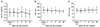 | Figure 1Long term changes of best corrected visual acuity (BCVA), central macular thickness (CMT), and intraocular pressure (IOP) before hemodialysis (HD), 1 month, 3 months, 6 months, 12 months, and 18 months after HD. (A) BCVA improved significantly at 18 months after HD compared with before HD. (B) CMT decreased significantly at 3, 12, 18 months after HD compared with before HD. (C) IOP did not show statistically significantly difference at 18 months after HD compared with before HD.
*p<0.05.
|
 | Figure 2The representative case of changes in spectral domain optical coherence tomography (SD-OCT) findings before and after hemodialysis (HD). A 47-year-old man with diabetic chronic kidney disease who was diagnosed with diabetes retinopathy 3 years previously before HD. He received intravitreal bevacizumab injection for the right eye due to diabetic macular edema three times before HD and twice after HD. (A) SD-OCT shows severe macular edema in his right eye before HD. (B) SD-OCT shows significantly reduced macular edema in his right eye at 18 months after HD.
|
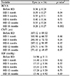
Table 1
Clinical characteristics of included patients
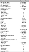
Values are presented as mean ± standard deviation or number (%) unless otherwise indicated.
HD = hemodialysis; DM = diabetes mellitus; DR = diabetic retinopathy; NPDR = non proliferative diabetic retinopathy; PDR = proliferative diabetic retinopathy; HTN = hypertension; BUN = blood urea nitrogen; GFR = glomerular filtration rate; VEGF = vascular endothelial growth factor; STTA = subtenon triamcinolone acetonide; VH = vitreous hemorrhage.
*p = 0.185; †p = 0.136; ‡p = 0.447, determined using the paired t-test.
![]()
References
1. Ciulla TA, Amador AG, Zinman B. Diabetic retinopathy and diabetic macular edema: pathophysiology, screening, and novel therapies. Diabetes Care. 2003; 26:2653–2664.
2. Nentwich MM, Ulbig MW. Diabetic retinopathy - ocular complications of diabetes mellitus. World J Diabetes. 2015; 6:489–499.

3. Klein R, Klein BE, Moss SE. Visual impairment in diabetes. Ophthalmology. 1984; 91:1–9.
4. Ticho U, Patz A. The role of capillary perfusion in the management of diabetic macular edema. Am J Ophthalmol. 1973; 76:880–886.
5. Sim DA, Keane PA, Zarranz-Ventura J, et al. The effects of macular ischemia on visual acuity in diabetic retinopathy. Invest Ophthalmol Vis Sci. 2013; 54:2353–2360.
6. Romero-Aroca P, Baget-Bernaldiz M, Pareja-Rios A, et al. Diabetic macular edema pathophysiology: vasogenic versus inflammatory. J Diabetes Res. 2016; 2016:2156273.
7. Klein R, Knudtson MD, Lee KE, et al. The Wisconsin Epidemiologic Study of Diabetic Retinopathy XXIII: the twenty-five-year incidence of macular edema in persons with type 1 diabetes. Ophthalmology. 2009; 116:497–503.
8. Ginsberg HN. The ACCORD (Action to Control Cardiovascular Risk in Diabetes) lipid trial: what we learn from subgroup analyses. Diabetes Care. 2011; 34 Suppl 2:S107–S108.
9. Leslie RD. United Kingdom prospective diabetes study (UKPDS): what now or so what. Diabetes Metab Res Rev. 1999; 15:65–71.
10. Lloyd CE, Klein R, Maser RE, et al. The progression of retinopathy over 2 years: the Pittsburgh Epidemiology of Diabetes Complications (EDC) study. J Diabetes Complications. 1995; 9:140–148.
11. Cruickshanks KJ, Ritter LL, Klein R, Moss SE. The association of microalbuminuria with diabetic retinopathy. The Wisconsin Epidemiologic Study of Diabetic Retinopathy. Ophthalmology. 1993; 100:862–867.
12. Jin DC, Yun SR, Lee SW, et al. Current characteristics of dialysis therapy in Korea: 2016 registry data focusing on diabetic patients. Kidney Res Clin Pract. 2018; 37:20–29.
13. Chelala E, Dirani A, Fadlallah A, et al. Effect of hemodialysis on visual acuity, intraocular pressure, and macular thickness in patients with chronic kidney disease. Clin Ophthalmol. 2015; 9:109–114.
14. Jung JW, Yoon MH, Lee SW, Chin HS. Effect of hemodialysis (HD) on intraocular pressure, ocular surface, and macular change in patients with chronic renal failure. Effect of hemodialysis on the ophthalmologic findings. Graefes Arch Clin Exp Ophthalmol. 2013; 251:153–162.
15. Azem N, Spierer O, Shaked M, Neudorfer M. Effect of hemodialysis on retinal thickness in patients with diabetic retinopathy, with and without macular edema, using optical coherence tomography. J Ophthalmol. 2014; 2014:709862.
16. Theodossiadis PG, Theodoropoulou S, Neamonitou G, et al. Hemodialysis-induced alterations in macular thickness measured by optical coherence tomography in diabetic patients with endstage renal disease. Ophthalmologica. 2012; 227:90–94.
17. Hwang H, Chae JB, Kim JY, et al. Changes in optical coherence tomography findings in patients with chronic renal failure undergoing dialysis for the first time. Retina. 2018; 08. 30. Accessed Aug 30, 2018. https://www.ncbi.nlm.nih.gov/pubmed/?term=Hwang+H%2C+Chae+JB%2C+Kim+JY%2C+et+al.+Changes+in+optical+coherence+tomography+findings+in+patients+with+chronic+renal+failure+undergoing+dialysis+for+the+first+time.
18. Wolf-Schnurrbusch UE, Ceklic L, Brinkmann CK, et al. Macular thickness measurements in healthy eyes using six different optical coherence tomography instruments. Invest Ophthalmol Vis Sci. 2009; 50:3432–3437.
19. Auyanet I, Rodríguez LJ, Bosch E, et al. Measurement of foveal thickness by optical coherence tomography in adult haemodialysis patients with diabetic nephropathy. Nefrologia. 2011; 31:66–69.
20. Yang SJ, Han YH, Song GI, et al. Changes of choroidal thickness, intraocular pressure and other optical coherence tomographic parameters after haemodialysis. Clin Exp Optom. 2013; 96:494–499.
21. Furushima M, Imaizumi M, Nakatsuka K. Changes in refraction caused by induction of acute hyperglycemia in healthy volunteers. Jpn J Ophthalmol. 1999; 43:398–403.
22. Diabetic Retinopathy, Browning DJ, Glassman AR, et al. Relationship between optical coherence tomography-measured central retinal thickness and visual acuity in diabetic macular edema. Ophthalmology. 2007; 114:525–536.
23. Landers MB 3rd, Stefansson E, Wolbarsht ML. Panretinal photocoagulation and retinal oxygenation. Retina. 1982; 2:167–175.
24. Nonaka A, Kiryu J, Tsujikawa A, et al. Inflammatory response after scatter laser photocoagulation in nonphotocoagulated retina. Invest Ophthalmol Vis Sci. 2002; 43:1204–1209.
25. Shimura M, Yasuda K, Nakazawa T, et al. Quantifying alterations of macular thickness before and after panretinal photocoagulation in patients with severe diabetic retinopathy and good vision. Ophthalmology. 2003; 110:2386–2394.
26. Gaucher D, Fortunato P, LeCleire-Collet A, et al. Spontaneous resolution of macular edema after panretinal photocoagulation in florid proliferative diabetic retinopathy. Retina. 2009; 29:1282–1288.
27. Tarakçioğlu M, Erbağci AB, Usalan C, et al. Acute effect of hemodialysis on serum levels of the proinflammatory cytokines. Mediators Inflamm. 2003; 12:15–19.
28. Kuo HL, Chou CY, Liu YL, et al. Reduction of pro-inflammatory cytokines through hemodiafiltration. Ren Fail. 2008; 30:796–800.
29. Hernández C, Fonollosa A, García-Ramírez M, et al. Erythropoietin is expressed in the human retina and it is highly elevated in the vitreous fluid of patients with diabetic macular edema. Diabetes Care. 2006; 29:2028–2033.
 XML Download