

 PDF
PDF ePub
ePub Citation
 Print
Print


Abstract
Purpose
To evaluate the changes in choroidal thickness and superficial vascular density of the macula and optic disc using optical coherence tomography angiography after laser photocoagulation.
Methods
We conducted a retrospective chart review of 25 eyes of diabetic retinopathy patients who underwent panretinal photocoagulation. The macula and optic disc were divided into nine areas, and the vascular density of each area was quantitatively measured using optical coherence tomography angiography. The changes in vascular density and choroidal thickness were analyzed before laser photocoagulation and at 1 week after, 1 month after, and 3 months after treatment.
Results
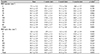
In the panretinal photocoagulation group, the average vascular densities of the macula were 13.5 ± 3.6 mm−1 before treatment, and 14.7 ± 3.1 mm−1 after 1 week, 13.7 ± 2.6 mm−1 after 1 month, and 12.8 ± 3.8 mm−1 after 3 months of treatment. The average vascular densities of the optic disc were 14.7 ± 5.2 mm−1 before treatment, and 14.1 ± 4.7 mm−1 after 1 week, 14.8 ± 5.3 mm−1 after 1 month, and 15.0 ± 4.7 mm−1 after 3 months of treatment. The average subfoveal choroidal thicknesses were 327.5 ± 57.9 µm before treatment, and 334.4 ± 52.5 µm after 1 week, 291.2 ± 52.9 µm after 1 month, and 286.3 ± 44.4 µm after 3 months of treatment.
Conclusions
The vascular density of the macula increased temporarily after 1 week of treatment but decreased afterwards. The vascular density of the optic disc decreased after 1 week of laser treatment but increased over time. The subfoveal choroidal thickness increased after 1 week of laser treatment but decreased afterwards.
Figures and Tables
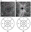 | Figure 1Early Treatment Diabetic Retinopathy Study grid subfield was applied to macular (A) and papillary area (B). ‘1’ means center. ‘2’ means nasal inner. ‘3’ means superior inner. ‘4’ means temporal inner. ‘5’ means inferior inner. ‘6’ means nasal outer. ‘7’ means superior outer. ‘8’ means temporal outer. ‘9’ means inferior outer. OD = oculus dexter; OS = oculus sinister.
|
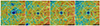 | Figure 2A case of panretinal photocoagulation in 71-year-old male. The superficial vascular density of macular area generally increased after 1 week but decreased afterwards.
|
 | Figure 3A case of panretinal photocoagulation in 69-year-old-male. The superficial vascular density of papillary area generally decreased after 1 week but increased afterwards.
|
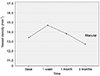 | Figure 4The calculated longitudinal changes of the average superficial vessel density of macular before and after panretinal photocoagulation. The vessel density of macular increased temporarily after 1 week but decreased afterwards.
|
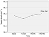 | Figure 5The calculated longitudinal changes of the average superficial vessel density of optic disc before and after panretinal photocoagulation. The vessel density of optic disc decreased after 1 week laser treatment, but increased over the time.
|

References
1. Rabiolo A, Gelormini F, Marchese A, et al. Macular perfusion parameters in different angiocube sizes: does the size matter in quantitative optical coherence tomography angiography? Invest Ophthalmol Vis Sci. 2018; 59:231–237.

2. Kohner EM, Oakley NW. Diabetic retinopathy. Metabolism. 1975; 24:1085–1102.
3. Ishibazawa A, Nagaoka T, Takahashi A, et al. Optical coherence tomography angiography in diabetic retinopathy: a prospective pilot study. Am J Ophthalmol. 2015; 160:35–44.
4. Al-Sheikh M, Akil H, Pfau M, Sadda SR. Swept-source OCT angiography imaging of the foveal avascular zone and macular capillary network density in diabetic retinopathy. Invest Ophthalmol Vis Sci. 2016; 57:3907–3913.
5. de Carlo TE, Chin AT, Bonini Filho MA, et al. Detection of microvascular changes in eyes of patients with diabetes but not clinical diabetic retinopathy using optical coherence tomography angiography. Retina. 2015; 35:2364–2370.
6. Ferris FL 3rd. Photocoagulation for diabetic retinopathy. Early Treatment Diabetic Retinopathy Study Research Group. JAMA. 1991; 266:1263–1265.
7. Okamoto M, Matsuura T, Ogata N. Effects of panretinal photocoagulation on choroidal thickness and choroidal blood flow in patients with severe nonproliferative diabetic retinopathy. Retina. 2016; 36:805–811.
8. Stefansson E. Oxygen and diabetic eye disease. Graefes Arch Clin Exp Ophthalmol. 1990; 228:120–123.
9. Spaide RF, Klancnik JM Jr, Cooney MJ. Retinal vascular layers imaged by fluorescein angiography and optical coherence tomography angiography. JAMA Ophthalmol. 2015; 133:45–50.
10. Lim HB, Kim YW, Kim JM, et al. The importance of signal strength in quantitative assessment of retinal vessel density using optical coherence tomography angiography. Sci Rep. 2018; 8:12897.
11. Nonaka A, Kiryu J, Tsujikawa A, et al. Inflammatory response after scatter laser photocoagulation in nonphotocoagulated retina. Invest Ophthalmol Vis Sci. 2002; 43:1204–1209.
12. Grunwald JE, Brucker AJ, Petrig BL, Riva CE. Retinal blood flow regulation and the clinical response to panretinal photocoagulation in proliferative diabetic retinopathy. Ophthalmology. 1989; 1518–1522.
13. Chey JH, Park JM. Retinal vascular caliber changes in diabetic retinopathy after panretinal photocoagulation and additive bevacizumab injections. J Korean Ophthalmol Soc. 2016; 57:917–923.
14. Yang HS, Kim JG, Cha JB, et al. Quantitative analysis of neural tissues around the optic disc after panretinal photocoagulation in patients with diabetic retinopathy. PLoS One. 2017; 12:e0186229.
15. Türk A, Esenülkü CM, Akyol N, et al. Pulsatile ocular blood flow changes after panretinal photocoagulation treatment in patients with proliferative diabetic retinopathy. Turk J Med Sci. 2014; 44:524–529.
16. Lee JC, Wong BJ, Tan O, et al. Pilot study of doppler optical coherence tomography of retinal blood flow following laser photocoagulation in poorly controlled diabetic patients. Invest Ophthalmol Vis Sci. 2013; 54:6104–6111.
17. Arvas S, Ocakoglu O, Ozkan S. The capillary blood flow in ischaemic type central retinal vein occlusion: the effect of laser photocoagulation. Acta Ophthalmol Scand. 2002; 80:490–494.
18. Lorusso M, Milano V, Nikolopoulou E, et al. Panretinal photocoagulation does not change macular perfusion in eyes with proliferative diabetic retinopathy. Ophthalmic Surg Lasers Imaging Retina. 2019; 50:174–178.
19. Kim JT, Lee DH, Joe SG, et al. Changes in choroidal thickness in relation to the severity of retinopathy and macular edema in type 2 diabetic patients. Invest Ophthalmol Vis Sci. 2013; 54:3378–3384.
20. Cho GE, Cho HY, Kim YT. Change in subfoveal choroidal thickness after argon laser panretinal photocoagulation. Int J Ophthalmol. 2013; 6:505–509.
21. Park BJ, Chung HW, Kim HC. Effects of diabetic retinopathy and intravitreal bevacizumab injection on choroidal thickness in diabetic patients. J Korean Ophthalmol Soc. 2013; 54:1520–1525.
 XML Download