

 PDF
PDF ePub
ePub Citation
 Print
Print


Abstract
Purpose
To investigate the clinical characteristics and histological features of tumors in caruncles.
Methods
We conducted a retrospective chart review of 126 eyes of 126 patients who underwent incisional or excisional biopsy of conjunctival masses between March 2008 and December 2016.
Results
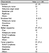
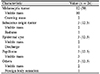
Twenty-four patients (19.0%) including 10 males and 14 females had a mass located on the caruncle. The mean age at diagnosis was 41.8 ± 13.4 years (range, 15–67 years). Most patients (75%) visited us for a cosmetically visible mass without other symptoms. The mean duration of symptoms was 73.8 ± 113.0 months (range, 1–240 months). The most common histological diagnosis was a melanocytic tumor (50%) followed by a sebaceous gland tumor (12.5%), an epidermoid cyst (12.5%), and papilloma (12.5%). The symptom duration of the melanocytic tumor was significantly longer than other types of tumors (153.6 ± 139.8 months, p = 0.025).
Figures and Tables
Figure 1
Representative photographs of caruncular tumors. (A) Melanocytic tumor, (B) sebaceous hyperplasia, (C) epidermal cyst, (D) papilloma.
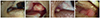
Figure 2
Representative histopathologic features of caruncular tumors. (A) Primary acquired melanosis. Increased number of melanocytes without defenite atypia is found at the basal layer of the epithelium (asterisk) (hematoxylin and eosin staining [H&E stain], ×400). (B) Sebaceous hyperplasia. Hyperplastic lobulesare found around a single duct (arrow) (H&E stain, ×12.5). (C) Epidermal cyst. Cyst wall consists of squamous epithelium and fills with keratinous materials (H&E stain, ×200). (D) Papilloma. The tumor shows papillary growth of squamous epithelium wrapped around fibrovascular cores (H&E stain, ×40).


References
1. Levy J, Ilsar M, Deckel Y, et al. Lesions of the caruncle: a description of 42 cases and a review of the literature. Eye (Lond). 2009; 23:1004–1018.
2. Evans WH. Tumors of the lacrimal caruncle. A study of 200 collected cases. Arch Ophthalmol. 1940; 24:83–106.
3. Kaeser PF, Uffer S, Zografos L, Hamédani M. Tumors of the caruncle: a clinicopathologic correlation. Am J Ophthalmol. 2006; 142:448–455.
4. Luthra CL, Doxanas MT, Green WR. Lesions of the caruncle: a clinicohistopathologic study. Surv Ophthalmol. 1978; 23:183–195.

5. Santos A, Gómez-Leal A. Lesions of the lacrimal caruncle. Clinicopathologic features. Ophthalmology. 1994; 101:943–949.
6. Serra GM. Tumors of the lacrimal caruncle. Clinical study and pathological-case history. Boll Ocul. 1928; 7:783–802.
7. Shields CL, Shields JA, White D, Augsburger JJ. Types and frequency of lesions of the caruncle. Am J Ophthalmol. 1986; 102:771–778.
8. Wilson RP. Tumours and cysts of the lacrimal caruncle. Trans Ophthalmol Soc N Z. 1959; 11:23–32.
9. Lee JW, Kim SY, Jung MS. Pathological classification and incidence of conjunctival tumors in korean patients: single-center study. J Korean Ophthalmol Soc. 2019; 60:119–125.
10. Jang SM, Lee H, Baek SH. Clinical characteristics of benign eyelid tumors. J Korean Ophthalmol Soc. 2016; 57:174–180.
11. Choi JH, Chi MJ, Baek SH. Clinical analysis of benign eyelid and conjunctival tumors. J Korean Ophthalmol Soc. 2003; 44:1268–1277.
12. Nasiri AM, Al-Akeel ES, Rayes NH. Differences in survival by race/ethnicity among cutaneous melanoma patients in the United States over a period from 1982 to 2011. Int J Adv Med. 2018; 5:5–10.
13. Wang Y, Zhao Y, Ma S. Racial differences in six major subtypes of melanoma: descriptive epidemiology. BMC Cancer. 2016; 16:691.
14. Shields CL, Fasiuddin AF, Mashayekhi A, Shields JA. Conjunctival nevi: clinical features and natural course in 410 consecutive patients. Arch Ophthalmol. 2004; 122:167–175.
15. Kao A, Afshar A, Bloomer M, Damato B. Management of primary acquired melanosis, nevus, and conjunctival melanoma. Cancer Control. 2016; 23:117–125.
16. Shields JA, Shields CL, Mashayekhi A, et al. Primary acquired melanosis of the conjunctiva: experience with 311 eyes. Trans Am Ophthalmol Soc. 2007; 105:61–67. discussion 71–2.
17. Miura-Karasawa M, Toshida H, Ohta T, Murakami A. Papilloma and sebaceous gland hyperplasia of the lacrimal caruncle: a case report. Int Med Case Rep J. 2018; 11:91–95.
18. Avisar I, Yassur I, Kremer I. Multiple conjunctival papillomas of eyelid margins in pemphigus vulgaris. Case Rep Ophthalmol Med. 2011; 2011:174912.
19. Yang JM, Lee JJ, Yoon KC. Clinical analysis of primary conjunctival malignant lymphoma. J Korean Ophthalmol Soc. 2014; 55:1298–1306.
20. Kaliki S, Arepalli S, Shields CL, et al. Conjunctival papilloma: features and outcomes based on age at initial examination. JAMA Ophthalmol. 2013; 131:585–593.
21. Ahn CJ, Kim NJ, Choung HK, et al. Clinical features and surgical treatment outcomes of conjunctival squamous papilloma. J Korean Ophthalmol Soc. 2016; 57:167–173.
 XML Download