Abstract
Orthodontic miniscrews have been widely used in various areas, because they are stable, easy to use, and inexpensive. Therefore, the aims of this study are to measure the palatal bone thickness, to analysis the correlation between the size of the alveolar arch and palatal bone thickness, and to discuss the skeletal structure of the hard palate for miniscrew placement. Twenty-four dry skulls in Koreans were used. The three different horizontal reference lines were established at first premolar and second premolar, between second premolar and first molar, and first molar. And then, a total of 12 points were set up in relation to each horizontal reference line by drawing a vertical reference line perpendicular to central incisor, between central incisor and lateral incisor, lateral incisor, between lateral incisor and canine. At each point, the palatal bone thickness, the width and length of the alveolar arch, and the width and length of the incisive foramen were measured directly with the bone caliper and the digital caliper. The correlation between the width and the length of alveolar arch and the palatal bone thickness was analyzed. The mean of palatal bone thickness based on the horizontal reference line was 11.4±3.2 mm in between first premolar and second premolar, 7.4±2.4 mm in between second premolar and first molar, and 5.2±1.5 mm in first molar, decreased posteriorly with statistically significant difference. The position between first premolar and second premolar showed a constant thickness, and thickened laterally from the median palatal suture due to the alveolar process, but no statistically significant difference. At the position between second premolar and first molar and the position in first molar, it were also constant, then became significantly thicker toward point between lateral incisor and canine due to the alveolar process and the palatal spine. The width of the alveolar arch was correlated with the length of the alveolar arch and the palatal bone thickness of between first premolar
and second premolar, but not with the length of the alveolar arch and the palatal bone thickness. These results can provide useful anatomical data on palatal bone thickness including skeletal structures of hard palate for orthodontic miniscrew placement.
Go to : 
References
1. Wehrbein H, Merz BR, Diedrich P. Palatal bone support for orthodontic implant anchorage – a clinical and radiological study. Eur J Orthod. 1999; 21:65–70.

2. Baumgaertel S. Quantitative investigation of palatal bone depth and cortical bone thickness for mini-implant placement in adults. Am J Orthod Dentofacial Orthop. 2009; 136:104–8.

3. Lai RF, Zou H, Kong WD, Lin W. Applied anatomic site study of palatal anchorage implants using cone beam computed tomography. Int J Oral Sci. 2010; 2:98–104.

4. Kim HJ, Yun HS, Park HD, Kim DH, Park YC. Soft-tissue and cortical-bone thickness at orthodontic implant sites. Am J Orthod Dentofacial Orthop. 2006; 130:177–82.

5. Park JT, Jeong RR, Kim KT, Kim SB, Hu KS, Kim HJ, et al. Maxillary soft tissue and cortical bone thickness for mini-im-plant placement. Korean J Phys Anthropol. 2008; 21:215–24.

6. Yadav S, Sachs E, Vishwanath M, Knecht K, Upadhyay M, Nanda R, et al. Gender and growth variation in palatal bone thickness and density for mini-implant placement. Prog Orthod. 2018; 19:43.

7. Chhatwani S, Rose-Zierau V, Haddad B, Almuzian M, Kirschneck C, Danesh G. Three-dimensional quantitative assessment of palatal bone height for insertion of orthodontic implants – a retrospective CBCT study. Head Face Med. 2019; 15:9.

8. Kang S, Lee SJ, Ahn SJ, Heo MS, Kim TW. Bone thickness of the palate for orthodontic mini-implant anchorage in adults. Am J Orthod Dentofacial Orthop. 2007; 131(4 Suppl):S74–81.

9. King KS, Lam EW, Faulkner MG, Heo G, Major PW. Vertical bone volume in the paramedian palate of adolescents: a computed tomography study. Am J Orthod Dentofacial Orthop. 2007; 132:783–8.

10. Bernhart T, Vollgruber A, Gahleitner A, Dörtbudak O, Haas R. Alternative to the median region of the palate for placement of an orthodontic implant. Clin Oral Implants Res. 2000; 11:595–601.

11. Kim YJ, Lim SH, Gang SN. Comparison of cephalometric measurements and cone-beam computed tomography-based measurements of palatal bone thickness. Am J Orthod Dentofacial Orthop. 2014; 145:165–72.

12. AlSamak S, Gkantidis N, Bitsanis E, Christou P. Assessment of potential orthodontic mini-implant insertion sites based on anatomical hard tissue parameters: a systematic review. Int J Oral Maxillofac Implants. 2012; 27:875–87.
13. Wang M, Sun Y, Yu Y, Ding X. Evaluation of palatal Bone thickness for insertion of orthodontic mini-implants in adults and adolescents. J Craniofac Surg. 2017; 28:1468–71.

14. Wang Y, Qiu Y, Liu H, He J, Fan X. Quantitative evaluation of palatal bone thickness for the placement of orthodontic miniscrews in adults with different facial types. Saudi Med J. 2017; 38:1051–7.

15. Suteerapongpun P, Wattanachai T, Janhom A, Tripuwabhrut P, Jotikasthira D. Quantitative evaluation of palatal bone thickness in patients with normal and open vertical skeletal con-figurations using cone-beam computed tomography. Imaging Sci Dent. 2018; 48:51–7.

16. Ryu JH, Park JH, Vu Thi Thu T, Bayome M, Kim Y, Kook YA. Palatal bone thickness compared with cone-beam computed tomography in adolescents and adults for mini-implant placement. Am J Orthod Dentofacial Orthop. 2012; 142:207–12.

17. Gündüz E, Schneider-Del Savio TT, Kucher G, Schneider B, Bantleon HP. Acceptance rate of palatal implants: a questionnaire study. Am J Orthod Dentofacial Orthop. 2004; 126:623–6.

18. Yu SK, Lee MH, Park BS, Jeon YH, Chung YY, Kim HJ. Topographical relationship of the greater palatine artery and the palatal spine. Significance for periodontal surgery. J Clin Periodontol. 2014; 41:908–13.

19. Yu SK, Lim JW, Cho YH, Kim HJ. Anatomical assessment of the palatal mucosa for connective tissue grafts. Oral Biol Res. 2018; 42:156–62.

20. Al-Amery SM, Nambiar P, Jamaludin M, John J, Ngeow WC. Cone beam computed tomography assessment of the maxillary incisive canal and foramen: considerations of anatomical variations when placing immediate implants. PLoS One. 2015; 10:e0117251.

21. Song WC, Jo DI, Lee JY, Kim JN, Hur MS, Hu KS, et al. Microanatomy of the incisive canal using three-di-mensional reconstruction of microCT images: an ex vivo study. Oral Surg Oral Med Oral Pathol Oral Radiol Endod.
22. Henriksen B, Bavitz B, Kelly B, Harn SD. Evaluation of bone thickness in the anterior hard palate relative to midsagittal orthodontic implants. Int J Oral Maxillofac Implants. 2003; 18:578–81.
Go to : 
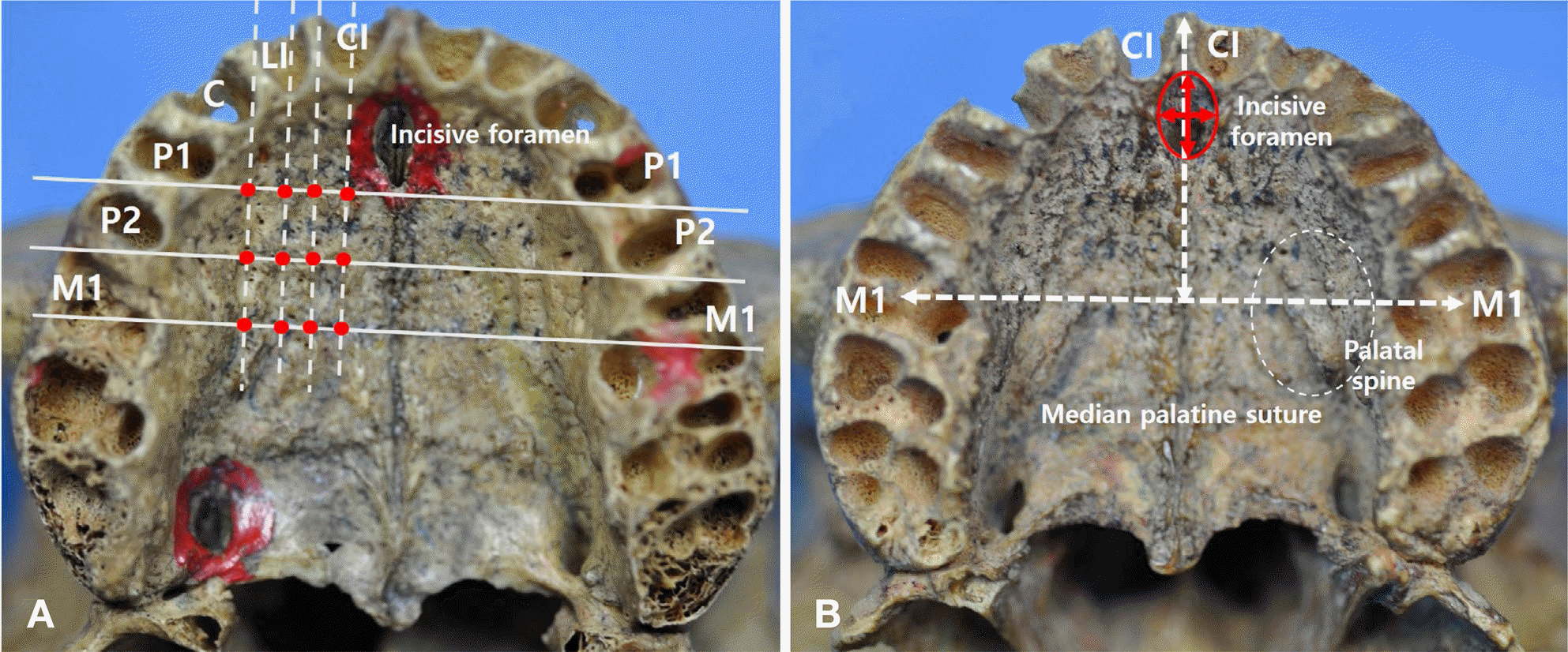
 | Fig. 1.Photographs showing the parameters of the palatal bone thickness, the width and length of the alveolar arch, and the width and length of the incisive foramen. A represents a total of 12 points (red dots) that are measurement points crossing between horizontal reference line (solid lines) and vertical reference line (dotted lines) at each tooth position. B represents the width and length of the alveolar arch (white dotted double arrows) and the width and length of the incisive foramen (red solid double arrows). CL, central incisor; LI, lateral incisor; C, canine; P1, first premolar; P2, second premolar; M1, first molar. |
Table 1.
Palatal bone thickness according to tooth position at a total of 12 measurement points
| Vertical reference 1 | Vertical reference 2 | Vertical reference 3 | Vertical reference 4 | Total | p | |
|---|---|---|---|---|---|---|
| (n = 48) | (n = 48) | (n = 48) | (n = 48) | |||
| P1-P2 | 11.1±3.2 | 10.9±3.1 | 11.3±3.3 | 12.3±3.3 | 11.4±3.2 | 0.160 |
| (4.8–17.0) | (4.0–16.0) | (4.3–17.0) | (5.3–18.0) | |||
| P2-M1 | 7.0±2.1 a | 6.9±2.1 b | 7.2±2.4 | 8.4±2.5 ab | 7.4±2.4 | 0.006∗ |
| (2.0–10.8) | (2.5–10.8) | (2.3–13.0) | (3.5–14.5) | |||
| M1 | 5.3±1.3 | 4.7±1.3 c | 4.9±1.5 d | 5.8±1.8 cd | 5.2±1.5 | 0.003∗ |
| (2.0–7.8) | (2.0–7.5) | (2.3–10.0) | (2.3–10.5) | |||
| Total | 7.8±3.3 | 7.5±3.5 | 7.8±3.7 | 8.8±3.7 | ||
| p | 0.000∗ | 0.000∗ | 0.000∗ | 0.000∗ |
Vertical reference 1, vertical extension line of central incisor; Vertical reference 2, vertical extension line of between central incisor and lateral incisor; Vertical reference 3, vertical extension line of lateral incisor; Vertical reference 4, vertical extension line of between lateral incisor and canine. P1, first premolar; P2, second premolar; M1, first molar.
Table 2.
Correlation coefficient between the width and the length of alveolar arch and the palatal bone thickness according to tooth position
| Alveolar arch width (n = 24) | Alveolar arch length(n = 24) | P1-P2 (n = 48) | P2-M1 (n = 48) | M1 (n = 48) | |
|---|---|---|---|---|---|
| Alveolar arch width | 1 | 0.46∗ | 0.42∗ | 0.05 | 0.02 |
| Alveolar arch length | 1 | 0.08 | – 0.02 | – 0.08 | |
| Alveolar arch length P1-P2 | 1 | 0.081 | 0.02 0.76∗∗ | 0.08 0.48∗∗ | |
| P2-M1 | 1 | 0.74∗∗ |


 PDF
PDF ePub
ePub Citation
 Print
Print


 XML Download