

 PDF
PDF ePub
ePub Citation
 Print
Print


INTRODUCTION
MicroRNAs (miRs) are small non-coding RNAs that typically consist of 18-25 nucleotides. They act as dynamic post-transcriptional regulators of gene networks and play a crucial role in regulating various biological processes such as cell signaling, biochemical pathways, tissue and organ development, and more. The innate functions of immune cells and their proliferation in response to pathogenic stimuli are also under the regulatory influence of miRs.1
There are 2 different classes of miRs, the intracellular and the extracellular, classified according to their topographical identification. The expression of specific intracellular miRs has tissue- and disease-specific patterns and therefore has been extensively studied as both prognostic and diagnostic biomarkers.2 In addition, there are extracellular miRs (ex-miRs, circulating miR), contained in extracellular vesicles (exosomes, microvesicles and apoptotic bodies). The ex-miRs are present and stable in a diverse array of extracellular body fluids3 and can be transferred between dendritic cells, from macrophages to epithelial cell lines, and between T cells and antigen-presenting cells in vitro and possibly in vivo.
4
The importance of miRs in regulating normal intracellular functions is becoming increasingly clear as more miR targets are discovered and the molecular mechanisms underlying miR gene regulation become unravelled.56 Modulation of miR expression has been studied in vitro as well as in vivo and has been used to ascribe key roles for miRs in epithelial function.789101112 Emerging studies implicate specific miRs in controlling epithelial cell processes such as regulation of cellular differentiation, determination of epithelial cell fate (cell death and proliferation), initiation and regulation of anti-microbial immunity, fine-control of inflammatory responses and activation of intracellular signaling pathways.910111314 Such control of epithelial cell functions is likely vital to fine-tuning of the epithelial immune response against infection. The roles of miRs in normal lung development and respiratory diseases have been extensively reviewed,15161718 including reviews on specific respiratory diseases such as asthma,19 chronic obstructive pulmonary disease,20 cystic fibrosis,21 idiopathic pulmonary fibrosis22 and lung cancer.2324
Evidently, miRs are critical for lung development and for maintaining disease-free lungs.252627 Comparisons of normal lung tissue and tissue from asthmatic patients reveal significant differences in miR profiles and suggest that miRs serve as a regulatory layer in the pathogenesis of asthma. Asthma is a heterogeneous disorder involving many cell types, such as alveolar macrophages, smooth muscle cells, innate lymphoid cells, epithelial cells and mast cells, making it difficult to discern underlying molecular mechanisms of the disease. Similar heterogeneity is observed in asthma phenotypes and the variations are observed among studies may be related to asthma severity (mild, moderate and severe). The results of studies regarding miR expression on asthmatic versus normal epithelium are sometimes contradictory, depending on the clinical characterization of subjects, sample location (nasal cavity or bronchi) and sample type (biopsies, swabs, lavage, primary cells and cell lines).
Viral acute respiratory infections (ARIs) are the most common cause of acute respiratory symptoms and many of these infections are linked to asthma exacerbations, which are in turn responsible for a considerable proportion of the morbidity and all the asthma-related mortality. The immune response against respiratory viruses, such as human rhinovirus (RV), respiratory syncytial virus (RSV) and influenza virus (IFV), has been associated with altered expression of several miRs. Changes in the expression profile of miRs in epithelial cells may contribute to the pathogenesis of acute and chronic respiratory diseases.28 For viral ARIs, miRs are involved in both the up and down regulations of the innate immune response. In recent years, miRs have been studied in order to discover anti-viral ARI drug targets as well as biomarkers for diagnosis, severity and prognosis.29
In the context of this review, we summarize recent findings on miR expression and function in asthma, and their role in the regulation of viral ARIs. The literature on miRs involved in asthma and ARI's was reviewed according to cell tissue specificity, independently for epithelium (cell lines, primary epithelial cells nasal or bronchial, nasal washes, bronchial lavage and sputum), and peripheral blood mononuclear cells (PBMCs). As studies on miRs seem to play an emerging role in asthma, we differentiate miR profiles according to asthma severity (mild, moderate and severe), highlighting the heterogeneity of this disease. We also identify miRs that may be involved in virus-induced asthma exacerbation, separately reviewing the main virus implicated in exacerbations (RV, RSV and IFV).30 Finally, using the above information, we attempt to link the miR profiles of asthmatic patients and the miR profiles that are induced by ARIs. The main goal/purpose of this linkage is to address the question of whether there might be a specific miR deficit in asthmatic subjects, making them more susceptible and/or reactive to ARIs.
miR EXPRESSION PROFILES IN THE EPITHELIUM OF MILD ASTHMATIC PATIENTS
The bronchial epithelium of mild asthmatics compared to healthy individuals has been shown to have up- and down-regulated miRs which are implicated in inflammation pathways, epithelium development and epithelial homeostasis (Table 1 and Supplementary Table S1).
Table 1
miRs up- or down-regulated in the epithelial cells of asthmatic subjects compared to healthy controls
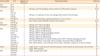
| Asthma profile | miR | Regulation in asthmatics | Role/pathway/target | Reference |
|---|---|---|---|---|
| Mild | let-7f | ↑ | - | 31 |
| Mild/severe | let-7b | ↓ | - | 40 |
| let-7a | ↓ | Pathways: JAK-STAT signalling, cytokine network and inflammatory response | 3340 | |
| Mild | let-7e | ↓ | - | 33 |
| miR-7-5p | ↑ | - | 35 | |
| miR-155 | ↓ | Pathway: IL-13 pathway in human macrophages determining the M2 phenotype | 33 | |
| miR-181c | ↑ | - | 31 | |
| Severe | miR-181b-5p | ↓ | Pathway: proinflammatory cytokines expression (IL-13, IL-1b, CCL11) | 34 |
| miR-19a | ↑ | Role: enhances cell proliferation of BEC in severe asthma | 3542 | |
| Mild | miR-19b-3p | ↑ | - | 35 |
| Mild/severe | miR-200b | ↓ | Pathway: CD4+ Th2 cell activation | 40 |
| miR-200c | ↓ | Pathway: CD4+ Th2 cell activation | 40 | |
| Mild | miR-34b-5p | ↓ | Pathway: ciliated cell differentiation | 36 |
| miR-34c-5p | ↓ | Pathway: ciliated cell differentiation | 36 | |
| miR-34 | ↓ | Pathway: IGFBP-3 mediated autophagy activation | 32 | |
| miR-449a | ↓ | Pathway: ciliated cell differentiation | 36 | |
| miR-449b-5p | ↓ | Pathway: ciliated cell differentiation | 36 | |
| miR-449 | ↓ | Pathways: differentiation of ciliated epithelial cells, IGFBP-3 mediated autophagy activation | 32 | |
| miR-203a | ↑ | Role: repress MEF2C, a transcription factor, leading to decreased cellular proliferation | 35 | |
| miR-3065-3p | ↑ | Role: repress MDGA1, a cell membrane anchor protein, resulting in suppression of cell-cell adhesion | 35 | |
| miR-221-5p | ↑ | - | 35 | |
| Severe | miR-221-3p | ↓ | Target: CXCL17 in asthmatics and chemokine suppress CCL24 (eotaxin-2), CCL26 (eotaxin-3) and POSTN | 44 |
| miRNA-221 | ↑ | Target: SIRT1 reduction | 43 | |
| Mild | miRNA-203 | ↓ | Target: aquaporin gene AQP4 | 31 |
| miR-487b | ↑ | - | 31 | |
| miR-487a | ↓ | - | 35 |
![]()
In primary bronchial epithelial cells cultured at the air-liquid interface, a miR microarray was performed, showing higher expression of miR-let-7f, miR-181c and miR-487b, but lower expression of miR-203 in mild asthmatics compared to healthy controls. Molecular network analysis indicated that putative targets of these miRs were principally involved in regulating the expression of inflammatory pathway genes and was also used to identify a novel asthma-associated gene.31 The miR-let-7 inhibits IL-13 expression, and silencing of miR-let-7 inhibits cytokine production and attenuates disease symptoms in an animal model of asthma.32 The miRs from this family (miR-let-7 and miR-let-7e) were down-regulated in nasal biopsies of asthmatic patients (with or without rhinitis).33 The members of the miR-181 group have been identified to be differentially expressed in asthmatics: miR-181b-5p has been reversely associated with eosinophilic asthma. Generally, miR-181b targets SPP1, which in turn regulates IL-13-induced proinflammatory cytokines IL1B and CCL11 expression.34 Also, the top-ranked predicted target of the highly down-regulated miR-203 in asthmatic cells was the aquaporin gene AQP4.31 On the contrary, in bronchial biopsy specimens from of mild-moderate asthmatics, miR-203a was up-regulated. It is thus suggested that it may repress MEF2C, a transcription factor, leading to decreased cellular proliferation. In addition, up-regulated miR-3065-3p may repress MDGA1, a cell membrane anchor protein, resulting in suppression of cell-cell adhesion. It has been proposed that aberrant regulations of miR-203a-MEF2C and miR-3065-3p-MDGA1 play important roles in airway epithelial homeostasis in asthma.35
Another genome wide profiling study of bronchial epithelial brushings also revealed 4 members of the miR-34/449 family (miR-34b-5p, miR-34c-5p, miR-449a, and miR-449b-5p) that were significantly suppressed in asthma.36 The miR-34/449 family is closely associated with regulation of epithelial cell proliferation and differentiation. Specifically, miR-449 is essential in regulating airway ciliated cells by targeting NOTCH1.37 Notch signaling triggers airway mucous metaplasia and inhibits alveolar development (ciliated cells).38 The low level of miR-449 in epithelial cells of asthmatics may therefore shift the fate of these cells toward more mucous production. One salient finding is that the members of the miR-34/449 family are highly repressed in vivo in asthma and repressed in vitro by IL-13 exposure, and that this repression persists despite corticosteroid treatment.36
Another miR which has been repeatedly associated with asthma and is involved in the regulation of allergic inflammation is miR-155. It is down-regulated in nasal biopsy specimens from asthmatics compared to healthy individuals and its expression in nasal mucosa in long-term asthmatics was similar in subjects with or without concomitant Ars.33 The miR-155 plays an important role in host defence and in the function of B and T lymphocytes, is down-regulated in asthmatic bronchial epithelial cells compared to cells from healthy donors, independent of asthma severity.39, 40 Plasma miR-155 levels are elevated in severe asthmatics when compared to non-asthmatics or mild-to-moderate asthmatics. The increased plasma miR-155 levels were also observed in asthmatics with cockroach allergy compared to controls, through regulation of the ROS-COX-2 gene axis that is related to cockroach allergen-induced oxidative stress.41
EPITHELIAL miR EXPRESSION PROFILES IN SEVERE ASTHMA
The bronchial epithelium is considered a key player in coordinating airway wall remodeling. In mild asthma, the epithelium is damaged, and fails to proliferate and repair, whereas in severe asthma, the epithelium is highly proliferative and thicker. Differences occur in the expression profiles of miRs in relation to asthma severity (Supplementary Table S1).
In the epithelium of severe asthmatics, miR-19a had higher expression when compared to cells from mild asthmatics and healthy controls, and these levels were not restored by corticosteroids. Functional studies suggested that miR-19a enhances cell proliferation of BEC in severe asthma through targeting the TGF-β receptor 2 mRNA. Moreover, repressed expression of miR-19a increased SMAD3 phosphorylation through TGF-β receptor 2 signaling and abrogated bronchial epithelial cell proliferation. This study uncovered a new regulatory pathway involving miR-19a that is critical to the severe phenotype of asthma and indicated that down-regulating miR-19a expression could be explored as a potential new therapy to modulate epithelium repair in asthma.42
Compared to healthy controls, miR-221 expression was significantly increased in bronchial epithelial cells from severe asthmatic subjects. In addition, miR-221 seems to be involved in airway epithelial injury by targeting SIRT1 mRNA, which regulates cell growth, cell differentiation, aging, energy metabolism and, importantly, inflammation.43 Furthermore, in the sputum of patients with eosinophilic asthma miR-221-3p was increased. Functional experiments have indicated that miR-221-3p suppresses anti-inflammatory cytokine (C-X-C motif) ligand 17 (CXCL17) expression and enhances CCL24, CCL26 and POSTN expression via the p38 MAPK pathway. Airway overexpression of miR-221-3p, in exacerbated airway eosinophilic inflammation, suppressed CXCL17 expression and enhanced CCL24, CCL26 and POSTN in house dust mite-challenged mice.44
Bronchoalveolar lavage of moderate-severe asthmatic patients revealed that miR-200b and miR-200c were significantly reduced in asthmatic patients compared to healthy controls. The reduction was validated in 2 independent models of allergen-induced allergic airway inflammation and further demonstrated to be inversely correlated with asthma severity as well as increased IL-33 production in asthmatic patients. In addition, the miR-200b and miR-200c binding sites in the 3' UTR of IL-33 mRNA were identified by bioinformatics analysis and reporter gene assay. Experimental assays showed that while inhibition of endogenous miR-200b and miR200c increased, the IL-33 expression was increased in lung epithelial cells. Exogenous administration of miR-200b to lungs of mice with allergic inflammation resulted in a decrease in IL-33 levels and resolution of airway inflammation phenotypes. In conclusion, miR-200b and miR-200c regulate the expression of IL-33, thus playing a potentially basic role in asthma.40
Last but not least, Let-7a, a miR extensively reviewed for its role in mild asthma, showed reduced expression in bronchial biopsy specimens from severe asthmatics compared to mild asthmatics.40, 45 The pathways of differentially expressed miRs are JAK-STAT signaling, cytokine/chemokine signaling pathway, ciliated cell differentiation, cell proliferation, CD4+ T helper 2 (Th2) cell activation and cell adhesion molecules pathway (Table 1). It is worthy to mention that many of the above differentially expressed miRs in asthmatic epithelium are implicated directly or indirectly with the IL-13 pathway, a pleiotropic Th2 cytokine that has been shown to be central to the pathogenesis of asthma.46 In summary, miRs may be sensitive markers for chronic inflammation in the airway epithelium due to their different patterns of expression according to asthma severity and may prove useful for phenotyping these patients.33
miRs PROFILES IN PBMCs OF PATIENTS WITH ASTHMA
Cells derived from peripheral blood include key cellular components in allergic inflammation and asthma. Several studies have compared the expression pattern of PBMC-derived miRs between asthmatic and healthy individuals (Table 2 and Supplementary Table S2). CD14+ and CD16+ intermediate monocytes were found to be increased in patients with bronchial asthma and were also found to express high levels of miR-124. The miR-124 serves as a regulator of the M2 polarization and its overexpression in asthmatic macrophages resulted in down-regulation of a number of M1 markers, such as MHC class II and CD86, as well as in up-regulation of M2 markers such as Fizz1 and Arg1.47 In another study, miR-145 from CD4+ T cells was found up-regulated in asthmatics compared to healthy controls, and RUNX3 expression was found to be suppressed in these patients, suggesting that there is a negative correlation between miR-145 and RUNX3.48 Up-regulation of miR-221 and miR-485-3p has been reported in children with asthma compared to healthy controls, together with down-regulated Spred-2, which is the predicted target of these miRs.49 Recently, studies have used CD4+ T cells from bronchoalveolar lavage and from PBMCs of steroid-naive and steroid-using asthmatics and compared them to healthy individuals. Additionally, miR-19a was up-regulated in all asthmatic patients compared to healthy individuals; this miR promoted TH2 cytokine production and amplified inflammatory signaling by directly targeting inositol phosphatase PTEN, the signalling inhibitor SOCS1 and the deubiquitinase A20.50
Table 2
miRs up- and down-regulated in PBMCs from asthmatic subjects compared to healthy controls
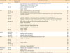
| Asthma profile | miR | Regulation in asthmatics | Role/pathway/target | Reference |
|---|---|---|---|---|
| Severe | miR-155 | ↑ | Role: associated with the expression of the Th2 cytokines IL-5 and IL-13 | 55 |
| Pathway: allergic inflammation in T-cells | ||||
| miR-126 | ↑ | Target: increased IL-4, reduced IFN-γ | 57 | |
| miR-192 | ↓ | Pathway: activation of Tfh cells | 58 | |
| Target: CXCR5 | ||||
| Mild | miR-145 | ↑ | Target: RUNX3 expression in asthma patients | 48 |
| miR-323-3p | ↑ | Role: up-regulated in IL-22-producing T cells | 51 | |
| miR-93 | ↓ | Pathways: leukocyte activation/extrinsic apoptotic signalling pathway/MAPK signalling pathway/cytokine production | 51 | |
| miR-181a | ↓ | - | 51 | |
| miR-26a | ↓ | Pathways: regulation of cell proliferation/MAPK cascade/Wnt signalling pathway | 51 | |
| miR-874 | ↓ | Pathways: response to cytokine stimulus/immune effector process/T-cell activation/ Apoptotic signalling pathway/MAPK signalling pathway/TNF signalling pathway/TNF signalling pathway | 51 | |
| miR-323-3p | ↑ | Role: down-regulated in IL-22- and IL-17-double-positive T-cells | 51 | |
| Target: IL-22 reduction | ||||
| miR-19a | ↑ | Role: promotes Th2 cytokine production | 50 | |
| Targets: PTEN, SOCS1, deubiquitinase A20 | ||||
| miR-15a | ↓ | Pathway: angiogenic or fibrotic processes | 52 | |
| Target: VEGFA | ||||
| miR-15b | ↓ | Target: VEGFA | 52 | |
| miR-20a | ↓ | Target: VEGFA | 52 | |
| Severe | miR-28-5p | ↓ | - | 59 |
| miR-146a | ↓ | Pathway: vitamin D pathway | 59 | |
| miR-146b | ↓ | Pathway: vitamin D pathway | 59 | |
| Mild | miR-21-5p | ↑ | Role: predicted to inhibit differential responses to HDM | 56 |
| miR-98 | ↑ | Target: TSP1 reduction | 61 | |
| miR-19a | ↓ | - | 53 | |
| miR-19b | ↓ | - | 53 | |
| miR-221 | ↑ | Target: Spred-2 | 49 | |
| miR-485-3p | ↑ | - | 49 | |
| miR-625-5p | ↓ | Pathways: Inflammatory cytokine | 49 | |
| Targets: CBL, PPARGC1B, ESR3 | ||||
| Mild/severe | miR-192 | ↓ | Pathway: cell cycle regulation of blood cells | 62 |
| Severe | miR-29c | ↓ | Target: B7-H3 | 60 |
| Pathway: Th cell differentiation | ||||
| Mild | miR-124 | ↑ | Role: regulator of the M2 polarization in various subsets of monocytes cells | 47 |
![]()
IL-22- and IL-17-positive T cells were sorted from PBMCs of patients with bronchial asthma and compared to healthy controls. The increased expression of miR-323-3p and reduced expression of miR-93, miR-181a, miR-26a and miR-874 were detected in IL-22-producing T cells. The differentially expressed miRs were proposed to play a role in the proliferation, differentiation and effector functions of T cells. Further analysis showed that miR-323-3p acts in a negative feedback loop to control the production of IL-22 in IL-22/IL-17-producing T cells and might thus impact the T cell responses in asthma.51 The 3 miRs, miR-15a, miR-15b, miR-20a — from CD4+ T cells binding to 3' UTR of VEGFA were also found down-regulated in paediatric atopic asthmatics, while their target demonstrated increased levels.52 T cells derived from bronchial biopsy samples of mild asthmatics showed decreased levels of miR-125b, miR-19a, miR-19b and miR-106b, and exhibited decreased airway-specific expression compared to healthy controls.53
Furthermore, 3 novel miRs, miR-22-3p, miR-513a-5p and miR-625-5p, were found to be significantly down-regulated in children with dust mite-induced asthma compared to controls, whereas the transcript levels of Cbl proto-oncogene, E3 ubiquitin protein ligase (CBL), peroxisome proliferator-activated receptor gamma, coactivator 1 beta (PPARGC1B) and estrogen receptor 1 (ESR1) that are targeted by these miRNAs were increased. These miRs may play a role in the regulation of the immune response and inflammatory cytokine pathways.54 In another study, miR-155 was up-regulated in CD4+ T cells from dust mite-allergic asthmatics, compared to allergic rhinitis and non-asthmatic individuals. It was also found that miR-155 is differentially expressed in allergic T cells exposed to dust mite extract, and it is inhibited by glucocorticoids. miR-155 is positively associated with the expression of the TH2 cytokines (IL-5 and IL-13) and thus contributes to allergic inflammation in T cells and could be an anti-inflammatory target of steroids.55 It has been indicated that miR-21-5p from PBMC of dust mite sensitized (HDM) atopics with current asthma are up-regulated and inhibit differential responses to HDM in asthmatics versus non-sensitized controls.56
EXPRESSION PATTERNS OF miRs FROM PBMCs IN ACUTE AND SEVERE ASTHMA
In PBMC from children with acute asthma, circulating miR-126 levels, the percentages of interleukin IL-4 levels, and Th17 cells were significantly higher than those in the control group, whereas the percentages of iIFN-γ levels and the CD4 + CD25 + Treg cells were significantly lower.57 In another acute asthma study, miR-192 was down-regulated and it was shown that it blocks the activation pathway of T follicular helper cells by targeting CXCR5.58 Three miRs miR-28-5p, miR-146a, miR-146b has been documented to be down-regulated in severe asthma associated with the activation of circulating CD8+ T cells, but not CD4+ T cells.59
In a recent study, miR-29c which was down-regulated in asthmatics has been found to bind directly to plasma B7-H3 molecules up-regulated. The miR-29c regulates Th2/Th17-cell differentiation and transfection with anti-miR-29c into macrophages enhanced ROR-γt and GATA-3 expression in co-cultured CD4+ T cells, and increase levels of IL-4 and IL-17 in the cell supernatants.60
Collectively, miRs are differentially expressed in PBMCs of asthmatic subjects and appear to be involved in basic allergic inflammatory and immune response pathways (Table 2). Their role in promoting Th2 cytokine production and regulating M2 polarization in various subsets of monocytes render them key players in asthma.
VIRUS-INDUCED miRs IN THE AIRWAY EPITHELIUM
ARIs are the most common cause of acute respiratory symptoms (e.g., flu and bronchitis), and these infections have been linked to the exacerbation of symptoms in chronic respiratory diseases, most notably asthma.
The host innate immune response is the first line of defence against all pathogens. A large variety of cells, including epithelial cells, dendritic cells, granulocytes, monocytes, macrophages and natural killer cells, play distinct roles in controlling infection.
Several miRs are induced during viral infection, modulating the function of each of these cell types (Table 3, Supplementary Table S3). Expression of miRs during respiratory infections is gaining attention in recent studies due to their potential in antiviral treatment.63
Table 3
miRs up- or down-regulated in epithelial cells of asthmatic subjects compared to healthy controls after virus infection (RV, RSV and IFV)
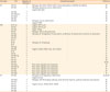
| Virus type | miR | Regulation in asthmatics | Role/pathway/target | Reference |
|---|---|---|---|---|
| RV | miR-155 | ↑ | Pathways: Akt, TGF-β, MAPK, STAT3, immune polarization, epithelial remodeling | 69 |
| miR-128 | ↑ | Pathways: apoptosis, cancer, inflammation | 69 | |
| Targets: BMI1, BAX | ||||
| miR-630 | ↑ | - | 69 | |
| miR-302d-3p | ↑ | - | 69 | |
| miR-320e | ↑ | - | 69 | |
| miR-612 | ↑ | - | 69 | |
| miR-23b | ↑ | Pathways: cancer, inflammation | 28 | |
| Targets: SPRY2 | ||||
| RSV | let-7c-5p | ↓ | Targets: HSP-70, p21 | 75 |
| miR-221 | ↓ | Targets: HSP-70, p21 | 94 | |
| miR-29a | ↑ | Target: IFNAR1 | 78 | |
| miR-24 | ↓ | Pathways: TGF-β, cell cycle arrest | 79 | |
| miR-29c | ↓ | Pathways: NF-κB signaling, Th1 polarization, proliferation of myeloid cells, dendritic cell maturation | 95 | |
| miR-27b | ↓ | - | 95 | |
| miR-155 | ↑ | - | 95 | |
| let-7d | ↑ | - | 95 | |
| let-7b | ↑ | Pathways: NF-κB pathway | 74 | |
| let-7i | ↑ | - | 74 | |
| miR-30b | ↑ | - | 74 | |
| miR-198 | ↓ | Targets: CCND1, DYRK2, ELF4, CCL7, SOCS3 | 77 | |
| let-7f | ↑ | - | 77 | |
| miR-24 | ↑ | - | 77 | |
| miR-339-5p | ↑ | - | 73 | |
| miR-45 | ↑ | - | 96 | |
| miR-574 | ↑ | - | 96 | |
| miR-744 | ↑ | - | 8496 | |
| miR-124a | ↑ | Role: down-regulates virus replication | 84 | |
| miR-542-5p | ↑ | Role: down-regulates virus replication | 97 | |
| miR-346 | ↑ | Role: down-regulates virus replication | 97 | |
| miR-452 | ↑ | Role: down-regulates virus replication | 97 | |
| miR-128a | ↑ | Role: down-regulates virus replication | 97 | |
| IFV | miR-29c | ↑ | Pathways: NF-κB signalling, inflammation | 606398 |
| Targets: BCL2L2 | ||||
| miR-146a | ↑ | Pathways: Toll-like receptor pathway, innate immune response, cytokine production and apoptosis | 92 | |
| miR-181c | ↑ | - | 98 | |
| miR-141 | ↑ | Targets: CXCL12, TGFB2, CRLF3, IFNAR1 | 91 | |
| miR-210 | ↑ | - | 91 | |
| miR-324 | ↑ | - | 79 | |
| miR-663 | ↑ | - | 79 |
![]()
RV
RV is the main cause of upper respiratory tract infections in children and adults, and it predominantly infects the epithelial cells of the respiratory tract.6465 RVs are single-stranded RNA viruses with icosahedral capsids and belong to the Picornaviridae family.6667 Bioinformatics software has been useful in predicting in silico whether certain miRs have viral sequences as targets, increasing or decreasing the viral replication rate.68 miR-128 and miR-155 were identified as possible regulators of the innate immune response against RV1B, since they target the RV genome. It has been demonstrated that gene silencing of these miRs increases RV replication by up to 50%.39 The potential biological relevance of the airway secretion of miR-155 using in silico models derived from gene datasets of experimental in vivo human RV infection confirmed that hsa-miR-155 targetome is an overrepresented pathway in the upper airways of individuals infected with RV. The secretory miRs from nasal washes of RV-infected children, identified that miR-630, miR-302d-3p, miR-320e and miR-612 are differentially expressed during infection. Then, an in vitro airway epithelium model using apical secretions from differentiated primary human bronchial epithelial cells (HBECs) verified miR-155 as the main change in the baseline miRNAome during RV infection in young children.69 Finally, miR-23b seems to be indirectly involved in the immune response against RV by down-regulating LPR5 and VLDLR transmembrane receptor expression.28
RSV
RSV contains a single strand of negative polarity70 that codes for 11 proteins (NS1, NS2, N, P, M, SH, G, F, M2-1, M2-2 and L) and it belongs to the Paramyxoviridae family.71 RSV is a common human pathogen that causes symptoms similar to those found in the common cold in adults and children. It generally affects the lower respiratory tract and is the respiratory virus most frequently isolated from infants hospitalized for bronchiolitis.72
RSV infection down-regulate, miR-221 expression in HBEC culture7374 and in HEp2 cell line.75 Another study suggested that RSV up-regulates the NGF-TrKA axis in human airways by silencing miR-221 expression and favors viral replication by interfering with the apoptotic death of infected cells.76
Other studies have determined that RSV induces miR expression in at least 2 different ways. First, in human monocyte-derived dendritic cells (MDDCs) and HBECs, the induction of let-7b and let-7i, respectively, is dependent on IFN-β.74 Secondly, in HBECs, miR-30b is induced independently of IFN, but dependently on NF-κB.74 expression of miR-30b and let-7i increases after 48 hours of RSV infection of HBECs in culture. Overexpression of miR-30b and let-7i is observed in normal HBEC line cultures infected with RSV that lack NS1 and NS2 proteins. NS1 and NS2 proteins may antagonize the up-regulation of let-7i and miR30b expression by inhibiting the production of type I IFNs and other cytokines involved in miRNA transcription.73 Also, miR let-7 is important for the induction of host genes during viral infection. In another study, it has been evidenced that the G protein of RSV increases the expression of let-7f which acts against CCND1 and DYRK2, allowing cell cycle to arrest in G1 and favoring viral replication.77
Among the miRs that appear to be deregulated during RSV infection are miR-27a, miR-339-5p, miR-453 and miR-574, which are all overexpressed.73
The miR-29a can directly target IFNAR1 3' UTR and down-regulate IFNAR1 expression. Also, RSV NS1 suppresses IFNAR1 expression at both RNA and protein levels in the human lung adenocarcinoma cell line A549. These results suggest that miR-29a up-regulated during RSV infection is a negative regulator of IFNAR1 and is critical for RSV NS1-induced virus replication.78 RSV NS1 interacts with KLF6 and modulates miR-24 expression and TGF-β, which facilitates RSV replication.79 There are 8 mimics that display a > 75% reduction in both RSV strains: miR-124a, miR-542-5p, miR-744, miR-155, miR-346, miR-452, miR-128a and miR-28. Of these, miR-155 has been well documented to regulate innate immunity, enhancing IFN-inducible gene expression.8081
Although this miR has antiviral properties, there are other miRs that may inhibit viral infection without stimulating an interferon response. The importance of p38 MAPK in RSV infection has previously been established, with p38 MAPK inhibitors causing a significant decrease in RSV replication.82 However, the role of downstream MK2 (MAPK activated protein kinase 2) is less clear, as studies have also shown that RSV sequesters phosphorylated p38 MAPK into cytoplasmic inclusion bodies upon infection, which might suggest that suppression of downstream kinases would be advantageous for the virus.83 Therefore, an analysis of the potential antiviral properties of direct MK2 suppression in RSV infection was conducted to determine whether this pathway could be responsible for a portion of the antiviral effects shown by miR-744, miR-124a and miR-24.84
IFV
IFV is a single-stranded RNA virus belonging to the Orthomyxoviridae family, in which there are 3 types: A, B and C. Type A (influenza A/IFV-A) viruses are subclassified according to the 2 proteins present on their surface, hemagglutinin and neuraminidase (H and N, respectively).85 There are 16 different types of hemagglutinin and 9 types of neuraminidase currently known.86, 87 The subtypes of IFV with the current highest circulation are influenza A (H1N1 or H3N2) and influenza B.88 Influenza is an acute and contagious viral respiratory disease characterized by fever, headache, myalgia, coryza, sore throat and coughing.89 IFV-A has a preference for the upper respiratory tract, but in severe cases it may affect the lower respiratory tract (lungs and bronchioles).90
There are many studies describing miR expression profiles after IFV-A infection, with some variability possibly due to differences in strains, cell line type, infection time points or experimental procedure (Table 3). An interesting study compared miR profiles after infection with 2 different strains of IFV-A (H1N1 and H5N1), at 4 time points after exposure (3, 6, 8 and 24 hours). Similar changes in miR profiles were observed in both strains. However, the magnitude of induction occurring in H1N1 infection was much lower than that in H5N1 infection. Moreover, there were many differences among time points, suggesting the importance of the time.
Among the listed profiles of differentially up-regulated miRs, miR-141, miR-181c, miR-210, miR29b, miR-324-5p and miR-663 were up-regulated at 3-hour post-infection with subtype H5 as compared to non-infected control cells. At this time point, only miR-141 was also found to be slightly induced by the H1 subtype. These findings indicate the importance of strain specificity effect in miR profiles and the time-dependent biological phenomenon. Evidently, miR-141 which is more highly induced by H5N1 than by H1N1 has an ability to suppress the expression of cytokine transforming growth factor (TGF-β2). This was supported by the observation that the inhibitory effect could be reversed by its antagonist. TGF-β2 can act as both an immunosuppressive agent and a potent proinflammatory molecule through its ability to attract and regulate inflammatory molecules. A previous report has shown that only seasonal influenza H1N1 (but not the other avian influenza subtypes) induces a persistent expression of TGF-β2. It can be speculated that the modulation of TGF-β2 expression by different influenza subtypes via miR-141 might be a critical step to determining the outcome of either normal or excessive inflammation progression.91
Results from 2 independent studies propose a functional role of miR-146a in influenza infection. The first study used miR global profiling in A549 cells infected by either H1N1 or H3N2. The only up-regulated miR in response to influenza infection was miR-146a. The functional analysis revealed 8 distinct biological processes strongly associated with these interactants: Toll-like receptor (TLR) pathway, innate immune response, cytokine production and apoptosis. Using a reporter assay and specific anti-miR-146a inhibitor, it was confirmed that infection increased the endogenous miR-146a promoter activity and that inhibition of miR-146a significantly increased viral propagation.92 The second study also supports that miR-146a reduces type I interferon responses by decreasing IFNB and IFN-stimulated gene (ISG) expression. IFN levels and IFV replication, regulated by miR-146a inhibitor, was partially reversed by depletion of IFNAR 1 or 2. Another significant finding was that miR-146a directly targets tumor necrosis factor receptor association factor 6 (TRAF6), which is involved in the production of type I IFN, and TRAF6, with overexpression reversing the replication-promoting effect of miR-146a on IAV. In vivo, inhibition of miR-146a alleviated IFV-induced mice lung injury and promoted survival rates by promoting type I antiviral activities. In conclusion, down-regulation of miR-146a inhibits IFV replication by enhancing type I IFN response through its target gene TRAF6 in vitro and in vivo, suggesting that miR-146a might be a potential therapeutic target 93.
Concluding the main findings the different virus infections of epithelial cells, it is noted that there are miRs directly binding to viruses and miRs acting indirectly through regulation of anti-inflammatory pathways (NF-κB, interferons, TGF-β and TLR pathway). It is also observed that different respiratory viruses (RV, RSV and IFV) induce different miR profiles in epithelial cells which are the main niche of virus propagation.
VIRUS-INDUCED miRs PROFILES IN PBMCs
RSV-infected children showed miR-26b expression and low TLR4 mRNA level in PBMCs. In vitro, miR-26b mimic markedly down-regulated TLR4 mRNA/protein expression and IFNB/CCL5 concentrations, while miR-26b inhibitor up-regulated these levels. Therefore, RSV infection inhibits TLR4 signaling via up-regulation of miR-26b, which provides a potential therapeutic target for preventing and treating RSV infection.99 In whole blood samples from RSV-infected infants, it was found that miR-106b-5p, miR-20b-5p, and miR-342-3p were upregulated with RSV infection, while miR-320e, miR-320d, miR-877-5p, miR-122-5p and miR-92b-5p were down-regulated. Pathway analysis indicated that the dysregulated miRs were involved in inflammatory and immune responses, including Wnt, TGF-β, insulin, and T and B cell receptor signaling. These results demonstrate that RSV infection is associated with a distinct miR fingerprint, inducing inflammatory responses in infants.100
In a previous study, miR-650 is identified as a novel pattern recognition receptor-responsive miR which is down-regulated upon stimulation of primary MDDCs, with a variety of different microbe-associated molecular patterns related to IFV. A comprehensive target search using in silico analysis, transcriptional profiling and reporter assays reveals that miR-650 regulates several well-known interferon-stimulated genes, including IFIT2 and MXA.101
It has been demonstrated that miR-451a is abundant in human serum extracellular vesicles. The presence of the latter miR in blood-circulating extracellular vesicles affects the innate immune responses of macrophages and dendritic cells to inactivated whole-virus vaccines against influenza.102 Interestingly, this miR has also been found to be up-regulated in murine dendritic cells when exposed to live IFV-A.103 Another study has evidenced that miR-26b is down-regulated in human macrophages in response to both H1N1 and H5N1 infection at 1, 3 and 6 hours post-infection. Other miRs have been shown to respond to only 1 strain of IFV infection; compared to mock infection, miR-3123 was up-regulated after H1N1 infection and down-regulated after H5N1 infection at 6 hours post-infection.104 A previous study based on human macrophages identifies that miR-342-5p is coupled to the antiviral IFN response via the IFN-induced transcription factor, IRF1. Interestingly, this study shows miR-342-5p targets mevalonate-sterol biosynthesis using a multi-hit mechanism suppressing the pathway at different functional levels: transcriptionally via SREBF2, post-transcriptionally via miR-33 and enzymatically via IDI1 and SC4MOL. These results reveal a previously unrecognized mechanism by which IFN may regulate the sterol pathway. The sterol pathway is known to be an integral part of the macrophage IFN antiviral response, and it is shown that miR-342-5p exerts broad antiviral effects against multiple, unrelated pathogenic viruses such as Cytomegalovirus and H1N1.105
The studies of miRs induced by PBMCs (or different subpopulations) are limited until now. A limited number of miRs induced by RSV and IFV infection in PBMCs are reported, involving regulation of inflammatory and immune response pathways (Table 4). Further studies are needed to elucidate the role of miRs in these cell populations.
Table 4
miRs are up- and down-regulated in PBMCs of asthmatic subjects compared to healthy controls after virus infection (RSV and IFV)
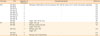
| Virus type | miR | Regulation in asthmatics | Role/pathway/target | Reference |
|---|---|---|---|---|
| RSV | miR-320e | ↓ | Pathways: inflammatory, immune response, Wnt, TGF-β, insulin, and T- and B-cell receptor signalling | 100 |
| miR-320d | ↓ | - | 100 | |
| miR-877-5p | ↓ | - | 100 | |
| miR-122-5p | ↓ | - | 100 | |
| miR-92b-5p | ↓ | - | 100 | |
| miR-106b-5p | ↑ | - | 100 | |
| miR-20b-5p | ↑ | - | 100 | |
| miR-342-3p | ↑ | - | 100 | |
| miR-26b | ↑ | Target: TLR4, IFN-β, CCL5 | 99 | |
| IFV | miR-26a | ↓ | Target: IFN-α/β | 106 |
| miR-650 | ↓ | Pathway: innate immunity | 101 | |
| Target: IFIT2, MXA | ||||
| miR-451a | ↑ | Pathway: type I IFN | 102 | |
| Target: IL-6 | ||||
| miR-3123 | ↑ | - | 104 | |
| miR-342-5p | ↑ | Pathway: sterol biosynthesis | 105 |
![]()
CONCLUSIONS
To date, only 2 publications have described the differential expression patterns of miRs in asthmatic patients after viral infection. The first one showed that the expression of TLR7, a crucial pattern-recognition receptor that responses to single-stranded RNA viruses, is significantly reduced in alveolar macrophages from patients with severe asthma in association with aberrant expression of miR-150, miR-152 and miR-375. Blocking the expression of these miRs can restore TLR7 expression and augment the expression of IFN responses to RV. These miRs represent a potential therapeutic target in this experimental setting.107
Many studies suggest that the expression pattern of miR-20a, 132 and 22 is similar in primary bronchial epithelial cells cultured in monolayer and differentiated air liquid interface conditions, which was not affected by asthma.39108109 When cells were cultured at air-liquid interface conditions and infected by H1N1 for 24 hours, the kinetics of miR-22 expression was different in asthmatics compared to non-asthmatics.108 The increased expression of miR-22 after IFV infection was associated with the suppression of CD147 mRNA, HDAC4 mRNA and protein expression in differentiated primary bronchial epithelial cells from non-asthmatics. However, in asthmatics miR-22 remained unchanged, while CD147 expression increased and HDAC4 remained unaffected. The study concluded that the different profile of miR-22 expression in differentiated epithelial cells from non-asthmatics may indicate a self-defence mechanism against aberrant epithelial responses through suppressing CD147 and HDAC4, which is limited in epithelial cells of asthmatics.39108
In the context of this review, combining the reports from the above comprehensive literature, we summarized several miRs potentially involved in virus-induced asthma (Supplementary Tables S2 and S3). Many miRs are increased after virus infection or involved actively in innate immunity, and are significantly depressed in asthmatic patients (Table 5). For example, miR-155 is induced in epithelial cells after RV infection,3969 while in other studies it seems to be reduced in asthmatic epithelium.3340 In addition, members of the let-7 family that seem to play a significant role in RSV infection through regulation of IFNB1 expression.737477 are reduced in nasal and bronchial biopsies of asthmatic patients compared to healthy individuals.334045
Table 5
List of respiratory virus-inducible miRs that are down-regulated in asthmatic subjects
| No. | Common miRs | Cell type | Virus type |
|---|---|---|---|
| 1 | miR-21 | Epithelium32 | IFV108 |
| 2 | miR-155 | Epithelium333940 | RV69, RSV99 |
| 3 | miR-24 | Epithelium3240 | RSV74 |
| 4 | miR-30a | Epithelium3240 | IFV92 |
| 5 | miR-146a | PBMCs (CD4+ T-cells)59 | RV69, IFAV92 |
| 6 | let-7b | Epithelium40 | RSV74 |
| 7 | miR-423-5p | Epithelium110 | IFV92 |
| 8 | miR-106b | PBMCs (CD4+ T-cells)53 | RV69, IFV92 |
| 9 | miR-126 | Epithelium33 | IFV92 |
| 10 | miR-29c | PBMCs (mononuclear macrophages and CD4+ T-cells)94 | IFV92111112 |
| 11 | miR-128 | Epithelium39 | IFV92 |
RV, rhinovirus; RSV, respiratory syncytial virus; IFV, influenza virus; PBMCs, peripheral blood mononuclear cells.
![]()
These findings point out a new field of study, involving the detailed exploration of miR profiles in asthmatics and non-asthmatic individuals, different levels of asthma severity or endotypes of asthma in relation to viral exposure. Such studies may not only unravel biomarkers for susceptibility but may also characterise the mechanisms that underpin the deficient innate immune responses of asthmatic patients. Further, functional experimental assays using potential miR-inhibiting or enhancing sequences and chemistries can evaluate suitable drug candidates for preclinical toxicity and pharmacokinetics studies prior to the onset of clinical trials. These assays may determine new molecules appropriate for therapeutic targeting according to patients' disease status supporting the evolution of personalised or precision medicine for chronic diseases such as asthma.
 XML Download