

 PDF
PDF ePub
ePub Citation
 Print
Print


INTRODUCTION
Hepatic steatosis is characterized by excess fat accumulation in hepatocytes in the form of lipid droplets.1 This intrahepatic accumulation results from abnormalities in lipid metabolism, such as increased whole body lipolysis, hepatic free fatty acid uptake and very low density lipoprotein synthesis, and also from reduced hepatic free fatty acid oxidation and triglyceride (TG) export.2 Obesity and insulin resistance are risk factors for non-alcoholic fatty liver disease (NAFLD) and contribute to lipid metabolism abnormalities.23 NAFLD is closely associated with insulin resistance-related diseases, such as obesity, dyslipidemia, type 2 diabetes, and metabolic syndrome3. However, it is uncertain whether the degree of fatty infiltration of the liver is associated with the incidence and/or severity of these insulin resistance-related diseases in patients with fatty liver.
Histologic examination of liver biopsy tissue is currently the standard method for accurate quantification of hepatic steatosis; however, this technique is invasive and occasionally associated with severe complications.4 Ultrasonography is reasonably accurate and the easiest to perform, but provides only a qualitative assessment of the fatty liver and cannot accurately assess the degree of fat accumulation.35 Unenhanced computed tomography (CT) techniques have been proposed to determine the degree of hepatic steatosis noninvasively: CT depicts fatty infiltration of the liver as a decrease in CT attenuation, and the reduction in attenuation has been shown to be directly correlated with the degree of fatty infiltration of the liver.5
We hypothesized that dyslipidemia is closely linked with hepatic steatosis. To test this hypothesis, we evaluated the association of dyslipidemia with fatty liver, as assessed by unenhanced hepatic CT imaging, and evaluated the differences in these associations according to the degree of hepatic steatosis.
MATERIALS AND METHODS
1. Subjects
This retrospective cross-sectional study included healthy Korean adults who underwent a comprehensive health check-up and abdominal CT scan between January 2010 December 2013 at the Samsung Changwon Hospital Healthcare Center. Among the 2,785 initial subjects, 323 individuals with any of the following were excluded: 1) positive serologic markers for hepatitis B (n=182) or C (n=20) virus, 2) findings of liver cirrhosis on CT scan (n=5), 3) splenectomy (n=7), and/or 4) absence of serum lipid concentration data (n=130). After applying the above exclusion criteria, 323 patients were excluded, and the total number of subjects eligible for the study was 2,462 (1,679 men and 783 women with a mean age of 46.1 years). Informed consent for this study was waived by the institutional review board because the researchers only accessed the database to obtain clinical data for the analyses, and personal information was not accessed. This study was approved by the Institutional Review Board at the Samsung Changwon Hospital (approval number: SMC201908003).
2. Measurements
Subject height and body weight were measured while barefoot and wearing light clothing. Body mass index (BMI) was calculated as the subject's body weight in kilograms divided by the square of their height in meters. Blood samples were collected from the antecubital vein after an overnight fast. Total serum cholesterol, TG, high-density lipoprotein cholesterol (HDL-C) and low-density lipoprotein cholesterol (LDL-C) were measured on a Roche Cobas 8000 Modular Analyser (Roche, Basel, Switzerland) using enzymatic colorimetric methods. All laboratory tests were performed in the same central, certified laboratory at the Samsung Changwon Hospital.
3. CT imaging
Hepatic steatosis was assessed as the degree of hepatic attenuation on non-contrast CT images as measured using Hounsfield units (HU). The CT scans of the abdomen were performed using a 64-detector row helical scanner (Somatom Definition AS 64; Siemens Healthcare, Forchheim, Germany). Contiguous transverse images were initially obtained from the dome of the diaphragm to the iliac crest with 5-mm slice thickness during a single breath hold without intravenous contrast agent administration prior to subsequent intravenous-contrast-enhanced CT examination. The unenhanced images were reviewed on a picture archiving and communication system (Marosis m-view; Marotech, Seoul, Korea) monitored by researchers (J.C.B., J.M.H., H.I.K., J.W.L., K.M.K., Y.J.L.) blinded to patient data. For each case, hepatic attenuation was measured by means of 12 circular regions of interest (ROIs) within three different transverse sections of the liver, with each section containing the confluence of the right hepatic vein, the umbilical portion of left portal vein, and the posterior branch of the right portal vein. At each representative level, the liver was divided into four sectors (right posterior, right anterior, left medial, and left lateral) and one ROI was randomly drawn inside each sector, avoiding the large vessels, biliary structure, and any focal lesions to represent liver parenchymal attenuation. The size of each ROI ranged from 1.0 to1.1 cm2. To provide an internal control, mean splenic attenuation was also calculated for three random ROIs measured within three different transverse sections of the spleen. With splenic attenuation acting as the control or reference value, the liver attenuation index (LAI), defined as the difference between mean hepatic attenuation and mean splenic attenuation, was used to assess the degree of hepatic steatosis.4
4. Definition of fatty liver disease and dyslipidemia
An LAI below 5 HU on an unenhanced hepatic CT image was defined as fatty liver disease in the present study. Individuals who met any of the following lipid abnormalities were defined as having dyslipidemia: 1) TG ≥200 mg/dL, 2) LDL-C ≥160 mg/dL, and 3) HDL-C ≤40 mg/dL.6
5. Statistical analyses
The results were expressed as the number of subjects and corresponding percentage (%) or as the mean measured value with standard deviation. The independent t-test or 1-way analysis of variance (ANOVA) were used to analyze normally distributed continuous variables. The Wilcoxon rank sum test and Kruskal-Wallis test were used to analyze non-normally distributed continuous variables. The independent t-test, Wilcoxon rank sum test, and Pearson's χ2 test were used to analyze statistical differences in the characteristics of the study subjects between the fatty liver and non-fatty liver groups. We estimated the odds ratios (ORs) for dyslipidemia according to the presence of fatty liver using a logistic regression model. The risk for each of the lipid abnormalities (TG ≥200 mg/dL, LDL-C ≥160 mg/dL, and HDL-C ≤40 mg/dL) according to the presence of fatty liver was also estimated using a logistic regression model. Study data from subjects with fatty liver (n=793; defined as LAI ≤5 HU on unenhanced hepatic CT images) were analyzed separately to evaluate the association of fatty liver severity with dyslipidemia. Subjects with fatty liver were categorized into tertiles based on their LAI values. One-way ANOVA and the Kruskal-Wallis test were used to assess statistical differences in clinical characteristics of the subjects with fatty liver as classified by the LAI value. We compared the prevalence of dyslipidemia between tertiles of LAI values using a logistic regression model. The data were analyzed using SPSS® version 22 (IBM SPSS Inc., Chicago, IL, USA).
RESULTS
1. Baseline characteristics of the study subjects
Overall, the mean age of the sunjects was 46.1±9.5 years, mean BMI was 24.1±3.1, and more subjects were male (68.2%, 1,649/2,462). We found that 32.2% of subjects had fatty liver. The prevalence of fatty liver was greater in men (38.5%) than women (18.6%). Hepatic attenuation ranged from 5.2 to 72.5 HU (mean, 54.8 HU). The LAI varied from −44.8 to 25.7 HU (mean, 7.0 HU). Subjects with fatty liver were significantly more obese (mean BMI, 25.6 kg/m2) than those without fatty liver (mean BMI, 23.3 kg/m2). In addition, LDL-C, TG, fasting glucose, and HbA1c concentrations were significantly greater in the fatty liver group compared with those of non-fatty liver group, while HDL-C concentrations were significantly lower in the fatty liver group (Table 1).
Table 1
Clinical characteristics of the study subjects
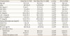
Data are presented as the mean±standard deviation or number (%).
BMI, body mass index; AST, aspartate aminotransferase; ALT, alanine aminotransferase; TC, total cholesterol; LDL-C, low density lipoprotein cholesterol; HDL-C, high density lipoprotein cholesterol; TG, triglyceride; CT, computed tomography; HU, Hounsfield units; LAI, liver attenuation index.
*Defined as HbA1c ≥6.5% or fasting glucose ≥126 mg/dL. †By independent t-test. ‡By Pearson's χ2 test. §By Wilcoxon rank sum test.
2. The risk of dyslipidemia in study subjects with fatty liver
Subjects with fatty liver had a 2.39-fold (95% confidence interval [CI], 1.99–2.86) greater risk for having dyslipidemia than did those without fatty liver after adjusting for age and sex (Model 1 at Table 2). Even after further adjustment for BMI and HbA1c, subjects with fatty liver had a greater risk of dyslipidemia, with an OR of 1.70 (95% CI, 1.40–2.08; Model 2 at Table 2). The risks of each of the lipid abnormalities (TG ≥200 mg/dL, LDL-C ≥160 mg/dL, or HDL-C ≤40 mg/dL) were also significantly greater in subjects with fatty liver than in those without fatty liver, with ORs of 1.65, 1.30, and 1.77, respectively (Table 2). When individuals with fatty liver were analyzed separately, LDL-C, TG, fasting glucose, BMI, and HbA1c concentrations increased, while HDL-C concentrations decreased with decreasing tertiles of the LAI value (Table 3).
Table 2
The risk for dyslipidemia according to the presence of fatty liver among study subjects
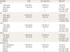
Model 1 was adjusted for age and sex; Model 2 comprised Model 1 with additional adjustment for BMI, and HbA1c. Values are presented as number of subjects (%) or odds ratio (95% confidence interval).
TG, triglyceride; TC, total cholesterol; LDL-C, low-density lipoprotein cholesterol; HDL-C, high-density lipoprotein cholesterol; BMI, body mass index; HbA1c, glycated hemoglobin.
*Individuals who met anyone of the following lipid abnormalities: 1) TG ≥200 mg/dL, 2) LDL-C ≥160 mg/dL, and 3) HDL <40 mg/dL.
Table 3
Clinical characteristics of study subjects with fatty liver
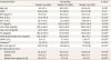
Data are mean±standard deviation or number (%).Tertiles 1, 2, and 3 are presented as ≤−3.1 HU, −3.0 to 2.9 HU, and ≥3.0 HU, respectively.
BMI, body mass index; AST, aspartate aminotransf-erase; ALT, alanine aminotransferase; TC, total cholesterol; LDL-C, low-density lipoprotein cholesterol; HDL-C, high-density lipoprotein cholesterol; TG, triglyceride; CT, computed tomography; HU, Hounsfield units; LAI, liver attenuation index; ANOVA, analysis of variance.
*Defined as HbA1c ≥6.5% or fasting glucose ≥126 mg/dL. †By one-way ANOVA. ‡By Pearson's χ2 test. §By the Kruskal-Wallis test.
As the LAI tertiles decreased, the risk for dyslipidemia increased with age- and sex-adjusted ORs of 1.47, and 2.24, respectively, compared with the upper tertile of LAI values (tertile 3, ≥3.0 HU using Model 1, as presented in Table 4). After further adjustment for BMI and HbA1c, this trend remained significant, although the ORs were reduced to 1.42 and 1.81, respectively (Model 2, as presented in Table 4). The risks for each of TG ≥200 mg/dL and LDL-C ≥160 mg/dL were greater in lower tertiles of LAI (tertile 1; ≤−3.1 HU) with adjusted ORs of 1.77 and 1.79, respectively, when compared with the upper tertile of LAI values (tertile 3; ≥3.0 HU). However, there was no difference in the risk of the lipid abnormality of HDL-C ≤40 mg/dL among the LAI tertiles. When we increased the cutoff value for low HDL-C from ≤40 mg/dL to ≤60 mg/dL, the severity of fatty liver became significantly correlated with subjects with this alternate definition of low HDL-C (Supplementary Tables 1 and 2). When we defined dyslipidemia as total serum cholesterol concentrations ≥240 mg/dL, there were no statistically significant differences in the prevalence dyslipidemia between the fatty liver and non-fatty liver groups (Table 2) or among LAI tertiles (Table 4).
Table 4
The risk for dyslipidemia by LAI tertile in subjects with fatty liver
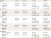
Adjusted for Model 1: age and sex; Adjusted for Model 2: Model 1+BMI, and HbA1c. Tertiles 1, 2, and 3 are presented as ≤−3.1 HU, −3.0 to 2.9 HU, and ≥3.0 HU, respectively. Values are presented as number of subjects (%) or odds ratio (95% confidence interval).
LAI, liver attenuation index; TG, triglyceride; TC, total cholesterol; LDL-C, low-density lipoprotein cholesterol; HDL-C, high-density lipoprotein cholesterol; BMI, body mass index; HbA1c, glycated hemoglobin.
*Individuals who met anyone of the following lipid abnormalities: 1) TG ≥200 mg/dL, 2) LDL-C ≥160 mg/dL, and 3) HDL <40 mg/dL.
DISCUSSION
In this study, the subjects with fatty liver had greater risk of dyslipidemia than those without fatty liver, and this association of dyslipidemia and fatty liver varied depending on the degree of hepatic fat accumulation.
Fatty liver represents the hepatic manifestation of metabolic syndrome. Patients with fatty liver disease often have dyslipidemia along with other metabolic risk factors, such as obesity, diabetes, and hypertension.78 Previous studies reported that dyslipidemia in patients with fatty liver was characterized by elevated serum TG and LDL-C, and decreased HDL-C concentrations.910 Our study also showed a similar pattern of lipid abnormalities in patients with fatty liver defined by liver attenuation index on CT (Table 1). Many imaging modalities are used to estimate of hepatic fat content, such as ultrasonography, CT, and magnetic resonance imaging.11 The LAI, as assessed by CT scanning, can be a relevant diagnostic tool, and has the advantages of non-invasiveness, accuracy, and reproducibility.
We demonstrated that patients with fatty liver had a greater risk of dyslipidemia than patients with non-fatty liver (Table 2). The prevalence of diabetes and morbid obesity has been reported to be 60 and 80%, respectively, in patients with fatty liver.12 Nonetheless, the greater risk of dyslipidemia in patients with fatty liver remained after adjustment for HbA1c concentrations and BMI as potential confounding factors. It is difficult to derive causal relationships from cross-sectional analyses, but fatty liver can be an independent risk factor for dyslipidemia. Insulin seems to be a key hormone in the regulation of lipid metabolism. Insulin resistance is the main etiopathogenic factor for dyslipidemia in fatty liver disease, which is related to hepatic overproduction of very low-density lipoprotein particles and dysregulated clearance of lipoproteins from the circulation.1314
The association between the presence of fatty liver and dyslipidemia has been demonstrated in many other studies21516; however, a direct association between the hepatic fat content of fatty liver patients and dyslipidemia has not been established. Instead of utilizing fatty liver scores and ultrasonography that may not reflect hepatic fat content accurately,1718 we adopted the LAI by CT imaging approach to measure hepatic fat accumulation in the current study.519 Interestingly, after classifying patients with fatty liver into tertiles according to the calculated LAI values, the patients' lipid profiles differed depending on the degree of hepatic fat accumulation: the lower the LAI tertile (i.e., the greater the hepatic fat content), the greater the proportion of the patients with TG ≥200 mg/dL, LDL-C ≥160 mg/dL, HDL-C ≤40 mg/dL, and dyslipidemia. The association between hepatic fat content and dyslipidemia was evident even after adjustment for age, sex, HbA1c, and BMI, each of which could have been possible confounding factors.20 This result strongly supports previous findings that fatty liver is associated with dyslipidemia. In addition, it implies that not only the presence of fatty liver, but also its severity as measured by hepatic fat accumulation, should be considered in fatty liver management: however, such recommendations should await further validation of our findings in future studies.
Our study had several limitations. Alcohol intake was not assessed, but may have a confounding factor in our study because moderate alcoholic consumption is major cause of hepatic steatosis, and hypertriglyceridemia is influenced by excessive drinking.2122 We also did not consider the use of medication for dyslipidemia, diabetes, or other drugs that can affect serum lipid concentrations or fatty liver. Although previously identified dyslipidemia or a history of lipid-lowering medication were not assessed in our study, study subjects with fatty liver and greater fat accumulation as identified on CT scanning were more obese and had more severe diabetes. As such, it is expected that study subjects with known dyslipidemia or taking lipid-lowering agents may have had greater fat content on CT or were more frequently diagnosed with fatty liver than patients without dyslipidemia. This suggests that our results are likely to be underestimated rather than overestimated. Subjects with steatohepatitis cannot be differentiated from those with simple steatosis on the basis of a CT scan, and additional analyses to detect the presence of steatohepatitis might have added greater detail to our study results. The omission of lifestyle factors, such as smoking and exercise, was also a limitation of this study.
Fatty liver was associated with dyslipidemia and this association varied according to the degree of hepatic steatosis, suggesting that not only the presence of fatty liver but also its severity by fat contents should be considered in the diagnostic evaluation and management of fatty liver.
 XML Download