

 PDF
PDF ePub
ePub Citation
 Print
Print


INTRODUCTION
Foreign body ingestion in children is common, affecting more than 100,000 patients annually in the United States [1]. Greater than 80% of cases occur in the pediatric population, the majority of which are accidental. Between 80 and 93% of foreign body ingestions that reach the stomach pass spontaneously and require no intervention [2]. Endoscopic retrieval is required in 10–20% of cases due to foreign body obstruction, but surgical intervention is only necessary in <1% of cases [3]. Surgery is indicated when the retained foreign body causes or is at risk of causing a bowel obstruction, perforation, abscess, or appendicitis. According to the American Society for Gastrointestinal Endoscopy guidelines, surgical intervention should be considered for short, blunt foreign bodies that are retained distal to the duodenum for a week or more without moving [4]. In the following report, we will discuss the case of an ingested foreign body which became retained within the appendix for a duration of ten months and ultimately required appendectomy.
CASE REPORT
A seven-year-old male with autism spectrum disorder and attention deficit hyperactivity disorder presented to the emergency room with a one-month history of intermittent non-bloody, non-bilious emesis and abdominal pain. The episodes of emesis occurred once or twice per week, generally at night time, and were accompanied by abdominal discomfort. He had not experienced any fevers, weight loss, headaches, diarrhea, or bloody stools, although his parents reported decreased oral intake and stool output. His medications included Vyvanse and Clonidine. Birth history and family history were unremarkable. He had no allergies and no recent travel history, and his vaccinations were up to date.
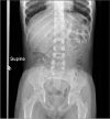
Upon examination, the patient was awake and alert in no acute distress. His vitals were all within normal limits. Abdominal exam revealed a soft, non-distended, non-tender abdomen with palpable stool. The remainder of the physical exam was unremarkable. A head computed tomography (CT) was negative for any intracranial process, and an abdominal x-ray showed significant fecal retention and a 7 mm radiopaque foreign body in the right lower quadrant (RLQ) (Fig. 1). As the foreign body was distal to the stomach and not causing an obstruction, it was predicted to pass spontaneously, and no intervention was performed. He was diagnosed with constipation, given an enema in the emergency room, started on a bowel regimen with daily MiraLAX (Bayer, Leverkusen, Germany), and instructed to follow-up with gastroenterology as an outpatient.
Fig. 1
Abdominal radiograph showing fecal retention and a 7 mm radiopaque foreign body in the right lower quadrant.
At one month follow-up, the patient continued to complain of nighttime emesis, intermittent abdominal pain, and decreased appetite. Abdominal exam again revealed palpable stool and was otherwise benign. Instructions for a second colon cleanout were given. A repeat abdominal x-ray two months later showed significant fecal burden and persistence of the foreign body within the RLQ, unchanged in location compared to his prior films.
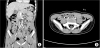
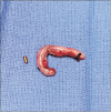
Despite two subsequent colon cleanouts, the foreign body did not move from the RLQ on repeat images. The patient otherwise remained asymptomatic, and his vomiting and abdominal pain resolved. A CT scan of the abdomen was performed five months later, which suggested the foreign body was within the appendix (Fig. 2). Colonoscopy with foreign body removal was planned. However, no foreign body was visualized within the terminal ileum, cecum, or appendix during colonoscopy. Due to the risk of developing appendicitis from an appendiceal foreign body, pediatric surgery elected to perform a diagnostic laparoscopy and laparoscopic appendectomy. The surgeon noted slight dilation in both the distal and proximal portion of the appendix, and the object was palpable within the appendix during the operation. Upon removal, a small metallic foreign body was found and sent to pathology (Fig. 3). Interestingly, Enterobius vermicularis were also identified in the specimen.
DISCUSSION
Foreign body ingestion is fairly common among children. It is rare, however, that surgical intervention is indicated, as most foreign bodies will spontaneously pass through the gastrointestinal tract after passing the stomach. Furthermore, most cases of foreign body retention are symptomatic. In the case of our patient, his symptoms had resolved despite retention of the foreign body within the appendix, and surgery was required due to concern that it could lead to the development of appendicitis.
Several cases of appendiceal retention of foreign bodies have been described in the literature. Presentation of appendiceal foreign body retention can range from completely asymptomatic to severe symptoms, such as abdominal pain, nausea, vomiting, and fevers. Balch and Silver [5] reported eight cases of appendiceal foreign bodies in 1971. One case reported a 20-year-old male presenting with abdominal pain, fever and vomiting who underwent appendectomy and was found to have a grossly inflamed appendix containing a corroded jackstone, which was encrusted with hard fecal material. Another case described a 15-year-old female who presented with two weeks of acutely worsening RLQ pain and a seven-year history of intermittent abdominal pain and was found to have several lead pellets within the lumen of her appendix upon appendectomy. Six of the cases presented with a history of chronic or acute abdominal pain, while two of the cases involved the incidental finding of retained appendiceal foreign bodies during appendectomies with no history of symptoms. Only two of the cases found appendiceal inflammation upon surgical inspection. Review of the literature by Balch and Silver [5] at the time revealed that 71% of patients found to have appendiceal foreign bodies were symptomatic, with 38% describing recurrent pain of more than one month's duration. The case described here demonstrates a unique scenario of a non-verbal child who may have been aware of the ingested foreign body but was unable to report it and may have been unable to accurately communicate symptoms, thus presenting a rare challenge in terms of clinical suspicion for retention and monitoring for its passage.
When foreign bodies become retained in the appendix, primary concerns include the development of appendicitis, abscess, and perforation. Objects that are heavier than intraluminal contents are at greater risk of becoming entrapped in the appendix as they tend to gravitate toward a dependent position. As such, those with retrocecal appendices are much less likely to develop retained appendiceal foreign bodies [6]. Objects which are long and sharp are more likely to cause appendiceal perforation, while blunt objects are more likely to become coated in fecal material and gradually enlarge, leading to partial or complete obstruction of the appendix and, thus, appendicitis [7]. However, as noted above, they may lay dormant in the appendix for years without causing any symptoms at all.
The foreign body in the case of our patient was small and blunt, and thus remained in the appendix for a duration of at least ten months, possibly longer, without causing significant signs or symptoms. Our patient did have symptoms of emesis associated with abdominal pain, but this was likely secondary to his chronic constipation as his complaints seemed to resolve after he had several bowel cleanouts. However, the presence of a foreign body in the appendix was felt to pose an increased risk for the future development of appendicitis and so an elective laparoscopic appendectomy was performed.
This case highlights the importance of persistent monitoring for passage in cases of foreign body ingestion. As the primary sites for entrapment in the GI tract are the proximal esophagus at the thoracic inlet, the mid esophagus at the aortic arch, and the distal esophagus at the lower esophageal sphincter [8], approximately 80–93% of foreign bodies that reach the stomach tend to pass spontaneously without complications. Therefore, simple observation at home for development of symptoms is often recommended for these patients [8]. This case demonstrates the importance of monitoring for passage of the object. After ingestion of a risky foreign body, an attempt should be made to retrieve it endoscopically. If endoscopic retrieval fails or is not possible, the patient should be followed clinically with serial abdominal radiographs (in the case of radiopaque objects). If the foreign body appears to be stagnant on repeat imaging and is visualized in the RLQ, colonoscopic removal should be attempted. If that is unsuccessful, laparoscopic retrieval should be considered, as in the case of our patient [9]. The case also speaks to the unique challenge presented by nonverbal, autistic, or developmentally delayed children who may be unable to communicate if they have ingested a foreign body or if they are having ongoing symptoms after ingestion.
 XML Download