

 PDF
PDF ePub
ePub Citation
 Print
Print


INTRODUCTION
Trimethylaminuria (TMAU), also known as fish odor syndrome, is a condition in which sufferers emit a body odor that resembles that of rotten fish despite good hygiene [1]. TMAU is characterized by the presence of trimethylamine (TMA), a tertiary amine, in the urine, sweat, breath, and reproductive fluids [2]. TMA is a volatile organic substance that has a characteristic fishy odor at low concentrations and an ammonia-like odor at higher concentrations. TMAU is a non-vital disorder with no associated morbidity or mortality by itself, although it has social, emotional, and psychiatric consequences, worsening patient quality of life and causing depression and even suicidal tendencies [3].
The most frequent presentation of this condition, primary TMAU, is caused by an inherited deficiency of the enzyme flavin monooxygenase 3 (FMO3), a vital enzyme in the metabolism of TMA [4].
In normal situations, dietary precursors (lecithin, choline) are metabolized to TMA by gut bacteria in the large intestine [2]. TMA is absorbed in the intestine by passive diffusion, transported to the liver by the mesenteric and portal veins, and converted into odorless trimethylamine n-oxide (TMAO) by FMO3 [2]. TMAO is finally excreted through bodily fluids.
In cases of inherited FMO3 deficiency, TMA is not efficiently converted to the non-odorous TMAO in the liver; rather, it accumulates and is excreted by bodily secretions and breath [24].
The incidence of primary TMAU is not well defined, but it has been suggested to range between one in 100 and one in 1,000 persons [5]. Secondary TMAU has also been described. Both portosystemic shunts [2] and severe liver disease [6] are related to secondary TMAU in a few cases thought to be due to decreased clearance of the absorbed TMA load.
The diagnosis of TMAU requires the measurement of TMA and TMAO in the urine collected after a meal overload of dietary precursors of TMA [2]. Results expressed as μmoL/mmoL creatinine can also be given as an ‘oxidizing ratio’ of TMAO/(TMAO+TMA)×100%. Unaffected individuals have a ratio >92% [2].
There is no systematic treatment for TMAU. Treatment consists of avoiding dietary precursors [27], oral antibiotics [2], probiotics [7], activated charcoal [27], low pH soaps [24] and, finally, psychological treatment [34].
Congenital portosystemic shunts (CPSS) are anomalous communications between the portal venous return and systemic venous system present at birth. CPSS occurs as a consequence of an anomaly during the embryonic development of the portal venous system. As a result, there is a direct derivation of the portal blood flow to the systemic venous system and consequently a reduction in the hepatic blood supply.
CPSS are extremely rare malformations. Two studies attempted to estimate the prevalence of CPSS using neonatal galactosemia screening results. The prevalence of all CPSS cases is 1:30,000 births, while that of persistent CPSS is 1:50,000 births [89].
CPSS can produce symptoms related to depletion of venous portal blood to the liver (liver atrophy [10] and benign [11] and malignant [12] liver nodes) and metabolic disorders including hepatopulmonary syndrome [13], portopulmonary hypertension [14], hepatic encephalopathy [15], postprandial hypoglycemia [16], hypothyroidism [16], and hyperandrogenism [16]. Other disorders such as gastrointestinal bleeding [17], nephrotic syndrome, and acute glomerulonephritis [18] are also reportedly related to CPSS.
In some cases, CPSS can regress spontaneously during the first 2 years of life. The patients who show regression without treatment usually presented with intrahepatic and distal shunts, whereas those with persistent CPSS presented with large and generally extrahepatic shunts. Treatment of CPSS includes a wide spectrum of strategies, from conservative management to liver transplantation, including surgical treatment and endovascular closure of the shunt [19].
Very few cases of congenital intrahepatic portocaval shunt associated with TMAU have been reported [20], and endovascular closure of CPSS has never been recognized as a treatment to resolve TMAU in affected patients.
CASE REPORT
Between November 2014 and April 2017, 15 patients (10 male; mean age, 129.42 months) with CPSS were enrolled in this prospective study to assess the efficacy of endovascular shunt closure and evaluate permanent changes in clinical symptoms, liver nodes, and intrahepatic portal flow development. Three patients presented clinical symptoms of TMAU; therefore, the urine test levels of TMA and TMAO were determined before and 1 year after endovascular treatment.
The statistical analyses were performed using SAS v9.3 (SAS Institute Inc., Cary, NC, USA). The urinary levels before and after endovascular closure were compared using the Wilcoxon signed-rank test. Values of p<0.05 were considered statistically significant. The study was approved by our institution's ethics committee and all patients provided consent to participate.
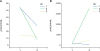
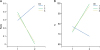
The clinical symptoms of TMAU disappeared after treatment in two (ID 2 and 3) patients and persisted in one (ID 1). Changes in TMA, TMAO, TMA/TMAO, and % of excretion of TMAO before and 1 year after endovascular treatment of CPSS are shown in Table 1 and Figs. 1 and 2.
Table 1
Changes in TMA, TMAO, TMA/TMAO ratio, and % of excretion of TMAO in the urine

DISCUSSION
One year after endovascular shunt closure, the TMA level in the urine decreased, TMAO levels increased, TMA/TMAO returned to normal, and % of excretion of TMAO increased in two patients (ID 2 and 3). The patients' clinical symptoms also disappeared. Ultrasonography in these two patients showed complete shunt occlusion and no new portosystemic communications. The results in these two patients supported the hypothesis of CPSS being a cause of secondary TMAU that endovascular closure could be used to treat. In contrast, one patient (ID 1) did not show clinical or analytical improvement of TMAU 1 year after treatment. Ultrasound scans in this patient showed complete shunt occlusion and the opening of new portocaval shunts nearby. These changes in and results of the third patient also reasonably support our hypothesis. Despite complete shunt occlusion, new portosystemic communications opened; the TMA absorbed in the intestine could bypass the liver through these new portosystemic shunts, reach systemic circulation, and be excreted in the urine. Retrospectively, the patient reported that the bad odor started in the first few months after shunt occlusion and recurred later. Urinary measurements were not obtained at that time because of the study protocol.
No statistically significant changes were observed. This is also reasonable due to the small number of patients included and the fact that CPSS and TMAU are both rare diseases.
No special clinical features but a fishy odor were observed in these three patients with TMAU versus the other patients with CPSS included in our study. The findings of the endovascular intervention demonstrated that all three patients presented with severe CPSS and very poor development of the intrahepatic portal branches before closure. This can be demonstrated during an occlusion test, in which a balloon is inflated in the shunt simulating closure and iodinated contrast is injected into the portal venous system.
Making the diagnosis of CPSS can be difficult in children due to variations in or a lack of clinical symptoms. Bad breath or a bad odor resembling rotting fish can alert clinicians to CPSS and could be useful in screening for this rare vascular anomaly.
 XML Download