

 PDF
PDF ePub
ePub Citation
 Print
Print


INTRODUCTION
Postoperative ileus (POI) refers to oral intolerance, abdominal distension, nausea, vomiting, and absence of bowel function following a surgical procedure. POI is most common after abdominal surgeries, particularly operations involving the colon, but can occur after retroperitoneal or extra-abdominal surgeries [12]. A physiologic POI is a multifactorial process, but is typically self-limiting, with most patients having return of bowel function within 48 to 72 hours postoperatively [1]. When a POI is prolonged (i.e. paralytic ileus), it may be indicative of a complication, such as infection or bowel leak and contributes to significant patient discomfort and prolonged hospital stays [34]. Though no consensus definition or timeline for prolonged POI exist, the incidence is thought to be between 3% and 32% in adult patients undergoing colorectal surgery [5]. Most studies on POI focus on adult colorectal patients, yet POI is a substantial problem in pediatric surgery, confounded by the inability of younger patients to self-report sign of POI. Kulaylat et al. [6] found that ileus or obstruction accounted for up to 25.5% of all pediatric thirty-day readmissions after a general or thoracic surgery.
There is no consensus definition or single objective test for determining complete return of bowel function postoperatively. Numerous signs and symptoms have been proposed and used, but each endpoint has its own limitations, particularly in the pediatric population. Bowel sounds are considered nonspecific for propulsive contractions and can be generated from the small or large intestines; flatus requires a conscious and cooperative patient who can reliably report its occurrence; bowel movements can be a sign of distal bowel emptying, but may not reflect function of the entire gastrointestinal tract [1]. Return of bowel function often dictates clinical decisions that affect recovery, including the initiation of feeding postoperatively, the use of nasogastric tubes, or need for further imaging. Relying on subjective symptoms for diagnosis is particularly difficult in children who often lack the language or cognitive ability to provide reliable information to providers.
Previous studies have utilized radiologic methods or radio-telemetry capsules measuring intraluminal pressures to objectively assess bowel function postoperatively [2]. Other studies have recorded and measured the electrical activity of the bowel (myoelectric activity) postoperatively using surgically implanted, transcutaneous electrodes [78]. While these studies have helped identify myoelectric pattern signals as potential means for differentiating physiologic POI from prolonged POI, the invasive nature of these tests is not practical for widespread clinical use. Cutaneous patches (G-Tech Medical, Fogarty Institute for Innovation, Mountain View, CA, USA) have recently been developed for continuous recording and measurement of intestinal myoelectric activity in humans. Using this technology, Dua et al. [9] showed gastric myoelectric activity measured by this technology could differentiate patients with longer or shorter times to a regular diet following a pancreaticoduodenectomy procedure while providing objective data to identify patients at risk for delayed gastric emptying, a major complication of the procedure. In another study, Navalgund et al. [10] noted the predictive value of colonic myoelectric activity in determining return of bowel function following surgery with the cumulative colonic activity at 36 hours predictive of first flatus as much as five days (±22 hours) in advance of its occurrence. The aim of this case series was to demonstrate the feasibility of using cutaneous patches in pediatric patients after major abdominal surgery as a proof of concept that myoelectric activities could be used to objectively assess bowel activity.
MATERIALS AND METHODS
Subject enrollment
After obtaining approval from Stanford University's Institutional Review Board (IRB Protocol # 44544), subjects undergoing abdominal surgery were enrolled to have their postoperative gastrointestinal myoelectric activity recorded. Patient demographics and characteristics, including height, weight, surgical history, procedure information, hospital length of stay and hospital course were obtained prospectively.
Technology
Myoelectric signals were acquired, digitized, and transmitted via Bluetooth Low Energy to a paired iPod Touch (Apple Inc., Cupertino, CA, USA) using a wearable patch (G-Tech Medical). Patches are placed directly on the skin and can be worn continuously for up to six days, as previously described [910]. The wireless patch has a diameter of 6.4 cm; multiple patches can be placed concurrently on different areas of the abdominal wall to capture location-specific signals. The variation in body size and tissue thickness between patients was addressed by a compensation factor which uses a patent-pending compensation algorithm utilizing specific aspects of the acquired data itself to arrive at a correction. A custom iPod Touch (Apple Inc.) application stores the myoelectric activity data and allows for providers to enter clinical information, including diet status, vomiting, and the return of bowel function.
Data analysis
Post-processing of the raw data was performed by a study engineer using a custom LabVIEW (LabVIEW Version 14.01, National Instruments, Austin, TX, USA) program that included removal of large amplitude artifacts and band-pass filtering, followed by Fourier transformation to frequency space over selected time intervals. Previous studies have documented frequency ranges for the three organs of interest; stomach with activity at three cycles per minute (cpm), small intestine with activity between five and 13 cpm, and for the colon, a wide range between zero to 40 cpm [1112]. For this study, peaks were identified and nominally assigned towards gastric if they were between two and four cpm, small intestine if they were between five and 13 cpm, and colon if frequencies were above 13 cpm to avoid overlap with small intestine and stomach. A waterfall spectral plot and corresponding peak frequency plot with spectra calculated in 10 minutes intervals and arranged chronologically was used to display trends in bowel activity.
RESULTS
Case 1
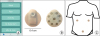
The first case is of a five-month-old female with a history of imperforate anus who underwent closure of a double-barrel colostomy and rectal dilation. Within two hours after surgery, one disposable wireless patch (G-Tech Medical) was placed on her anterior abdominal wall to transcutaneously record myoelectric signals from the gastrointestinal tract (Fig. 1). The patch was worn continuously until discharge on postoperative day (POD) three. Given this patient's small abdominal wall surface area, only one patch was placed.
Fig. 1
(A) Mobile app interface for entering clinical information postoperatively. (B) Cutaneous myoelectric patches. (C) Schematic of cutaneous myoelectric patch placement on the anterior abdominal wall.
GI: gastrointestinal, BM: bowel movement.
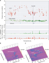
The patient's postoperative course was uncomplicated, with diet starting on POD1. She had a bowel movement late on POD1 and her feeds were advanced to goal over the next day. She was discharged home on POD3. The myoelectric intestinal and colonic activity correlated with her clinical course, demonstrating a gradual increase in signal intensity from the small intestine after POD1, represented as darker green on the scatter plot and higher peaks on the waterfall spectral plot (Fig. 2). Within each scatter plot, the colors are assigned to a given organ based on frequencies (red=colon; green=small intestine; blue=stomach). The darkness/intensity of the color is correlated to the relative intensity of the signals from that organ (i.e. darker color represents higher amplitude peaks). The colon also demonstrated a gradual return to normal activity, which correlated with the passing of gas and stool recorded by the caregiver using the app (Fig. 1). Just prior to bowel movement, there are more frequent and more intense signal emanating from the colon seen on the waterfall spectral plot. After the initial bowel movement, steady activity is seen throughout POD2 until the patch was removed.
Fig. 2
Intestinal and colonic myoelectric activity from wireless patch system on the five-month-old patient. (A) Frequency peak plots over POD0 to POD3. More intense/darker color represents increased amplitude of peaks. Organ-specific plots demonstrate relative signal intensity over time. Waterfall spectral plots for (B) small intestine and (C) colon in the immediate postoperative period. Increase in small intestine and colon activity prior to first BM.
POD: postoperative day, BM: bowel movement.
Case 2
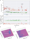
The second case was a four-year-old female who underwent resection of a benign retroperitoneal mass. The procedure started as a laparoscopic case but was converted to open due to the size of the mass (15 cm diameter). Postoperatively, two patches were placed on the anterior abdominal wall in the upper right and upper left abdominal region (Fig. 1). She was started on a clear diet on POD1 and advanced to a regular diet on POD2. She had return of bowel function (stool) on POD2 and was started on a bowel regimen (Docusate, PO) on POD3. Regarding her myoelectric activity, the scatter plot demonstrated a sharp rise in colonic activity and signal intensity just prior to her first bowel movement towards the end of POD2 (Fig. 3). She was discharged home on POD4 without complications.
Fig. 3
Myoelectric activity from the seven-year-old patient demonstrating (A) frequency peak plots by organ of signal origin, as well as waterfall spectral plots for (B) the stomach, small intestine and (C) colon. Spectral plots are calculated in 10 minute intervals and arranged chronologically.
BM: bowel movement.
Case 3
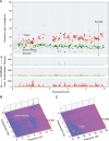
The third case was a 16-year-old female with a history of Crohn's disease complicated by ileal stricture and ileo-ileal fistula who underwent a laparoscopic ileocecectomy with primary anastomosis. This patient had three abdominal patches placed on the anterior abdominal wall, as shown in Fig. 1, a typical configuration used for adults. The patient had return of bowel function (flatus) on POD4 and was given a regular diet on POD5. No bowel regimen was needed for this patient. The patient was discharged home on POD6. The patches recorded myoelectric intestinal and colonic activity that showed relative quiescence on POD1, and gradual return to more frequent activity over the postoperative course. The waterfall spectral plot demonstrated increased colonic myoelectric activity in the hours prior to her initial bowel movement (Fig. 4).
Fig. 4
Myoelectric activity from the 16-year-old patient. (A) Frequency peak plots demonstrate gradual increase in signal amplitude leading up to first BM on POD4. Waterfall spectral plots show steady signal from (B) the stomach and small intestine, with (C) increased signal in the colon prior to first BM.
BM: bowel movement, POD: postoperative day.
DISCUSSION
In this case series, cutaneous patches were used to continuously monitor and record gastrointestinal myoelectric activity in pediatric patients after abdominal surgery. Organ-specific signals were identified based primarily on signal frequencies and, to a lesser extent, on patch location. The patterns of myoelectric activity differ in all three patients, though there seems to be an increase in activity and intensity just prior to return of bowel function (stool) postoperatively. The application was able to capture myoelectric activity from multiple patches and record relevant clinical activity without interfering in the patients' postoperative care. Of note, while frequencies for gastric and small intestine were consistent with ranges previously observed in adults [1012], colonic frequencies were observed across a much larger range particularly in the younger patients with frequencies as high as 50 cpm observed in the five-month-old female.
As discussed, this technology has recently been used to measure continuous myoelectric activity in adult patients following abdominal surgery [910]; however, the signals recorded using cutaneous patches have not been directly compared to signals measured directly on the bowel wall. The risks of embedding electrodes in the gastric or intestinal wall would likely outweigh the benefits. Therefore, we have attempted to validate the signals observed from the cutaneous patches using historic frequency references and direct comparison with pressure measurements [7812].
Reducing the occurrence and duration of POI is an important part of decreasing hospital stays and associated costs [13]. Enhanced recovery after surgery programs have been utilized in both adult and pediatric patients to improve postoperative care through emphasis on early enteral feeding, opioid sparing pain medication, and early ambulation postoperatively [1415]. While there is some evidence that these protocols reduce hospital length of stay and hasten return of bowel function, up to 24% of patients fail to tolerate feeds and require reinsertion of nasogastric tube for decompression [14]. The non-invasive technology used in this series could be used to augment treatment protocols by identifying patients at greater risk of POI using objective and personalized myoelectric pattern recognition. Recent studies using this technology have demonstrated that gastrointestinal myoelectric activity measured by the system can reliably predict return of gastrointestinal function following complex abdominal surgical procedures [910]. Though larger studies are needed to establish benchmarks within pediatric patients, this technology could be particularly useful in pediatric surgical patients for deciding when to initiate feeding postoperatively. Timing of feeding is critically important in this population, and no objective test currently exists to determine the ideal timing of postoperative feeding.
This study, as a proof of concept, is limited by sample size. The increased myoelectric activity varied greatly in each patient prior to their return of bowel function; there is no simple or consistent linear progression of signals from all organs prior to return of bowel function. More data from pediatric patients is needed to thoroughly examine the various patterns of bowel activity that may predict return of bowel function postoperatively. It is unclear from this limited data set which signals and which organ activity will be most important clinically. The patches can be used in infants, though abdominal surface area was a limitation in this series. The use of only one patch in the five-month-old in a location further away from the stomach is thought to possibly explain the lack of gastric myoelectric activity signal in the data. Smaller size patches could be developed specifically for use in neonatal patients as a way to monitor bowel function in high-risk patients.
Future work includes expanding enrollment for pediatric patients undergoing both laparoscopic and open abdominal surgeries to measure gastrointestinal myoelectric activity non-invasively. More data is needed to examine the patterns of myoelectric activity postoperatively, and further analysis is needed to correlate these signals with clinical outcomes. However, this small sample of preliminary data supports the notion that recorded myoelectric patterns in pediatric patients could be used to predict return of bowel function or detect ileus. A randomized trial is also being considered where feeding regimens would be initiated based on myoelectric measurements versus standard of care.
In conclusion, gastrointestinal and colonic myoelectric activity recording is feasible in pediatric patients using a non-invasive cutaneous patch. Initial results suggest a possible correlation between signal intensity/activity and clinical return of bowel function. This method provides objective, personalized data on gastrointestinal function that could be used to optimize postoperative feeding management, though more data is needed to better understand the significance of these signals.
 XML Download