

 PDF
PDF ePub
ePub Citation
 Print
Print


INTRODUCTION
Metal-ceramic restorations have been successfully used in fixed partial dentures (FPDs) for many years because of their favorable long-term clinical performance.1 Accurate impression making is one of the most important stages in fabricating FPDs,2 and insufficiently detailed impressions may result in incompatible marginal adaptation of the restorations.3 Conventional impression techniques require high technical skills, and restorations may deform during the fabrication stages in the laboratory.45 Factors such as the change in ambient temperature, the length of time between impression and casting, the type and/or quality of the cast material, the storage conditions of the impression and cast material, surface wettability of the impression material, and disinfection procedures are technical issues of the conventional impression method, and these factors directly affect the compatibility of the restorations. Fabrication processes such as the application of the die spacer as well as laboratory steps for prosthesis production such as waxing, investing, casting or pressing process can result in dimensional error and affect the fit of the definitive restoration. Contrary to these disadvantages, the conventional impression technique is inexpensive than the digital technique.6
Digital impression systems were developed as an alternative to conventional impression systems.789 However, scanning with powder, low-resolution, and insufficient computing power caused restorations with unacceptable values of marginal misfit.10 The CEREC 1 system (Sirona Dental Systems GmbH, Bensheim, Germany) was the first commercially available intraoral impression system and was developed in 1987.1112 With the development of fabrication technology and engineering, highly accurate scanners, more advanced software, and several digital intraoral impression systems have become available.11121314 With these systems, crowns or FPDs can be fabricated on the images of the prepared teeth captured by the intraoral scanning device.111215 As a consequence, several problems of conventional impression making can been eliminated.1116 Nowadays, The CEREC Omnicam (DentsplySirona, Bensheim, Germany),11 the Lava chair side oral scanner (Lava COS; 3M ESPE, St. Paul, MN, USA),91117 iTero (Align Technology, Inc., San Jose, CA, USA),2 Trios3 (3Shape A/S, Copenhagen, Denmark),918 and E4D PlanScan (d4d, Planmeca, Richardson, TX, USA)19 systems are frequently used to fabricate dental prostheses.
The Trios3 intraoral scanning system (3Shape A/S, Copenhagen, Denmark) is the first generation scanner and the system works on the principle of ultrafast optical section and confocal microscopy.20 It uses a quick scanning speed and captures up to 3000 figures per second. As the effect of relative movement between the scanning probe and teeth is reduced, contrast spraying is unnecessary.18 The Cerec Omnicam (DentsplySirona, Bensheim, Germany) works on the principle of active triangulation. It uses a white light to capture the image and captures data continually in color, again without the need for contrast spraying.17
The digital impression is a noninvasive technique, which involves the use of an intraoral scanner of small dimensions that does not create discomfort for the patient. The scanner records a series of snapshots of the oral cavity of the patient, which are transferred onto a computer where they will be processed and a virtual model will be obtained.12 In this way, all the requirements expected from the restoration can be examined from the data obtained and the desired restoration planning can be realized. This technique has significant advantages in eliminating operator dependent variability, reducing impression time and treatment cost, and improving patient compliance for rehabilitation over conventional impression technique.21 Contrary to these superior features, the digital impression technique may show particular short-comings in transferring subgingival margins to the system. In this technique, the full reflection of the gingival margin to the system is possible only by providing ideal isolation. Today, retraction cords and rubber dams are used for this purpose. Otherwise, it is inevitable that the produced restorations will fail clinically.12
Several factors affect the success of dental restorations22 and it has been reported that the incidence of biological complications is greater than mechanical complications in metal-ceramic restorations.23242526272829 Accurate adaptation seems to be one of the most important factors for the longevity of a restoration.252627 The restoration boundary should be perfectly adapted to the finish line of the prepared tooth. In practice, it is impossible to achieve perfect adaptation. The misfit of a complete coverage restoration can cause luting agent dissolution, leakage, the risk of secondary caries,28 hypersensitivities, and periodontal disease.13
Numerous studies evaluating the marginal and/or internal adaptation of single-unit dental restorations fabricated with digital impression systems are available in the literature,21314151630 but a small number of studies are about marginal misfit of three-unit metal frameworks fabricated with different impression techniques.1829 The number of studies evaluating the marginal misfit of three-unit frameworks fabricated with different impression techniques is small and the results of the studies are incompatible.182931 In some studies, it was reported that there is inconsistency between the marginal misfits of different abutment teeth in the frameworks fabricated with the same impression technique.233132 However, incompatible data are insufficient to reach consensus on this issue.2332 Because of these shortcomings, the primary purpose of this in vitro study was to evaluate the marginal misfit of three-unit frameworks fabricated with two digital and one conventional impression technique. The secondary purpose was to evaluate whether the marginal misfits differentiate according to the type of abutment teeth supporting the same three-unit framework specimens. The primary null hypothesis was that there was no difference between digital and conventional impression techniques on the marginal misfit of three-unit laser-sintered frameworks. The secondary null hypothesis was that there was no difference between the marginal misfit values of different abutment teeth supporting the same framework.
Go to : 
MATERIALS AND METHODS
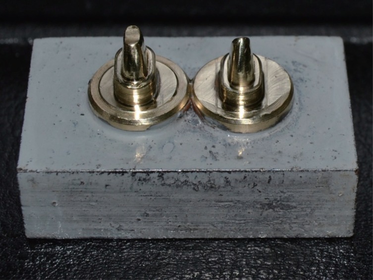
Artificial left maxillary canine and second premolar teeth (ANKA-4 Z, Frasaco GmbH, Tettnang, Germany) were used for fabrication of master dies. The abutment teeth were prepared as follows: 6 degrees of convergence angle, anatomic occlusal preparation of approximately 2 mm, axial preparation of approximately 1 to 1.5 mm, and a shoulder circumferential finish line of 1.0 mm in depth. The teeth were prepared by using a paralellometer (Paraskop, Hebst GmbH & Co., Bremen, Germany) to eliminate any undercuts. Thirty canine and 30 second premolar brass abutments were duplicated (Sarbak Metal, Sarbak Co., Istanbul, Turkey) from the prepared master teeth by using a computer numerical control (CNC) machine YM 64DV jig borer (Victor, Taipei, Taiwan).33
According to power analysis (95% power and P < .05), thirty study specimens were obtained by assembling these brass abutments (Fig. 1) and they were divided into 3 equal groups (n = 10) for fabrication of cast dies with conventional and digital impression techniques.30
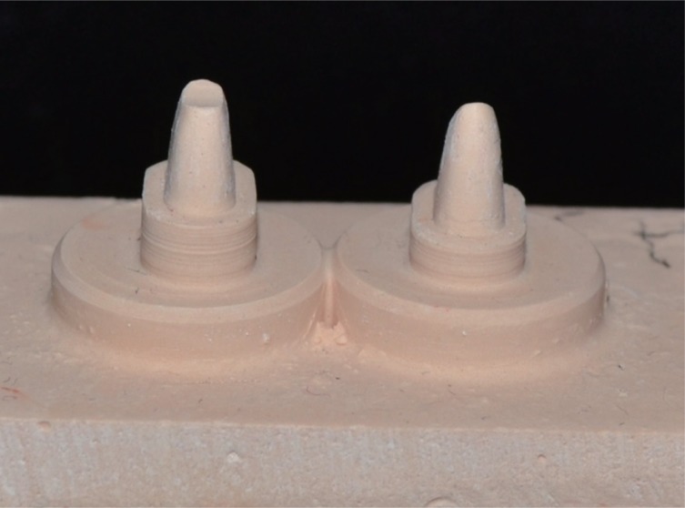
In Group Ci, the frameworks were fabricated with conventional impression techniques, in which two-stage impressions were made with a polyvinyl siloxane (PVS) impression material (Variotime Easy Putty and Light Flow, Heraeus Kulzer GmbH, New York, NY, USA) and a custom tray. The conventional two-stage impression process was performed by applying finger pressure for 5 minutes at room temperature on the custom tray and the impression was taken for each study specimens in this group by the same experienced operator each time. After finishing the impression process, the same operator examined the quality of all impression about the tears and voids and connection between the custom tray and impression material. When an impression was assessed as having a critical defect, the impression was retaken.32132 After the impressions had been removed from the abutments, they were disinfected for 10 minutes (Impresept, 3M ESPE, St. Paul, MN, USA). Type IV dental stone was poured into the impressions (Fujirock EP, GC Corp., Tokyo, Japan) according to the manufacturer's instructions to obtain casts. Ten cast die models were obtained from each of the Group Ci specimens and then sent to the dental laboratory for the fabrication of the three-unit LS frameworks (Fig. 2).
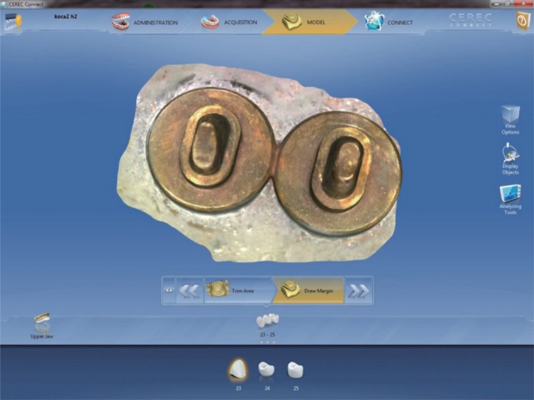
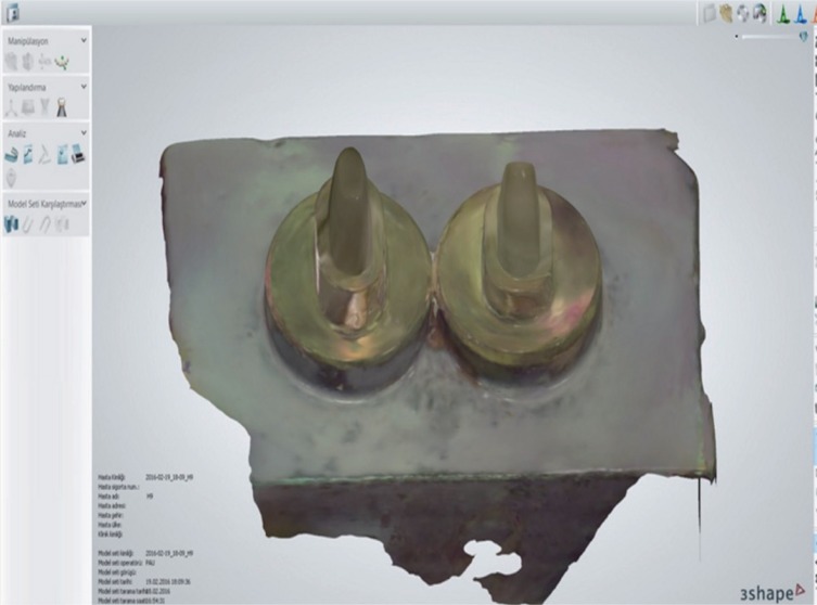
In Groups Cdi and Tdi, the frameworks were fabricated with digital impression techniques, in which study specimens were scanned with a CEREC Omnicam (DentsplySirona, Bensheim, Germany) and a 3 shape TRIOS-3 (3Shape A/S, Copenhagen, Denmark) intraoral scanning systems, respectively (Fig. 3 and Fig. 4).
The digital scanning was performed by the same experienced operator (H.K.) and the scan was continuously assessed during the scanning procedure. If a critical defect was observed, the scan was corrected by rescanning the flawed area. Specific scanning was performed for each study specimens in Group Cdi and Tdi. In total, 10 scan files were obtained for each digital measurement technique. After the scan was finished successfully, the scan files were converted to STL image format and then sent to a dental laboratory for the fabrication of the there-unit LS frameworks.
The frameworks were designed by an experienced dental technician with a CAD program (Dwos, Dental Wings, Montreal, Canada) with a 0.5 mm thick abutment framework, a 3 mm pontic width, a 3 mm connector height, a 3 mm connector width, and a 20 µm cement space (0.5 mm from the margin) for the abutment of the working cast. The frameworks were then fabricated with a prototyping technology. With this technology, a high precision (EOSINT M270, EOS GmbH, Krailling, Munich, Germany), high-energy laser (yb-fiber laser of approximately 200 W with compressed air of 7000 hPa) was used to melt a controlled deposition of 20 µm thick full dense CoCrMoW alloys made from a commercial alloy powder (EOSINT M EOS CobaltChrome SP2, EOS GmbH). After this sintering procedure was finished, the frameworks were separated from the metallic base and the fabrication phase was terminated (Fig. 5).
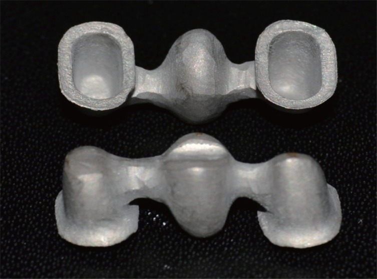
According to the manufacturer's instructions,34 a thermal stress relieving process was performed and then all frameworks were evaluated for internal surface defects that could prevent their complete placement. They were then cleaned ultrasonically (Elmasonic S100H, Elma GmbH&Co KG, Singen, Germany) in 95% methyl alcohol for 15 minutes and left to dry. The marginal misfits were measured by using a stereomicroscope (Nikon SMZ 1500, Nikon Corp., Melville, NY, USA) at 30× magnification on the frameworks.726 The microscope images were captured by a digital camera (Clemex; Clemex Technologies Inc., Quebec, Canada) that was connected to the microscope (Fig. 6) and transferred to a computer.
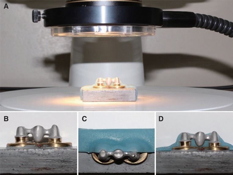
Measurements were made for each surface (buccal, mesial, distal, and palatal) of each abutment (canine and premolar) from the edge of the shoulder finish line of the dies to the inferior edge of the frameworks (Fig. 7).3536
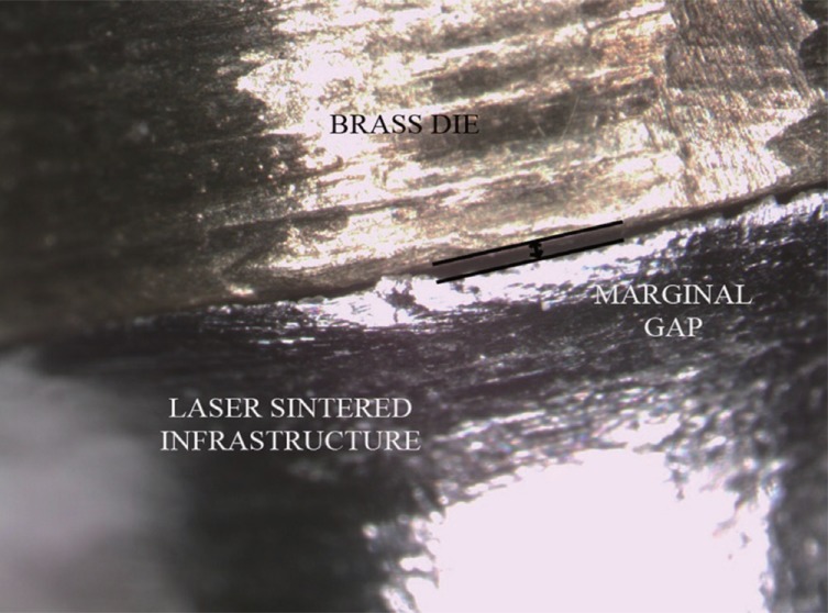
Marginal discrepancy measurements were made four times from each side of each abutment using a computer program (Vision Lite, Clemex Technologies Inc., Quebec, Canada).36 In total, 480 measurements were made for each abutment. Average values of these measurements were calculated for each abutment separately and the average total marginal misfit values of frameworks were recorded.
The recorded misfit data of each abutment and total marginal misfit data were subjected to the Kolmogorov-Smirnov test to examine whether the data were normally distributed (α = .05). One-way ANOVAs was performed because the data in all groups were consistent with normal distribution. The HSD Tukey post hoc tests were applied to evaluate marginal misfit values among each abutment groups. Possible marginal agreement differences between abutment teeth were analyzed by independent simple t tests (α = .05). Statistical analyses were performed by using a software program with a 95% confidence interval (SPSS 20.0 V, SPSS Inc., Chicago, IL, USA).
Go to :
RESULTS
Table 1 shows one-way ANOVA values of canine and premolar teeth and total average values. Table 2 shows mean and standard deviation (SD) values of the abutment teeth and total average marginal misfits values. As shown in Table 2, statistically significant differences were found between the framework groups produced with conventional and digital measurements in terms of marginal discrepancy (P < .001). According to the Tukey multiple comparison test, Group Ci revealed larger marginal misfits than those of Group Cdi and Group Tdi in both abutment teeth (P = .000 for average; P = .001 for canine; P < .001 for premolar). However, no statistically significant difference was found between Group Tdi and Group Cdi in either abutment tooth (P > .05) (Table 2).
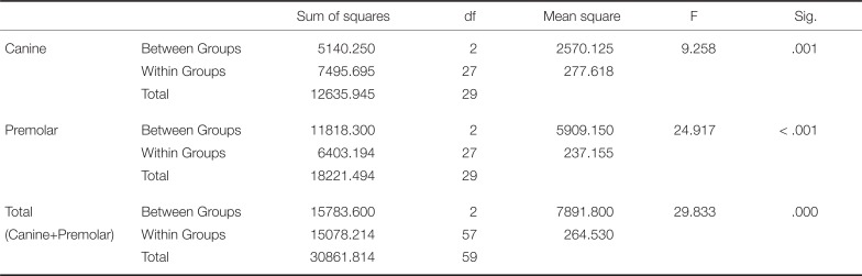
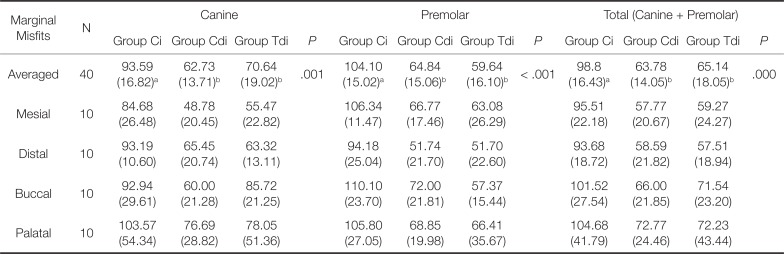
Table 1
One-way ANOVAs of canine and premolar teeth and total average values
![]()
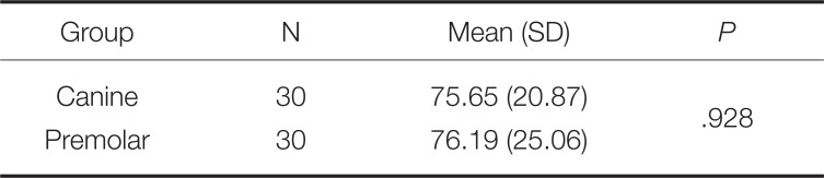
Table 2
Mean and standard deviations (±SD) values of the abutment teeth and total marginal misfit values (µm)
![]()
The results of the t-test examining the possible marginal discrepancy differences between the abutment teeth supporting the same independent framework specimens showed that there was no statistical difference between the abutment teeth in terms of marginal discrepancy (P > .05) (Table 3).
Go to :
DISCUSSION
In this study, the marginal misfit of three-unit laser-sintered (LS) frameworks fabricated by different digital impression techniques was compared with that of the frameworks fabricated by the conventional impression technique. According to the data obtained, the primary null hypothesis that there were no differences between digital and conventional impression techniques on the marginal misfit of three-unit laser-sintered metal frameworks was rejected. In the study, the total mean marginal misfit values of Group Cdi and Group Tdi specimens were found to be significantly smaller than Group Ci (P = .000). In addition, the marginal misfits in Group Cdi and in Group Tdi were also significantly smaller than those of Group Ci in either canine or premolar teeth (P = .001 and P < .001, respectively). In Group Ci, the obtained mean marginal misfit values of frameworks were 93.59 µm (± 16.82) for canine, 104.10 µm (± 15.02) for premolar abutment teeth and 98.84 µm (± 16.43) for the average of both abutments. These results were found to be consistent with similar studies.11313537
Euán et al.7 compared single unit zirconia copings fabricated with digital and conventional impression techniques and reported that the restorations fabricated with the digital impression technique had lower marginal misfits than those of the conventional impression technique. The marginal misfit values in that study were lower than the present study. This difference in the results may be due to the fact that the intraoral scanner, restoration material, and restoration type used in this study are different from the current study.
Haddadi et al.20 compared lithium disilicate crowns fabricated with digital and conventional impression techniques and reported that the restorations fabricated with the digital impression technique had lower marginal misfits than those of the conventional impression technique. The marginal adaptation data of the specimens acquired by the digital measurement technique were found to be similar with the present study. It can be thought that this similarity is due to the fact that the same digital scanner was used in both studies.
In another study using digitized version of the impression replica technique (dual scan technique), the adaptation of single crown fixed dental prostheses was evaluated. In the study, presintered zirconia, hot isostatic pressed yttriatetragonal zirconia polycrystalline ceramic, lithium disilicate reinforced glass ceramic, milled cobalt-chromium, and lasersintered cobalt-chromium crowns were used. For the control group, the single-unit frameworks were produced with the lost wax metal casting technique with cobalt-chromium by using conventional impression techniques. According to the study results, the researchers reported that laser sintered cobalt-chromium crowns fabricated with the digital impressions technique with the help of Trios intraoral scanner showed significantly better marginal fit than all other digital technique groups and the control group using the conventional technique.8 Similarly, in the present study, the measured marginal fit values of the restorations produced with conventional impression was found to be greater than those produced by the digital impression. On the other hand, there was no significant difference in the marginal fit between the digital measurement methods (P > .05).
Keul et al.29 evaluated the adaptation of four-unit milled base metal alloy and zirconia frameworks fabricated with the digital impression technique and conventional impression technique. The study results showed that milled base metal alloys fabricated with the digital impression technique showed significantly better marginal adaptation than those metal alloys fabricated with the conventional technique, and this finding supports the current study.
Su and Sun18 compared the marginal and internal fit of 3-unit zirconia frameworks fabricated with either a conventional or digital impression technique. They reported that the zirconia frameworks fabricated with digital impression systems had better marginal adaptation than those of conventional impression. The results for marginal adaptation were in accordance with the present study. It can be thought that the similar results are due to the fact that the same intraoral scanner and the impression material were used in both studies.
In this study, after the different impression methods, the compatibility of the frameworks produced with CAD/CAM supported LS technology was examined. LS technologies have overcome many of the disadvantages of the conventional casting. However, this technology has variables that can influence the results of the manufacturing process, such as scanning protocols, software design, and material processing. For this reason, inconsistencies may be seen among the results of studies that test the marginal misfits of CAD/CAM supported frameworks.31 On the other hand, some authors have reported that molar abutments in digitally fabricated frameworks have greater misfits than premolar abutments in the same specimen tested. The authors associated these findings with differences in abutment size and reported that molar teeth may exhibit greater misfits due to their larger surface area.323738 In multi-unit frameworks, although the average marginal misfit values of the abutments are within acceptable limits relative to the clinical marginal misfit threshold values reported in the literature, this is not always sufficient for the clinical acceptability of the restoration. Because, when each abutment in the framework is evaluated separately, the marginal misfit values determined may be outside these threshold value.
In multi unit restorations, if the marginal accordance of one of the abutments is insufficient, this causes the final restoration to fail.35 The secondary purpose of this study was to evaluate the marginal misfit of each abutment separately. In the present study, when the marginal compatibility of each abutment tooth was examined, it was found that there was no difference in the marginal misfit between canine or premolar abutments in the same framework. Thus, the second null hypothesis of this study was accepted. Similar to the present study, in another study using premolar and molar teeth as the abutment, the marginal misfits of LS 3-unit frameworks were evaluated and numerical differences were found about the marginal misfits between the abutments. However, these differences were not statistically significant.31
Colpani et al.39 reported that a marginal discrepancy can be considered acceptable when it is visually indistinct or cannot be identified with a dental explorer. Up to this time, no consensus exists on clinically acceptable marginal misfit values. McLean and von Fraunhofer reported that restorations with marginal misfits less than 120 µm were clinically acceptable.25 Other studies reported that misfit values below 100 µm were acceptable.2627 In the present study, the marginal adaptation of LS FDPs in both the digital and conventional impression groups were clinically acceptable.25 Larger marginal misfit values were obtained in Group Ci, in both canine and premolar teeth. The better marginal adaptation of the digital impression groups may be explained by error caused during the conventional impression making process and by the result of the multiple production steps or impression procedures in group Ci. The casts might be deformed during the polymerization or adaptation of the restorations. A recent study reported that Type IV dental stone had a linear expansion of between 0.06% and 0.5%.40 Corso et al.41 reported that PVS impression material contracted by 17 µm in the horizontal line and 2 µm in the vertical line after storage at room temperature. The results obtained in these studies could explain the marginal discrepancy differences between the digital and conventional impression groups in present study.
There has been an increase in the number of studies on the marginal fit of restorations produced by laser-sintered technology in dentistry. In most of these studies, although researchers have evaluated different frameworks and manufacturing techniques, superior marginal adaptation of Co-Cr specimens produced by laser-sinter is thought to be from preventing the distortion of casting methods.8
In previous studies, several materials such as dentoform teeth,1431 cobalt-chromium,2 and zirconia42 have been used for master dies. In this study, brass dies were used as the master dies because of their high deformation resistance.43
This standardized study evaluated the marginal misfit of three-unit LS frameworks fabricated with different impression techniques. All the frameworks were fabricated with the same software and design, with the same anatomy, and with the same laser sintering machine software. The single variable was the impression technique. All conventional and digital impression making procedures and the marginal discrepancy measurements were performed by the same operator (H.K.). The main drawback of LS crown fabrication is the need for trained technicians to evaluate subgingival margins.25 For this reason, in this study, all set-up and production procedures of the frameworks were performed by the same trained technician using the same settings and the same equipment.8
Several techniques for measuring marginal misfit values have been reported, including the silicone replica technique,30 3-dimensional measurements,14 the cross-sectional technique,17 direct techniques with cone beam computed tomography (CBCT) evaluation,4 and the silicone weight technique.25 The cross-sectional technique damages the frameworks and master dies, and both CBCT and 3-dimensional measurement techniques require advanced technology.9 In this study, a direct view method was used to measure the marginal misfit of the frameworks10 because it was straight-forward, inexpensive, and reproducible, and did not damage the frameworks or dies.
This study, which evaluated the marginal misfit of frameworks fabricated with different impression techniques, has some limitations. The effect of the artificial aging process, veneering ceramic, and intraoral conditions on the marginal misfit were not evaluated. This study should be developed by examining the effect of artificial aging process, veneering ceramic material and veneering process on marginal adaptation. In addition, it is important to examine the effect of intraoral conditions on marginal adaptation and to support present study with clinical studies. In this study, only the frameworks produced by LS fabrication technique were examined. Further research is necessary to validate the present results and to compare LS fabrication technique with various other types of fabrication techniques.
Go to :
CONCLUSION
Within the limitations of this in vitro study, the following conclusions were obtained. In this study, it has been found that, the frameworks fabricated with digital impression techniques had achieved a better marginal fit compared to those fabricated with conventional impression technique. There was no significant difference between the intraoral scanners of the digital systems used in this study. Regardless of the impression technique used, the marginal misfits of the frameworks in all conventional or digital impression groups were clinically acceptable. No significant difference was found between the canine and premolar abutment teeth in terms of marginal misfits in the same framework design.
Go to :
 XML Download