

 PDF
PDF ePub
ePub Citation
 Print
Print


Introduction
Fetal growth restriction (FGR) is known as an intrauterine pathological condition that is associated with diseases later in life [1]. Chronic placental insufficiency is an underlying cause of fetal malnutrition and deterioration. Early-onset FGR is a severe condition characterized by disturbed uteroplacental and/or umbilical hemodynamics [2]. Fetal distress could be a satellite of FGR and is an indication for cesarean delivery [34].
Fetal heart rate variability (HRV) is a prospective approach to monitoring fetal well-being and neurological maturation [56]. Cardiotocography (CTG) is based on the investigation of the mechanical events of ventricular blood ejection [7]. Non-invasive fetal electrocardiography (NI-FECG) is feasible to detect true cardiocycles in the initial bioelectrical processes. Therefore, NI-FECG is more applicable for the evaluation of fetal autonomic function than CTG [28]. The validity of the amplitude of mode (AMo), stress index (SI), short-term variations (STV), and long-term variations (LTV) in fetal distress detection was demonstrated in a previous study [9]. The AMo is the number of intervals corresponding to the mode value in a percentage of the sample size. The AMo and SI reflect the activity of sympathetic regulation [5]. The fetal autonomic brain age score (fABAS) is a descriptor of fetal neurodevelopment [11011]. It is based on a systems biology approach that applies universal principles of evolution and self-organization to describe the functional development of the fetus. The fABAS estimates the gestational age by means of a multivariate linear model based on fetal HRV indices. The coefficients of fABAS are the result of a learning set of around 500 magnetocardiography recordings of normally developing fetuses between 19 and 40 weeks of gestation from the Jena Study Centre [11]. In this paper, we focus on NI-FECG as a supplementary method to be used in combination with ultrasound Doppler technologies to diagnose fetal distress in early-onset FGR. For this purpose, we present 3 cases wherein NI-FECG provided more precise information about autonomic control for the detection of fetal distress.
Case report
Case 1. A primigravida woman aged 26 years at 26 weeks of gestation was admitted to the division of maternal–fetal medicine. FGR with disturbed uteroplacental and umbilical hemodynamics was diagnosed via ultrasound. No maternal comorbidities were noted. Diastolic flow in the umbilical artery (UA) was absent. A pulsatile index >95th percentile and an absent a-wave in the ductus venosus (DV) were noted. Pulsations of the umbilical vein (UV) were detected. The conventional CTG pattern was non-reactive. A course of betamethasone was initiated. NI-FECG tracing was obtained from the maternal abdominal wall using Cardiolab Babycard equipment (the KhAI Medica Scientific Research Centre, Kharkiv, Ukraine) [18]. The sampling rate was 500 Hz. The study protocol was approved by the Bioethics Committee of the Kharkiv Medical Academy of Postgraduate Education (registration No. 0105U002865). The NI-FECG data was rather surprising. The tracing of beat-to-beat variations (an analog of the ultrasound CTG curve) demonstrated a reactive pattern in the nonstress test and normal LTV (=34.4 ms) and STV (=6.0 ms) variables (Fig. 1). The levels of HRV markers of fetal distress were 943 (SI) and 66% (AMo). These descriptors were normal. The calculation of the fABAS gave a value of 0.03 standard deviations (SDs) of weeks of gestation below the mean of the normal population from the Jena Study Centre. This value indicated that the autonomic maturation was within the normal range. Observation and fetal monitoring were prolonged to 30 weeks of gestation. A male baby weighing 820 g, 35 cm in length, with a 26-cm head circumference and Apgar score of 5→6 was delivered via cesarean section. The newborn was passed to the neonatal resuscitation unit and was discharged from the hospital 36 days later.
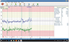
Fig. 1
The tracing of beat-to-beat variations (an analog of the ultrasound cardiotocography curve) in case 1. Blue line is fetal beat-to-beat variatiations curve and green one is maternal. The blue line crossing the fetal heart rate pattern shows the estimated basal heart rate (baseline). The gray shaded areas show the detected accelerations and decelerations.
Case 2. A pregnant woman aged 30 years was admitted to the division of maternal–fetal medicine at 26 weeks of gestation. She had attended irregular antenatal visits, which revealed no abnormalities. She had a blood pressure of 140/90, a pulse of 82 per minute, and unremarkable laboratory findings with a trace of proteinuria in urinalysis. She had no prodromes suggestive of arterial hypertension. She had mild pre-eclampsia and early-onset FGR. No maternal medical diseases were known. The reversed diastolic component in the UA, absent a-wave in the DV, and UV pulsations were found. Non-reactivity was detected on the CTG record, but the fetal heart rate was normal. An investigation of beat-to-beat variations using NI-FECG demonstrated fetal non-reactivity with a decreased value of STV (=1.2 ms) and a lack of accelerations. Two decelerations were found on this tracing. The HRV variables were also pathological: SI, 3102; AMo, 85%. The fABAS value of 3.89 SD of weeks of gestation below the mean of the normal population indicated a clear reduction in autonomic maturation/activity outside the normal range. Fetal antenatal death occurred the next day. Labor was induced. She delivered a stillborn male baby weighing 410 g, 27 cm in body length, and with a 19-cm head circumference.
Case 3. A 26-year-old pregnant woman was admitted to the department of maternal–fetal medicine at 26 weeks of gestation. Early FGR, reversed diastolic umbilical blood UA flow, reversed DV a-wave, and UV pulsations were found. Conventional CTG tracing was non-reactive. No further clinical or instrumental pathological findings were detected in this case. An assessment of fetal HRV by NI-FECG revealed non-reactivity with low STV (=1.8 ms) and lack of accelerations. The HRV descriptors were pathological: SI, 2104; AMo, 81%. The estimated value of the fABAS was 3.85 SD of weeks of gestation below the mean of the normal population, which indicated fetal autonomic malfunction. Furthermore, antenatal fetal death occurred in several days. She delivered a stillborn male baby weighing 440 g, 29 cm in body length, and with a 19-cm head circumference.
Discussion
Timely and precise diagnosing of fetal distress is an issue in perinatology. FGR is a condition detected and monitored using ultrasound techniques. Thus, the estimated fetal weight and amniotic fluid index are markers of FGR [23]. Hemodynamic Doppler waveform analysis is very popular nowadays, but the specificity and sensitivity of absent or reversed blood flow in the UA, DV, or UV pulsations are not maximal [2]. For this reason, it remains unclear whether it is preferable to prolong observation (especially before 30 weeks of gestation) to achieve better fetal maturity or to perform urgent cesarean section and continue treatment in the neonatal resuscitation unit. Based on this, cesarean section is indicated in cases of FGR, according to a protocol adopted in Ukraine. Some variables of CTG are used for the detection of fetal distress in growth-restricted fetuses [16].
The results of these cases support the possible prospect of NI-FECG clinical implementation. NI-FECG could provide higher temporal resolution and could be used for the investigation of PQRST morphology [3891213]. The data of the studies that have been reported and are available on PubMed from the last decade are promising (Table 1) [312131415]. The quality of fetal heart rate patterns registered by NI-FECG from 24 to 28 weeks of gestation was found to be very good [1]. These terms were described as a period of neurological maturation. Therefore, NI-FECG variables could be sensitive markers of fetal well-being. Previous findings reflected the considerable false-positive fetal distress diagnoses based on hemodynamic Doppler monitoring and conventional CTG [47].
Table 1
Recent studies of the non-invasive fetal electrocardiography (NI-FECG) clinical feasibility

| Study author | Study title | Conclusion |
|---|---|---|
| Fuchs et al. [14] | Fetal heart rate monitoring using maternal abdominal surface electrodes in third trimester: can we obtain additional information other than CTG trace? | Higher values of T/QRS ratio in FGR pregnancies with normal and reduced cerebro-placental ratio than in control group regardless of the result of CTG examination may indicate minimal worsening of intrauterine fetal well-being in growth retarded fetuses. |
| Fuchs [3] | Values of T/QRS ratio in pregnancies complicated by intrauterine growth restriction. | T/QRS ratio may be useful in assessing fetal well-being in FGR pregnancies; however, future studies are needed to determine typical ranges of T/QRS ratio in pregnancies complicated by IUGR. |
| Lakhno [8] | Fetal non-invasive electrocardiography contributes to better diagnostics of fetal distress: a cross-sectional study among patients with preeclampsia. | The results suggest that fetal NI-FECG monitoring is more objective than conventional CTG. |
| Reinhard et al. [13] | Comparison of non-invasive fetal electrocardiogram to Doppler cardiotocogram during the 1st stage of labor. | This non-invasive NI-FECG presents an alternative, reliable and accurate assessment for fetal well-being during the 1 (st) stage of labor. |
We have demonstrated for the first time the possible use of fetal HRV variables obtained by NI-FECG in the detection of fetal deterioration. The first case reflected that, despite the disturbed UA, UV, and DV hemodynamics, the fetal HRV variables were normal. This indicated the compensatory ability of the autonomic mechanisms of regulation. Since all variables detected via ultrasound were abnormal, we found a mosaic of evidence. Therefore, the inclusion of NI-FECG in the program of management helped to achieve better fetal maturity in the first case. The possibilities of fetal distress detection using NI-FECG were shown in the other 2 cases. The second case demonstrates that fetal HRV values have been very useful in the detection of fetal compromise. Fetal HRV parameters were good predictors of stillbirth in the third case.
We could conclude that STV, LTV, AMo, and SI are very valuable biophysical markers of fetal distress, as determined previously [9]. The calculation of the fABAS reflected the absence of the evolutionarily determined process of the neurological maturation in FGR and fetal distress in these cases. This case series demonstrated that fetal autonomic tone investigation could be of great practical use in early-onset FGR. Fetal autonomic regulation is involved in the fetal biophysical activity and deteriorated in fetal compromise. The usage of NI-FECG in early FGR is a novel diagnostic option that requires further trials.
In conclusions, NI-FECG could be a prospective method for the detection of fetal distress in early FGR. The variables of beat-to-beat variations, fetal HRV, and the fABAS should be investigated as biophysical markers of fetal deterioration.
 XML Download