

 PDF
PDF ePub
ePub Citation
 Print
Print


I. Introduction
Due to its relative rarity, metastatic carcinoma in the oral region can be difficult to treat. Metastatic carcinoma accounts for approximately 1% of cancer in the oral region1. The most common region of primary tumors leading to mandibular metastasis is the lung in men and breast in women2. Metastasis of tumors to the jaw bone can be especially difficult to detect because they commonly appear as an osteolytic lesion, which can be difficult to discriminate from odontogenic cysts. According to the literature, in 30% of cases with metastasis to the jaw, the primary lesion was asymptomatic and was not detected before the metastatic lesion3.
This report presents a case of oral carcinoma metastasized from follicular thyroid carcinoma. Follicular cell cancers are the second most common thyroid cancer after papillary thyroid carcinoma. These tumors are derived from follicular cells of the thyroid region and are more likely to be found among females 40 to 60 years old. They account for 10% of all thyroid malignancies4. They are usually asymptomatic and are initially detected via palpation of a solid nodule, multinodular goiter, or cervical lymphadenopathy5. In this report, we describe a case of follicular thyroid carcinoma in which metastasis to the mandibular anterior-posterior region was detected prior to that of the primary thyroid carcinoma.
II. Case Report
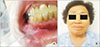
In October 2017, a 67-year-old female was referred to the Department of Oral and Maxillofacial Surgery of Dankook University Dental Hospital (Cheonan, Korea) for evaluation of gingival and facial swelling. Prior to the patient's first visit to our hospital, the swelling had been attributed to follicular carcinoma originating from the thyroid region via fine needle aspiration performed at a local hospital for mandibular asymmetry evaluation. The patient was a non-smoker and suffered from hypertension; she had no family history of thyroid disease. The mandibular lesion had been detected approximately 5 months prior in a local dental clinic, and the patient was recommended to visit a higher-tier hospital for further evaluation of a suspected mandibular cyst. However, no further visits to the hospital were made before September, when the patient started to suffer from facial swelling and paresthesia of the right mandibular region. At that time, she visited another local hospital, where fine needle aspiration biopsy was performed. The patient was finally referred to our hospital for further evaluation and treatment. Gingival swelling of the right mandibular area was observed upon initial intra-oral examination, and extra-oral examination revealed facial asymmetry with swelling and paresthesia of the right mandibular area.(Fig. 1)
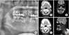
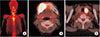
X-ray radiographs (panoramic radiograph, computed tomography [CT], and magnetic resonance imaging [MRI]) were collected for further diagnosis.(Fig. 2) Axial CT and sagittal MRI images revealed a large (52 mm×44 mm×34 mm) mass with a lobulated margin in the right mandibular body. An identical mass in the corresponding region was observed on panoramic view. There was no evidence of a lesion effect one the adjacent teeth—such as resorption of roots or occlusion change. A whole-body bone scan and positron emission tomography-CT (PET-CT) were performed for further analysis (Fig. 3), revealing a mass in the same region and additional identification of the primary lesion in the thyroid area. The primary thyroid lesion was referred to the ENT (ear, nose and throat) department for further management, and additional fine needle aspiration of this lesion was performed for additional diagnosis, revealing follicular thyroid carcinoma.
Partial mandibulectomy from the right mandibular angle to the left mandibular central incisor and supraomohyoid neck dissection were performed. A fibular free flap was used to restore mandibular stability and function. Results of the excisional biopsy revealed that the lesion was metastatic carcinoma originating from follicular thyroid carcinoma of the thyroid gland.(Fig. 4) Total thyroidectomy was performed in the ENT department one month later. The patient received subsequent radioactive iodine therapy in March 2018, and no specific signs were observed on follow-up radiographic images. The patient is currently using a temporary denture, and the mandibular operation field remains in a stabilized state. (Fig. 5)
III. Discussion
Distant metastasis of thyroid carcinoma is rare, with bone (vertebrae, pelvis, and ribs) and lung as its most common metastatic sites6. Distant metastasis to the oral region from thyroid carcinoma accounts for approximately 3% of all oral metastatic carcinomas7. The vast majority of metastatic tumors of the jaw is clinically presents with intraoral or extraoral swelling accompanied by chin paresthesia and pain68. Although thyroid carcinomas in general affect women more often than men, equal incidence between the sexes has been found for metastasis9. The mandible is more commonly affected than the maxilla, with the posterior region being the most frequent site of metastasis2. Because follicular thyroid carcinomas are more prone to hematogenous spread in advanced forms, the posterior part of the mandible, located directly along the main blood stream of the inferior alveolar vessel, can be easily affected89. Furthermore, the vast amount of hematopoietic bone marrow situated in this particular region allows for much greater hematopoietic activity of this region compared to other mandibular areas. Thus, the posterior mandible can serve as a favorable niche for attracting metastatic tumor cells89. Moreover, not only does this type of carcinoma spread through a hematogenous route, but it also has a tendency to grow to an extremely vascularized form of tumor in its advanced stages. This high vascularity with pulsation may lead to incorrect diagnosis, such as arteriovenous malformation10. Therefore, consideration of bleeding control is necessary. The literature has recommended preoperative angiography and selective embolization or external carotid artery ligation for intraoperative bleeding control911. However, because thyroid carcinomas are more likely to spread hematogenously, their propensity for lymphatic spread is relatively small9. Neck dissection may not be necessary for cases without lymphadenopathy. Selective excision of suspicious lymph nodes along the internal jugular vein and frozen sectioning may be considered for “staging” and determination of the necessity of neck dissection9. For the case herein, no sign of tumors was found among the lymph node groups of the supraomohyoid region.
Complete treatment of metastatic carcinoma requires addressing both the primary lesion and metastatic site. Treatment for the primary thyroid lesion often involves high-dose radioiodine therapy or thyroidectomy, as it did in this case of follicular thyroid carcinoma. However, for cases with bone metastasis, sufficient concentration of radioiodine cannot be easily achieved via intravenous administration, and external beam radiation therapy can be considered as an alternative12. Most metastatic carcinoma cases are dealt with via surgical resection, radiation therapy, or a combination. Treatment of our patient was carried out in a similar way via partial surgical resection of the mandible and reconstruction with an osteocutaneous fibular free flap. A previous study reported a similar case of mandibular metastasis of papillary thyroid carcinoma treated through surgical resection and bone-impacted fibular free flap reconstruction, which was followed up for 17 months13.
The prognosis and survival rate of follicular thyroid carcinoma have been reviewed in several studies. The prognosis of follicular thyroid carcinoma depends on age, size, and extent of vascular invasion and distant metastasis. An overall 5-year survival rate of 40% and 10-year survival rate of 27% for bone metastasis of differentiated thyroid carcinoma have been reported1415. More recently, in a systematic review literature of 59 cases of thyroid cancer with facial skeleton metastasis, the overall survival rate was 96% at 2 years and 59% at 5 years. Among them, 22 cases were treated with surgery and postoperative radioactive iodine, yielding a survival rate of 100% at 2 years and 71% at 5 years16.
Because of their rarity, metastatic follicular thyroid carcinoma of the mandible has an unknown exact incidence, and treatment of this type of carcinoma can be a clinical challenge. Early diagnosis and adequate surgical and radiological treatment are vital to long-term survival of patients suffering from this type of carcinoma. It is therefore recommended that the maxillofacial surgeon collaborate with relevant specialists for successful treatment of metastatic follicular thyroid carcinoma.


 XML Download