

 PDF
PDF ePub
ePub Citation
 Print
Print


I. Introduction
Current literature defines dental implants shorter than 8 mm as short dental implants (SDIs)1234. SDIs were considered to have a lower success rate than standard length implants45. However, no distinct linear relationship between implant length and survival rate has been identified46, and recent studies have shown that SDIs have comparable success rates78910. In some situations, the mechanical stress on a shorter implant might be lower than that on a longer implant111213.
In some patients, pathologic conditions lead to insufficient residual bone quality. Planning implant therapy in these patients needs careful consideration to gain predictable results and avoid complications.(Fig. 1) In patients with insufficient bone volume, several procedures can be used such as maxillary sinus elevation, guided bone regeneration or edentulous ridge expansion; but they all involve prolonged healing time, higher morbidity, and high cost5. Recently, SDIs have been considered to be an alternative resolution to those conditions.
While the success and survival of SDIs has been widely investigated, studies on the survival rate of SDIs in medically compromised patients are limited. The purpose of this study was to determine the survival rate of SDIs in medically compromised patients. There are several systemic disorders that were approved to have the significance influence on dental implant treatment success. Although some authors did not find the negative effect of diabetes mellitus to implantation outcomes14, other studies found statistically significant relationship of implant failure and diabetes mellitus (controlled and uncontrolled)15. The compromised condition of gingival microvascular in diabetes patients may affect wound healing and increasing the risk of infection15.
The effect of uncontrolled hypertension condition on survival rate of dental implant is under controversy16. The risk of cardiovascular complications and renal failure in uncontrolled hypertension patients are well established17. These complications may affect the ossteointegration and change of alveolar bone level. Result of some studies show that patients with cardiovascular disease had increased peri-implant bone loss and peri-implantitis18.
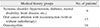
In this retrospective study, patients who had special medical histories were categorized into 3 groups: systemic disorders, such as uncontrolled or controlled diabetes mellitus, mental disability, and uncontrolled hypertension; oral cancer ablation with reconstruction, with or without radiotherapy; diverse osteomyelitis, such as osteoradionecrosis and bisphosphonate-related osteonecrosis of the jaw (BRONJ). Most of these patients have insufficient residual bone quality due to mandible atrophy or sinus pneumatization. Marginal bone loss (MBL) is a generally accepted parameter for evaluating bone response around a dental implant19. Therefore, we evaluated MBL of SDIs on panoramic radiographs taken at implant installation, 3 month, 1-year and 2-years follow-up visits.
Thirty-three patients with forty-seven implants that were 7-mm long were examined during the last 4 years. The implant diameters were 4.0 (n=38), 4.5 (n=8), and 5.0 mm (n=1). We analyzed SDI survival rate in the 3 patient groups, evaluated MBL, and discussed clinical implications.
II. Materials and Methods
1. Patients data
Thirty-three patients with SDIs placed from January 2015 to March 2018 at the Department of Oral and Maxillofacial Surgery at Seoul National University Dental Hospital (Seoul, Korea) were evaluated in this study.
Sample was chosen according to the following inclusion criterions: (1) medically compromised patients that belong to at least one of 3 groups: systemic disorders, such as diabetes mellitus (controlled or uncontrolled), mental disability, and uncontrolled hypertension; oral cancer ablation with reconstruction that associated to implantation sites, with or without radiotherapy; and diverse osteomyelitis such as osteoradionecrosis or BRONJ (Table 1); (2) patients were treated with the installation of internal submerged tapered Luna (Shinhung, Seoul, Korea) and internal non-submerged Stella (Shinhung) sand blasted and acid etched (S&E) SDIs; and (3) patients didn't receive any bone augmentation at the implantation site. All the selected patients have insufficient residual bone quality due to mandible atrophy or sinus pneumatization.
All implants were placed through 1- or 2-stage procedures with a 3- to 6-month interval. Under local anesthesia, implants were installed according to the Luna and Stella implant surgical protocol by a single maxillofacial implant surgeon. All implants initially achieved good primary stability. A panoramic radiograph was taken of all cases after implant surgery. This retrospective data analysis was approved by the Institutional Review Board of Seoul National University (S-D20180022).
2. Marginal bone loss evaluation
MBL was determined from panoramic radiographs and expressed as the distance from the implant shoulder to the most coronal bone-to-implant contact on the mesial and distal sides of the implant. The relationship between the implant shoulder and marginal bone was measured mesially and distally by using reference lines including a line along the longitudinal implant axis, a horizontal line at the most coronal level of the implant shoulder, and two horizontal lines at the most coronal level of bone-to-implant contact mesially and distally20. MBL was evaluated on panoramic radiographs taken at implant placement, and at 3 months, 1 year, and 2 years follow-up visit. The MBL was measured at the same magnification on all installation and follow-up radiographs. Each aspect was measured 3 times, and the average was recorded.(Fig. 2) The change in MBL from installation at follow-up visits and changes between consecutive visits were calculated. A failed implant was considered as a lost or mobile implant or severe peri-implantitis that required prompt removal.
3. Statistical analysis
The collected data included descriptive and quantitative data. IBM SPSS Statistics software (ver. 25.0; IBM Corp., Armonk, NY, USA) was used for statistical analyses. Descriptive statistics were used to analyze and calculate the distributions of qualitative variables. For analyzing quantitative variables to assess MBL, mean and standard deviation were calculated. We evaluated MBL data using the Shapiro–Wilk normality test. Data review and statistical analysis were performed by a single researcher (T.T.H.N.).
III. Results
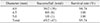
Among the 33 patients, 11 were male, and 22 were female. Patient ages at installation ranged from 30 to 82 years and averaged 62 years. In total 47 implants were installed with diameters of 4.0 (n=38), 4.5 (n=8), and 5.0 mm (n=1). Of the 47 implants, 6 were Stella implants, and 41 were Luna implants. Nineteen implants were installed in the maxilla and twenty-eight in the mandible.(Table 2) The follow-up periods ranged from 7 to 36 months with an average of 15 months.
In total 45 success implants, there were 19 implants supporting single crown restorations, 22 implants supporting multiple fixed prostheses, 4 implants supporting removable overdentures.(Table 3)
1. Survival rate
Among the 47 implants placed, 2 implants failed before the last follow-up. The survival rate of 7-mm SDIs was 95.74% from stage I surgery to the last follow-up. The survival rates of 4.0-mm-diameter implants was 94.74%, 4.5-mm-diameter implants was 100%, and 5.0-mm-diameter implants was 100%.(Table 4) Both failed implants were 4.0 mm in diameter in a patient who had oral cancer and underwent reconstruction. Survival rates for the three diameters did not differ significantly (P=0.069; P>0.05).
2. Marginal bone loss
The mean MBL between implant installation and 1 month on the mesial and distal aspects was 0.34±0.47 mm and 0.53±0.57 mm, between installation and 1 year on the mesial and distal aspects was 0.53±0.58 mm and 0.67±0.56 mm, respectively. It was 0.58±0.60 mm and 0.71±0.60 mm, respectively, between implant installation and 2 years. Between 3 month and 1 year, the mean MBL on mesial and distal aspects increased by 0.19±0.24 and 0.14±0.02; between 1 and 2 years, it was 0.05±0.12 and 0.04±0.05 mm, respectively. The mean MBL at 3 months and 1 and 2 years was significantly higher than at implant installation. The mean MBL at 1 year was also significantly higher than at 3 months. The MBL at 1 and 2 years did not differ significantly (P<0.05).(Table 5)
IV. Discussion
Using the longest possible implants was considered conventional therapy based on the principle that longer implants would have higher survival rates and a more favorable prognosis21. However, in many clinical conditions, long implants were limited or unfavorable due to insufficient bone volume, maxillary sinus pneumatization, and inferior alveolar nerve canal position.
The indication for SDIs remains controversial because of challenges such as less bone-to-implant contact due to reduced implant surface, more crestal bone resorption due to a smaller surface over which to distribute forces, and increased crown-to-implant (C/I) ratio1119. The technique for installing SDIs involves some considerations. First, the direction is easily distorted when drilling because the hole made for SDIs is shallower than for longer implants. Therefore, placing an SDI requires more skill. Second, the hole made by the counterbore should not be too deep because of the short fixture. Third, the implant-supported restoration should not be too large.
Despite these considerations, SDI has many advantages to both the patient and surgeon. Using SDI avoids bone grafting and nerve transposition, reduces donor site morbidity for autogenous bone grafting, reduces nerve damage for nerve transposition, and, therefore, reduces treatment time and cost and patient discomfort22. SDI can help decrease the possibility of contact with adjacent tooth roots, lower the risk of surgical paresthesia, reduce bone overheating, and lower the risk of bone graft exposure, which brings significant advantages to implant therapy for medical compromised patients.
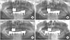
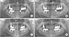
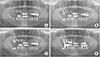
We present 3 cases of SDI placement, one from each medical condition group. Case 1 was a 76-year-old female who had squamous cell cancer and was treated with maxillary mass resection and radiotherapy on the left, and then underwent radical neck dissection and radiotherapy due to right neck metastasis. An implant was planned for the edentulous region of the right posterior mandible. After considering mandible atrophy and proximity to the inferior alveolar nerve, a 4.5 mm×7 mm Stella implant was placed. The implant achieved good stability and bone integration after loading and showed acceptable MBL at 3 years.(Fig. 3) Case 2 is a 72-year-old male with osteomyelitis and a history of hypertension and diabetes. A 4 mm×7 mm Stella implant was installed in the 37 tooth site. The implant showed good stability and low MBL after loading and at 1 year.(Fig. 4) Case 3 is a 67-year-old female who had hypertension and osteomyelitis in the posterior right mandible. A previous implant installed in the 37 tooth position failed due to bone resorption. In addition, the edentulous posterior of the right maxilla also had insufficient bone and sinus pneumatization. Therefore, in planning the implant therapy, a 4 mm×7 mm Luna implant was chosen for the 16 position, and a 4.5 mm×7 mm Stella implant was chosen for the 47 tooth position after removal of the failed implant. The two implants showed good stability and acceptable MBL on follow-up examination.(Fig. 5)
Of the 47 implants, two failed, and the survival rate was 95.74%. The two failed implants belonged to a patient with oral cancer who was treated with mandibular resection and reconstruction. The postoperative bone had insufficient volume and unfavorable quality. Dental implant treatment has few absolute contraindications, and the impact of health risks on implant outcome remains unclear due to the scarcity of prospective studies19. However, studies have shown a negative impact of bisphosphonates on implant success19. In oral cancer patients, a lack of residual bone following resection makes placing implants in an ideal position difficult2324. Considering that all patients were medically compromised, including cancer and BRONJ, the survival rate of 7-mm-long implants in the general population would be higher than in this study. The failed implants in this study were placed adventurously in alveolar bone, which had insufficient height and had been involved in cancer treatment and reconstruction surgery. Most implant failures were reported early, during the healing phase at abutment connection2526272829.
To guarantee long-term clinical results, maintaining stable marginal bone is more critical with SDIs18. MBL is a generally accepted parameter to evaluate the bone response around a dental implant. Originally, a mean MBL of ≥1.5 mm in the first year and an MBL of ≥0.2 mm per year afterward was considered a threshold for implant success2030. Randomized, controlled studies31 on SDIs in the posterior maxilla had an MBL from 1.02 to 0.1 mm. In this study, the MBL results on the mesial and distal aspects after 1 year were 0.53±0.58 mm and 0.67±0.56 mm, respectively, and 0.58±0.60 mm and 0.71±0.60 mm, respectively, after 2 years. These MBL results are within the success threshold20; however, long-term follow-up is needed.
V. Conclusion
The present study showed comparable survival rates of SDIs in medically compromised patients to the conventional implants in a healthy population. In addition, the stability of marginal bone around an SDI in these patients was acceptable in comparison with MBL in healthy patients. The results suggest that placing an SDI is a reliable treatment option, especially for medically compromised patients, and can be an alternative when sinus lifting or vertical bone grafting should be avoided. Further, long-term follow-up and evaluation of SDIs in these patients is needed.


 XML Download