

 PDF
PDF Citation
 Print
Print


INTRODUCTION
Curcumin, a chemical product from Curcuma longa, is a diarylheptanoid that has numerous interference properties in inflammation, cancer, diabetes, and colitis, among others (12345). Recent studies have emphasized its role in the regulation of cancer via an increase in tumor immunity; however, most of these studies were focused on the anti-inflammatory action of curcumin via inhibition of ROS and inflammatory cytokine production (67891011). This compound is emerging as an anticancer agent because it not only targets tumor cell growth or metastatic functions but also regulates tumor-immunity by increasing Th1 and inhibiting Treg cells (111213). According to previous reports, curcumin is also known to regulate the NF-κB pathway of various immune cells such as dendritic cells (14), macrophages (151617), NK cells (18), T cells (19), and B cells (20), suggesting its potent immune modulatory action on proliferative moieties of immune cells.
A very old study demonstrated that dietary curcumin enhances IgG Ab production in rats (21); however, little is known about its cellular mechanism of action on humoral immunity, such as Ab production and isotype class switching. During Ag exposure, B cells can generate high affinity Abs with different isotypes by cross-talk between B and T cells in the germinal center (GC) (222324). T follicular helper (TFH) cells, which express the CXCR5 and key transcription factor B-cell lymphoma 6 (Bcl-6), are localized in the GC in draining lymph nodes (25). TFH cells provide critical costimulatory signals and cytokines to B cell cognates, resulting in somatic mutation for affinity maturation and isotype-class switch (26272829).
Here, we attempted to examine curcumin function in mice to assess whether it is involved in Ab production with regard to the TFH and GC response. We selected intra-peritoneal administration due to the low stability of curcumin via the oral route; moreover, we opted for NP-ovalbumin (NP-OVA) immunization with the complete adjuvant method to induce a significant amount of TFH and GC B cells in the draining lymph nodes. The results revealed that curcumin causes in a significant increase in CXCR5+Bcl6+ TFH and CD95+GL-7+ GC B cells, with elevated total Ab production as well as high affinity Ag specific IgG1 and IgG2b Ab production.
MATERIALS AND METHODS
Mice
C57BL/6 female mice (6–8 weeks old) were purchased from Orient Bio (Daejeon, South Korea) and maintained at the Hanyang University mouse facilities under pathogen-free conditions with ad libitum feeding. All animal protocols were approved by the Animal Experimentation Ethics Committee of Hanyang University, and the experiments were performed according to the guidelines of the Institutional Animal Care and Use Committee of Hanyang University (2016-0141).
NP-OVA immunization and curcumin administration
NP-OVA (50 μg, Bioresearch Technologies, Novato, CA, USA), diluted in Complete Fruend's Adjuvant (Chondrex, Redmond, WA, USA) was subcutaneously injected into the dorsal region of mice. Seven days after immunization, the mice were sacrificed, and the inguinal lymph node cells were isolated and analyzed by flow cytometry. Curcumin was purchased from Abcam (141921; Cambridge, England) and dissolved in DMSO. Curcumin (200 μg) was injected intra-peritoneally (i.p.) daily following the indicated experimental scheme.
Flow cytometry for TFH cells and GC B cells
To analyze the TFH cells and GC responses, the inguinal lymph node cells were isolated and stained with anti-mouse CXCR5-biotin for 30 min at 4°C, followed by anti-mouse CD44-Ag-presenting cell (APC)/Cy7 and CD4-Percp/Cy5.5 and streptavidin-APC staining. After fixation and permeabilization using a Foxp3 Staining Kit (eBioscience, San Diego, CA, USA), intracellular Bcl-6 and Foxp3 was stained using anti-mouse Bcl-6-PE (BD Bioscience, San Jose, CA, USA) and Foxp3-FITC (eBioscience) for 40 min at room temperature. To analyze GC B cells, the isolated single cells were stained with anti-mouse B220-Percp/Cy5.5, GL-7-FITC, and CD95-PE for 30 min at 4°C. Then, the cells were examined using a FACSCanto II system (BD Bioscience); the data were analyzed using FlowJo software (Treestar, Ashland, OR, USA).
ELISA for Ab measurement
The total Ab production in mouse serum was measured using the IgM ELISA kit (eBioscience) and IgG ELISA kit (Abcam). To measure low and high affinity Abs, NP30-conjugated BSA or NP7-conjugated BSA (Biosearch Technologies, Petaluma, CA, USA) were coated, respectively. For the isotype Ab analysis, HRP-conjugated goat anti-mouse IgG1, IgG2b, IgG2c, and IgG3 Abs (Southern Biotech, Birmingham, AL, USA) were used to detect the Abs.
RESULTS AND DISCUSSION
Curcumin administration increases Bcl-6+CXCR5+ TFH cells
To examine curcumin function in the TFH and GC response to a specific Ag, 6-week-old female C57BL/6 mice were subcutaneously immunized with NP-OVA in complete adjuvant on day 0, followed by daily administration of 200 μg curcumin by i.p. injection until day 6 (Fig. 1). At day 7, inguinal lymph node cells were isolated and analyzed by flow cytometry. As shown in Fig. 2A, NP-OVA immunization, with DMSO vehicle, led to a significant increase in Bcl-6+CXCR5+ TFH cells compared to non-immunized group. Curcumin treatment significantly increased Bcl-6+CXCR5+ TFH cells, which was gated from CD4+CD44+ cells in comparison with the DMSO vehicle control group. The CD44 expression level of CD4 T cells (Fig. 2B) and percentage of Bcl-6+Foxp3+ T follicular regulatory cells (Fig. 2C) was comparable between the two groups. The expression level of TFH cell marker molecules such as Bcl-6 or CXCR5 (Fig. 2D) was significantly increased, collectively suggesting that curcumin administration significantly induces TFH cells in the draining lymph nodes during Ag immunization.
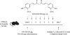 | Figure 1Experimental scheme of NP-OVA immunization and curcumin administration. Six to eight-week-old female C57BL/6 mice were immunized with NP-OVA in complete adjuvant, and 200 µg of curcumin was i.p. injected daily until day 6. The mice were sacrificed and the draining lymph node cells were analyzed on day 7.CFA, complete freund's adjuvant.
|
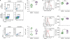 | Figure 2Curcumin administration increases Bcl-6+CXCR5+ TFH cells. (A) Flow cytometric analysis of CD4+CD44+CXCR5+Bcl-6+ TFH cells in the inguinal lymph nodes from NP-OVA-immunized mice treated with DMSO or curcumin. (B) The expression level of CD44 on CD4+ cells (C) The proportion of CD4+CD44+CXCR5+Bcl-6+Foxp3+ TFR cells. (D) The level of Bcl-6 and CXCR5 on CD4+CD44+ cells. Each dot represents an individual sample. Data are presented as the mean±standard deviation of 2 independent experiments (n=7–8).MFI, mean fluorescence intensity; SSC-A, standard scrapie cell assay; TFR, T follicular regulatory; NS, not significant.
*p<0.05, ***p<0.001.
|
Curcumin administration increases GL-7+CD95+ GC B cells.
Due to the observed increased proportion of TFH cells and the results of a previous study that showed enhanced B cell function in sheep RBC immunized mice (27), we hypothesized that there would be elevated levels of GC B cells. The inguinal lymph node cells were analyzed by flow cytometry. With no difference in the proportion of B cells (Fig. 3A), NP-OVA immunization of the DMSO-treated group led to an increase in the GL-7+CD95+ cells, whereas curcumin administration significantly increased the proportion of GC B cells in the draining lymph nodes (Fig. 3B). In addition, there was an increase in the high affinity total IgG and IgM Abs in the serum upon curcumin treatment (Fig. 3C), suggesting that increased TFH cells by curcumin administration could result in elevated GC B cells and Ab production.
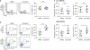 | Figure 3Curcumin administration increases GL-7+CD95+ GC B cells. Flow cytometric analysis of (A) B220+ B cell and (B) B220+GL-7+CD95+ GC B cells in the inguinal lymph nodes. (C) Total IgM and IgG Ab level in the serum of NP-OVA-immunized mice. Each dot represents an individual sample. Data are presented as the mean±standard deviation of 2 independent experiments (n=7–8).SSC-A, standard scrapie cell assay; NS, not significant.
*p<0.05, ***p<0.001.
|
Curcumin administration increases production of high affinity IgG1 and IgG2b Ab
As TFH cells are key players in affinity maturation and isotype class-switching by the stimulation of GC B cells, we examined various NP-specific isotypes of the Abs in the serum to confirm the consequences of increased TFH and GC responses by curcumin administration. Fig. 4A and B shows the significant increase in IgG1 and IgG2b Ab production upon curcumin treatment, whereas no significant differences were observed for other isotypes or low affinity Abs. These results suggest that curcumin treatment stimulates high affinity Ab production with isotype class-switching.
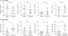 | Figure 4Curcumin administration increases the production of high affinity IgG1 and IgG2b Abs. (A) NP30-(low affinity) or (B) NP7-(high affinity) specific Abs to measure IgG1, IgG2c, IgG2b, and IgG3 in the serum of NP-OVA-immunized mice. Each dot represents an individual mouse. Data are presented as the mean±standard deviation of 2 independent experiments (n=7–8).NS, not significant.
*p<0.05.
|
Curcumin, one of the most widely studied natural compounds, has diverse biological functions in attenuating chronic inflammatory diseases, including cancer, asthma, inflammatory bowel disease, and rheumatoid arthritis (3031). These therapeutic functions of curcumin have been mainly associated with the suppression of the production of inflammatory cytokines TNF, IL-1, IL-6, and IL-8 (1832). However, recent studies have reported that curcumin can directly regulate T cells by inhibiting IL-2 signaling and NF-κB activation (3334). Moreover, curcumin inhibited Th1 differentiation by blocking JAK-STAT signal activation (3536). Here, we demonstrate for the first time, to our knowledge, the positive biological function of curcumin in TFH cell differentiation and GC B cell formation. We found that curcumin administration in vivo increases Bcl-6+CXCR5+ TFH cells and GL-7+CD95+ GC B cells, with high affinity Ab production. As curcumin has a direct effect on the biological functions of T cells, we further examined curcumin treatment in T cell differentiation and found that curcumin appears to specifically inhibit Th1 and Th2 differentiation rather than Th17 (Supplementary Fig. 1). Collectively, these results indicate that curcumin modulates multiple molecular targets and has potent anti-inflammatory activities regulating effector T cell functions (TFH, Th1, and Th2 cells). Direct regulation of the NF-κB and JAK-STAT signaling pathway by curcumin might be the clue to elucidating the potential mechanisms involved in the generation of TFH cells and GC responses. Furthermore, considering previous reports that curcumin can enhance B cell function (2137), dissecting the mechanism underlying the Ab production increasing effect of curcumin should be addressed in further studies.
In conclusion, this study is the first to report that the administration of curcumin increases humoral immunity by Ab production, which is presumably mediated by increased TFH cells in the draining lymph nodes. Interestingly, curcumin also contributes in the production of high affinity Abs of the IgG1 and IgG2b isotypes during immunization. Although the molecular mechanisms of curcumin's action on the TFH response should be further evaluated in detail, we believe that curcumin could be an advantageous supplement, to enhance protective immunity via increased Ab production, in the treatment of infectious diseases or cancer.
 XML Download