

 PDF
PDF ePub
ePub Citation
 Print
Print


Seong-Min Kim, Kyu-Bok Lee
Abstract
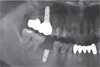
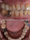
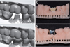
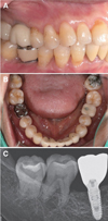
Accurate impression taking for the success of implant prosthesis is a very important process. Methods of taking implant impression include the conventional method using impression coping and impression material, and the digital method using an intraoral scanner and scanbody. However, the impression coping or the scanbody must install after remove healing abutment. Because of this, the dentist must repeat the process of removing and installing the healing abutment, the impression coping or the scanbody several times. In addition, the impression coping or the scanbody rises higher than the occlusal surface, so the patient has the inconvenience of constantly maintaining the open state. Recently, a scannable healing abutment, which can be scanned by a intraoral scanner directly, without the need to remove the healing abutment by applying a scannable part of the scanbody to the healing abutment, was introduced. We present a case of single posterior implant prosthesis using a scannable healing abutment.
Figures and Tables
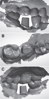
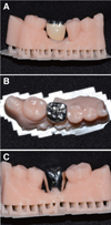
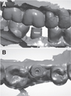
Fig. 4
Design of retrievable cement type slot (RCS) type customized abutment. (A) Buccal, (B) Occlusal, (C) Lingual side with lingual slot.
References
1. Del'Acqua MA, Arioli-Filho JN, Compagnoni MA, Mollo Fde A Jr. Accuracy of impression and pouring techniques for an implant-supported prosthesis. Int J Oral Maxillofac Implants. 2008; 23:226–236.
2. Wee AG. Comparison of impression materials for direct multi-implant impressions. J Prosthet Dent. 2000; 83:323–331.

3. Del'Acqua MA, Chávez AM, Compagnoni MA, Mollo Fde A Jr. Accuracy of impression techniques for an implant-supported prosthesis. Int J Oral Maxillofac Implants. 2010; 25:715–721.
4. Nayyar N, Yilmaz B, McGlumphy E. Using digitally coded healing abutments and an intraoral scanner to fabricate implant-supported, cement-retained restorations. J Prosthet Dent. 2013; 109:210–215.
5. Kim YG, Lee YI. The relationship between dental treatment and temporomandibular disorder. J Korean Dent Assoc. 2008; 46:308–314.
6. Grossmann Y, Pasciuta M, Finger IM. A novel technique using a coded healing abutment for the fabrication of a CAD/CAM titanium abutment for an implant-supported restoration. J Prosthet Dent. 2006; 95:258–261.
7. Hategan SI, Ionel TF, Goguta L, Gavrilovici A, Negrutiu ML, Jivanescu A. Powder and powder-free intra-oral scanners: Digital impression accuracy. Prim Dent J. 2018; 7:40–43.
 XML Download