

 PDF
PDF ePub
ePub Citation
 Print
Print


Ye-Ji Kim , Young-Gyun Song
, Young-Gyun Song
Abstract
In patients with severely resorbed alveolar bone, it is difficult to gain retention in denture. Lack of retention makes denture unstable and lead to trouble in using denture. Suction denture seals the entire denture border with movable mucosa and this sealing mechanism forms negative pressure beneath the denture and produce higher retention and stability to denture. In this case, 4 edentulous patients visited for lack of retention with dentures. Considering their high expectation with retention, suction denture concept was used to fabricate retentive and stable denture. The purpose of this case report is to compare and analyze the considerations of suction denture restorations in edentulous patients.
Figures and Tables
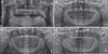 | Fig. 1Panoramic radiographs (A) Patient No. 1, (B) Patient No. 2, (C) Patient No. 3, (D) Patient No. 4.
|
 | Fig. 2Intraoral examination of patient No. 1. (A) Maxillary occlusal view, (B) Mandibular occlusal view.
|

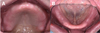
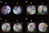 | Fig. 6Intraoral photos taken with bronchoscope. Note that buccal mucosa has filled the posterior space of the denture in patient No. 1, 2, and 3, but not in patient No. 4. (A) Patient No. 1, (B) Patient No. 2, (C) patient No. 3, (D) Patient No. 4. (R: right, L: left, BM: buccal mucosa, UD: upper denture, LD: lower denture, T: tongue).
|
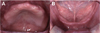 | Fig. 7Intraoral examination of patient No. 2. (A) Maxillary occlusal view, (B) Mandibular occlusal view.
|
 | Fig. 8Border of the denture was modified with modeling compound and functional impression was taken with tissue conditioner.
|
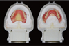
 | Fig. 9Sublingual border of the final impression (A), wax denture (B), and final denture (C) in patient No. 2.
|
 | Fig. 10Intraoral examination of patient No. 2. (A) Maxillary occlusal view, (B) Mandibular occlusal view.
|
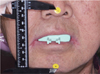
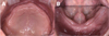
 | Fig. 11Intraoral examination of patient No. 4. (A) Maxillary occlusal view, (B) Frontal view, (C) Mandibular occlusal view.
|

References
1. Jacobson TE, Krol AJ. A contemporary review of the factors involved in complete denture retention, stability, and support. Part I: retention. J Prosthet Dent. 1983; 49:5–15.

2. Zarb GA, Bolender CL, Eckert SE, Fenton AH, Jacob RF, Mericsko-Stern R. Prosthodontic treatment for edentulous patients: Complete dentures and implant-supported prostheses. 13th ed. St. Louis, Mosby: 2012. p. 23–25.
3. Abe J, Kokubo K, Sato K. Mandibular Suction-effective denture and BPS: A complete guide. . Tokyo: Quintessence Publishing Co. Ltd;2012. p. 46–78.
4. Tyson KW, McCord JF. Chairside options for the treatment of complete denture problems associated with the atrophic (flat) mandibular ridge. Br Dent J. 2000; 188:10–14.
5. Azzam MK, Yurkstas AA, Kronman J. The sublingual crescent extension and its relation to the stability and retention of mandibular complete dentures. J Prosthet Dent. 1992; 67:205–210.
6. von Krammer R. Principles and technique in sublingual flange extension and complete mandibular dentures. J Prosthet Dent. 1982; 47:479–482.
7. Lawson WA. Influence of the sublingual fold on retention of complete lower dentures. J Prosthet Dent. 1961; 11:1038–1044.
8. Wright CR, Muyskens JH, Strong LH, Westerman KN, Kingery RH, Williams ST. A study of the tongue and its relation to denture stability. J Am Dent Assoc. 1949; 39:269–275.
9. Rajeshwari K, Kohli S, Mathew XK. Evaluation of resting tongue position in recently extracted and long term completely edentulous patients: A prospective interventional study. J Clin Diagn Res. 2017; 11:ZC61–ZC63.
10. Beresin VE, Schiesser FJ. The neutral zone in complete dentures. J Prosthet Dent. 1976; 36:356–367.
11. Klein P. Piezography: dynamic modeling or prosthetic volume. Actual Odontostomatol (Paris). 1974; 28:266–276.
 XML Download