

 PDF
PDF ePub
ePub Citation
 Print
Print


Hyejin Choi , Jaehoon Lee
, Jaehoon Lee
Abstract
Increased anterior teeth mastication following posterior teeth loss leads to greater anterior occlusal force. It may cause greater attrition of anterior teeth, traumatic force occlusion (TFO), also often followed by antagonist extrusion and occlusal disharmony. This clinical report describes the treatment for a 67-year-old female patient diagnosed with loss of both maxillary and left mandibular posterior teeth, severe attrition of maxillary and mandibular anterior teeth and extrusion of multiple teeth. A diagnostic cast was mounted on articular in centric relation (CR) position to evaluate vertical dimension (VD) and interspace. To provide adequate space for the prosthetic reconstructions, VD was increased by 3 mm on the anterior pin. And then diagnostic wax-up was completed upon that VD. Wax-up was converted to provisional restorations and verified in the patient's mouth and the final restorations were delivered. Clinical follow up examination held 3 months after temporary restoration owing to changes in vertical dimension revealed proper function in mastication without evidence of temporo-mandibular joint (TMJ) disorders. This clinical report presents successfully restoring severe attrition case with increasing vertical dimension resulting in satisfaction in esthetics and function.
Figures and Tables
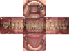
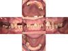 | Fig. 1Intraoral photograph before treatment. (A) Maxillary occlusal view, (B) Right lateral view, (C) Frontal view, (D) Left lateral view, (E) Mandibular occlusal view.
|
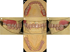 | Fig. 3Diagnostic wax up model with new occlusal vertical dimension. (A) Maxillary occlusal view, (B) Right lateral view, (C) Frontal view, (D) Left lateral view, (E) Mandibular occlusal view.
|
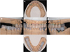 | Fig. 4Full contour wax up and wax milling model for final restorations. (A) Maxillary occlusal view, (B) Right lateral view, (C) Frontal view, (D) Left lateral view, (E) Mandibular occlusal view.
|

References
1. Mehta SB, Banerji S, Millar BJ, Suarez-Feito JM. Current concepts on the management of tooth wear: part 1. Assessment, treatment planning and strategies for the prevention and the passive management of tooth wear. Br Dent J. 2012; 212:17–27.

2. Johansson A, Johansson AK, Omar R, Carlsson GE. Rehabilitation of the worn dentition. J Oral Rehabil. 2008; 35:548–566.
3. Jaeggi T, Lussi A. Prevalence, incidence and distribution of erosion. Monogr Oral Sci. 2006; 20:44–65.
4. Dawson PE. Functional occlusion: from TMJ to smile design. . St. Louis: MO: Mosby;2007. p. 345–348. p. 429–452.
5. Turner KA, Missirlian DM. Restoration of the extremely worn dentition. J Prosthet Dent. 1984; 52:467–474.
6. Vig RG, Brundo GC. The kinetics of anterior tooth display. J Prosthet Dent. 1978; 39:502–504.
7. Carlsson GE, Johansson A, Lundqvist S. Occlusal wear. A follow-up study of 18 subjects with extensively worn dentitions. Acta Odontol Scand. 1985; 43:83–90.
8. Lynch CD, McConnell RJ. Prosthodontic management of the curve of Spee: use of the Broadrick flag. J Prosthet Dent. 2002; 87:593–597.
9. Berry DC, Poole DF. Attrition: possible mechanisms of compensation. J Oral Rehabil. 1976; 3:201–206.
 XML Download