

 PDF
PDF ePub
ePub Citation
 Print
Print


Jin-Yong Park1 , Yuan-Kun Wang1, Kwang-Yeob Song1, 2, Ju-Mi Park1, 2, Jung-Jin Lee1, 2
, Yuan-Kun Wang1, Kwang-Yeob Song1, 2, Ju-Mi Park1, 2, Jung-Jin Lee1, 2
Abstract
A patient who went through maxillectomy can have soft palate defects including oronasal fistulas and suffer from dysphagia and dysarthria due to velopharyngeal insufficiency. This defect causes the food to enter nasal cavity and creates hypernasal sound which debilitates a quality of life. An obturator can rehabilitate the substantial oral tissue defects. The maxillary obturator separates the nasopharynx from the oropharynx during speech and deglutition by closing of the defect. For edentulous obturator patient, it is difficult to obtain proper retention due to reduced peripheral sealing. Therefore, the contours of the defects must be used to maximize the retention, stability, and support. Hollow type obturator can improve physiologic function by reducing weight than the traditional obturator. This case report describes a patient with hemi-maxillectomy who recovers mastication, speech, deglutition, and appearance with a maxillary obturator using physiological border molding of the velopharyngeal area and double-processing method.
Figures and Tables
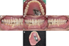
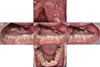 | Fig. 1Intraoral photo at first visit. (A) Maxillary occlusal view, (B) Right lateral, (C) Frontal view, (D) Left lateral, (E) Mandibular occlusal view.
|
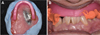
 | Fig. 3(A) Paraffin wax was added on stock tray to adjust to defect, (B) Preliminary impression with irreversible hydrocolloid material, (C) Preliminary stone cast for individual tray.
|
 | Fig. 4(A) Border molding of edentulous and velopharyngeal area with modeling compound, (B) Final impression with polyvinyl siloxane, (C) Stone working cast.
|
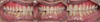
 | Fig. 5(A) Metal framework, (B) 1.0 mm baseplate wax was applied for wall of bulb. Internal space was filled with silicone putty and record base was fabricated, (C) First base of obturator was cured.
|


References
1. Beumer J, Marunick MT, Esposito SJ. Maxillofacial rehabilitation: prosthodontic and surgical management of cancerrelated, acquired, and congenital defects of the head and neck. Quintessence Pub;2011. p. 175–181.
2. Zarb GA, Hobkirk J, Eckert S, Jacob R. Prosthodontic treatment for edentulous patients: complete dentures and implantsupported prosthesis. Elsevier;2013. p. 351–366.
3. Brown KE. Peripheral consideration in improving obturator retention. J Prosthet Dent. 1968; 20:176–181.

4. Desjardins RP. Obturator prosthesis design for acquired maxillary defect. J Prosthet Dent. 1978; 39:424–435.
5. Schwartzman B, Caputo AA, Beumer J. Gravity-induced stresses by an obturator prosthesis. J Prosthet Dent. 1990; 64:466–468.
6. Minsley GE, Nelson DR, Rothenberger SL. An alternative method for fabrication of a closed hollow obturator. J Prosthet Dent. 1986; 55:485–490.
7. Matalon V, LaFuente H. A simplified method for making a hollow obturator. J Prosthet Dent. 1976; 36:580–582.
8. Oh WS, Roumanas ED. Optimization of maxillary obturator thickness using a double-processing technique. J Prosthodont. 2008; 17:60–63.
9. Seaver EJ, Dalston RM, Leeper HA, Adams LE. A study of nasometric values for normal nasal resonance. J Speech Hear Res. 1991; 34:715–721.
10. Aramany MA. Basic principles of obturator design for partially edentulous patients. Part I: classification. J Prosthet Dent. 1978; 40:554–557.
11. Eckert SE, Desjardins RP, Taylor TD. Clinical maxillofacial prosthetics. 1st ed. Quintessence Pub;2000. p. 125–131.
12. Wu YL, Schaaf NG. Comparison of weight reduction in different designs of solid and hollow obturator prostheses. J Prosthet Dent. 1989; 62:214–217.
13. Murata H, Hamada T, Nagasiri R. Stabilizing record bases for edentulous obturator prostheses with silicone resilient relining material. J Prosthet Dent. 1999; 82:366–368.
14. Kim SI, Baik JA, Shin HK, Kim OH. Study of nasalance for normal Korean adults using nasometer II. Speech Science. 2000; 7:219–228.
 XML Download