

 PDF
PDF Citation
 Print
Print


INTRODUCTION
During episodes of asthma exacerbation, respiratory viral infection can be detected in nearly 80%–85% of school-age children [1]. Respiratory syncytial virus (RSV) is a main cause of bronchiolitis in children and may increase susceptibility to the development of asthma [2]. Eosinophil activation results in the eosinophil extracellular traps (EETs) formation which is capable of killing bacteria in the extracellular space [3]. In this context, our group described that EETs are released in bronchoalveolar lavage fluid (BALF) and lung tissue of asthmatic mice [4567]. Currently, there are no available data in the literature regarding the production of EETs induced by RSV. Therefore, we investigated whether RSV in vitro induces EETs in BALF cells in a murine model of asthma.
MATERIALS AND METHODS
Sensibilization and challenge
Six- to eight-week-old specific pathogens-free female BALB/cJ mice weighing approximately 20 g from the Center for Experimental Biological Models (CeMBE, PUCRS) were used in the experiments. Mice were sensitized with 2 subcutaneous injections of ovalbumin (20 μg) (OVA - Grade V, Sigma, St. Louis, MO, USA) on days 0 and 7, followed by intranasal challenges with OVA (100 μg) on days 14, 15, and 16 of the protocol [5]. The control group received only Dulbecco's phosphate-buffered saline.
Bronchoalveolar lavage fluid
On day 17 of the protocol, the animals were anesthetized with ketamine (0. 4 mg/g) and xylazine (0.2 mg/g) and the trachea was cannulated with a blunt needle. BALF was performed with phosphate-buffered saline (PBS) 2% fetal bovine serum (FBS) (1 mL). BALF was centrifuged, and the pellet was resuspended in PBS 2% FBS. For the confirmation of the experimental model of asthma induction, total cell counts were determined in a Neubauer chamber (BOECO, Hamburg, Germany) and differential cell counts of BALF was determined by cytospin preparations stained with H&E (Panótico Rápido - Laborclin, Brazil).
Virus culture and eosinophils stimulation
The production of RSV A2 strain (kindly donated by Dr. Fernando Polack, Vanderbilt University School of Medicine, Nashville, TN, USA) was obtained in VERO cells (ATCC CCL-81, ATCC, Manassas, VA, USA) with 2% FBS at 37°C under 5% CO2. BALF cells (2 × 10⁵ /mL) were stimulated with RSV (103 PFU/mL) for 3 hours at 37°C under 5% CO2 while others remained unstimulated. The mentioned RSV concentration was used because higher concentrations (104–106 PFU/mL) did not increase extracellular DNA traps. Thus, we showed that RSV induces EETs release in a concentration-dependent manner. We thought that higher concentrations are cytotoxic to eosinophils.
Quantification of extracellular DNA traps
Extracellular DNA traps were quantified in BALF cells supernatants stimulated with RSV (103 PFU/mL) or unstimulated using Quant-iT ds DNA HS (Invitrogen, Carlsbad, CA, USA) and measured in the Quibit 2.0 fluorimeter (Invitrogen), according to the manufacturer's recommendations. Visualization of EETs by fluorescence microscopy BALF cells (2 × 10⁵/mL) were stimulated with phorbol-12-myristate-13-acetate (50 nM). After that, cells stimulated with RSV (103 PFU/mL) and unstimulated ones were incubated for 3 hours, fixed with 4% paraformaldehyde and stained with anti-eosinophil peroxidase (EPO) and anti-histone H2B (1:250; Santa Cruz Biotechnology, Dallas, TX, USA) for 45 minutes. Later, cells were incubated with fluorescein isothiocyanate (1:100; Santa Cruz Biotechnology) and alexa fluor 633 (1:100; Invitrogen) for 30 minutes. Next, the cells were stained with Hoechst 33342 DNA dye (1:2,000; Invitrogen) for 4 minutes. Confocal images were taken on a Leica TCS-SP8 exciter microscope (Leica Microsystem, Wetzlar, Germany).
Determination of cell death
Cell death in BALF was analyzed by annexin V and propidium iodide (PI) (BD Pharmingen, BD Biosciences, San Jose, CA, USA) according to the manufacturer's instructions. Cells were incubated with annexin V and PI and analyzed by flow cytometry (FACS Canto II, BD Bioscience). Data were analyzed using FlowJo software version X.0.7 (TreeStar, Woodburn, Oregon, USA).
Cytokine levels
Interleukin-4 (IL-4) and interferon-ɣ (IFN-ɣ) levels in culture supernatant were measured by the multiplex technique (MAGPIX TECHNOLOGY, MILLIPLEX MAP, Merck Millipore, Burlington, MA, USA) and analyzes were performed on the Magpix equipment (Millipore, Darmstadt, Germany). Results were analyzed using the xPONENT Solutions software (Luminex Corp., Austin, TX, USA).
EPO activity
EPO activity was measured in culture supernatant and was determined through colorimetric assay based on the oxidation of O-phenylenediamine (Sigma) in the presence of hydrogen peroxide [8].
Statistical analysis
Data were expressed as mean ± standard deviation. Results were analyzed using GraphPad Prism ver. 5 (GraphPad Software Inc., La Jolla, CA, USA). One-way analysis of variance followed by the Tukey post hoc test, or Student t test was used. A p value less than 0.05 was considered statistically significant.
RESULTS
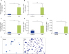
Firstly, we evaluate BALF cells in order to experimental asthma model establishment. We observed that OVA group had a significant increase in the total cell counts, as well as an increase in eosinophils, neutrophils, macrophages, and lymphocytes cell counts compared to the control group (Fig. 1A-E). There are more than 70% of eosinophils in BALF in OVA group.
Fig. 1
Ovalbumin-induced airway eosinophilic inflammation. (A) Total cell counts in BALF. (B-E) Differential cell counts in BALF (eosinophils, macrophages, neutrophils, and lymphocytes. (F) Representative illustration of BALF cells stained with H&E (×400, arrows indicate eosinophils). Results are expressed as mean ± standard deviation, for 7–10 animals in each group, of 3 independent experiments, **p < 0.01, ***p < 0.001. BALF, bronchoalveolar lavage fluid; OVA, ovalbumin.
Later, we investigated whether RSV in a concentration dependent manner (103–106 PFU/mL) would induce the release of EETs in BALF cells of asthmatic mice. RSV was able to stimulate EETs only in RSV 103 PFU/mL concentration (Fig. 2A). Thus, we decided to stimulate BALF cells from asthmatic mice with RSV 103 PFU/mL.
Fig. 2
RSV in vitro induces EETs colocalized with EPO in BALF cells from asthmatic mice. (A) Effect of different concentrations of RSV (103–106 PFU/mL) in vitro in extracellular DNA concentration. (B) Extracellular DNA concentration in BALF cells from asthmatic and control mice stimulated with RSV (103 PFU/mL) in vitro or unstimulated. (C) EETs release in BALF cells from asthmatic and control mice stimulated with RSV (103 PFU/mL) in vitro or unstimulated (×630, arrows indicate EETs formation). (D) Analysis of annexin V binding and PI uptake in BALF cells from all groups. Results are expressed as mean ± standard deviation, for 7–10 animals in each group, of 3 independent experiments, *p < 0.05, **p < 0.01, ***p < 0.001. BALF, bronchoalveolar lavage fluid; DPBS, Dulbecco's phosphate-buffered saline; EPO, eosinophil peroxidase; OVA, ovalbumin; PFU, plaque-forming unit; PI, propidium iodide; RSV, respiratory syncytial virus; FITC, fluorescein isothiocyanate.
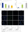
Eosinophils from BALF-asthmatic mice stimulated with RSV increased extracellular DNA in the culture supernatant when compared to the OVA group (Fig. 2B). In confocal microscopy, we observed multiple extracellular DNA extrusions in eosinophils colocalized with EPO but not colocalized with histone H2B in BALF cells in the OVA group stimulated with RSV (Fig. 2C). EETs release with RSV stimulation was not due to apoptosis showing that most cells were negative for annexin V and PI (Fig. 2D).
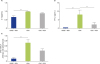
Finally, we showed that in culture supernatant from BALF cells stimulated with RSV from asthmatic mice there was no alteration in IL-4 levels (Fig. 3A). On the other hand, we showed a decrease in IFN-ɣ levels when compared to the OVA group (Fig. 3B). In BALF cells from the OVA group there was an increase in EPO activity but we could not see any alteration in this parameter when stimulated with RSV in vitro (Fig. 3C).
Fig. 3
Effect of RSV in BALF cells from asthmatic mice in cytokines levels and EPO activity. (A) IL-4, (B) IFN-ɣ, and (C) EPO activity in from BALF cells (2 × 10⁵ /mL) from asthmatic mice stimulated with RSV (103 PFU/mL) in vitro or unstimulated. Results are expressed as mean ± standard deviation, for 5 animals in each group, of 3 independent experiments, **p < 0.01. BALF, bronchoalveolar lavage fluid; EPO, eosinophil peroxidase; DPBS, Dulbecco's phosphate-buffered saline; IL-4, interleukin-4; IFN-ɣ, interferon-ɣ; OVA, ovalbumin; RSV, respiratory syncytial virus.
DISCUSSION
EETs are web-like networks of expelled DNA covered with microbicide and cytotoxic proteins [3] which are able to efficiently kill bacteria through DNA traps and may also contribute to lung dysfunction. The first evidence of EETs formation in asthma was in endobronchial biopsies from human atopic asthmatic patients [9]. Moreover, studies have demonstrated that viruses such as the RSV are capable of inducing neutrophil extracellular traps formation [10]. Therefore, we demonstrated for the first time that RSV in vitro can induce EETs colocalized with EPO in BALF cells from asthmatic mice. Previous studies of our group have shown that BALF cells produced EETs without showing sign of apoptosis [4567]. We showed that RSV stimulation in vitro did not induce cell death. Furthermore, we demonstrated that EETs were not colocalized with histone H2B, which suggests that DNA released by EETs is of mitochondrial origin. RSV infection enhances T helper type 2 (Th2) cytokine in lung tissue [11]. IL-4 levels were elevated in the OVA group when compared to the control group while that RSV stimulation in vitro did not alter IL-4 levels in BALF cells in asthmatic mice. Moreover, RSV in vitro reduces IFN-ɣ levels, a T helper type 1 (Th1) cytokine when compared to the OVA group. Aberle et al. [12] showed that severe RSV bronchiolitis is associated with decreased mRNA IFN-ɣ expression in peripheral blood. Sanz et al. [13] showed that EPO serum levels are significantly higher in asthmatic patients than in healthy controls. In addition, EPO activity in BALF cells was elevated in the OVA group when compared to the control group, but the RSV stimulation in vitro did not alter this parameter. Taken together our results showed, for the first time, that RSV in vitro increases EETs release in BALF cells from asthmatic mice, which probably contributes to airway obstruction and tissue damage in asthma.
 XML Download