

 PDF
PDF ePub
ePub Citation
 Print
Print


INTRODUCTION
Peri-implantitis is characterized by loss of the supporting bone with the presence of inflammation in the mucosa surrounding an osseointegrated implant in function [1]. Gram-negative anaerobes were proven to be predominantly associated with peri-implant diseases [2] and the colonization of bacteria on the implant surface seems to be influenced by surface properties such as surface roughness, surface free energy, and chemical composition [34]. In a series of studies on bacterial adhesion to titanium surfaces, a surface roughness of up to 0.2 µm was suggested as a threshold value below which no further reduction in the amount of adhering bacteria could be observed [3567].
The prevalence of peri-implantitis ranges from 15.4% to 43.3% of implants [1]. Hence, instrumentation of exposed implant surfaces has become an inevitable part of periodontal maintenance procedures. Although various surface decontamination methods such as scaling with metal, plastic or ultrasonic instruments, rubber cup polishing, air-powder abrasive systems, and brushing with a conventional or a rotating brush have been introduced, there is no consensus on the best method for implant surface decontamination [8910111213141516].
The implant surface may be affected by any type of mechanical instrumentation. Previous studies have reported alterations of surface topography following instrumentation [13141516171819202122]. Most studies have evaluated the surface alterations in terms of Ra (mean roughness at midline: the arithmetic mean of the departure of the profile from the mean line) and Rz (maximum height: the average of the 5 largest peak-to-valley heights in the assessment length). Following instrumentation, smooth surfaces showed unchanged or increased roughness, while rough surfaces exhibited unchanged or decreased roughness depending on the instruments used [23]. However, the impact of these alterations on clinical factors has not been elucidated.
Surface topography can be measured by scanning electron microscopy (SEM), profilometry, and confocal microscopy. Although SEM and profilometry have been widely used to evaluate surface topography, confocal microscopy has been proven to be useful and reliable for evaluating surfaces [9111423]. Confocal microscopy reconstructs surface topography from optical sections, by either reflection or fluorescence imaging [2425]. Confocal microscopy has advantages over the other methods in that it does not require specimen preparation, as is the case for SEM, or require a stylus, as is the case for a surface profilometer, the use of which may limit the detection threshold.
For the decontamination or maintenance of surfaces affected by peri-implantitis, 2 instruments for implant surface decontamination have become available. One is a nylon brush with bristles that is connected to a handheld instrument, and the other is a low-speed rotary bur with stainless steel bristles at the end. In the present study, titanium surface alterations following instrumentation with those 2 instruments were evaluated using confocal microscopy.
MATERIALS AND METHODS
Specimens and experimental groups
A total of 27 titanium discs (Neobiotech Co., LTD., Seoul, Korea) with dimensions of 10 mm in diameter and 3 mm in thickness were used for the present study. The discs included 3 surfaces: 9 machined (M) surface discs, 9 sandblasted and acid-etched (SA) surface discs, and 9 titanium discs surface-treated with resorbable blast media (RBM). Among the 9 discs of each surface type, 3 discs were instrumented with a nylon brush (GingiBrush, Neobiotech Co., LTD.) and 3 other discs were instrumented with a metal brush (iBrush, Neobiotech Co., LTD.). The remaining 3 discs in each group served as controls. Consequently, there were 9 experimental groups: M surfaces without any instrumentation, M surfaces instrumented with a nylon brush, M surfaces instrumented with a metal brush, SA surfaces without any instrumentation, SA surfaces instrumented with a nylon brush, SA surfaces instrumented with a metal brush, RBM surfaces without any instrumentation, RBM surfaces instrumented with a nylon brush, and RBM surfaces instrumented with a metal brush.
Instruments and instrumentation
The instruments used for the present study are shown in Figure 1. The nylon brush was shaped similarly to a tapered interproximal brush and should be used with equipment provided by the manufacturer. This nylon brush was designed for removal of submucosally deposited plaque adhering onto the implant/abutment surface during periodic maintenance procedures. The metal brush is a low-speed rotary bur with laterally protruding stainless steel bristles at its end, and it is to be used for decontamination of implant surfaces affected by peri-implantitis during surgical procedures.
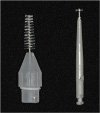 | Figure 1The instruments used in the present study. The nylon brush (left) had a shape similar to a tapered interdental brush and it should be used with an equipment (not shown in this picture) provided by the manufacturer. The metal brush (right) is a low-speed rotary bur with laterally protruding stainless steel bristles at its end.
|
The nylon brush was instrumented for 40 seconds with irrigation by a single operator (YK). Instrumentation with the metal brush was performed at 1,000 rpm under copious irrigation for 40 seconds by another single operator (JP). No special attempt to standardize instrumentation protocols, including the force and angle of instrument application was made, in order to simulate clinical situations. However, calibrations were performed before the experiments so that the average force applied in the present study was approximately 10 × g.
Surface alteration investigations with confocal microscopy
Gross surface changes of the titanium discs were investigated with the naked eye and photographed. Detailed observations were conducted with a confocal laser microscope (LSM5 Pascal, Zeiss, Jena, Germany). With the confocal microscope, the surface parameters of the arithmetic mean value of a linear profile (Ra), maximum height of a linear profile (Rz), skewness of the assessed linear profile (Rsk), arithmetic mean height of a surface (Sa), maximum height of a surface (Sz), developed interfacial area ratio (Sdr), skewness of a surface profile (Ssk), and kurtosis of a surface profile (Sku) were measured. Measurements were taken at 5 random areas on each disc. Each measured area had dimensions of 460.7 μm × 460.7 μm, and a Gaussian filter was used to determine the parameters. Values were determined at a cut-off length of 0.04 mm in 50 sections and the stack size of z-sections was 0.80 μm. Roughness measurements were calculated using proprietary software (Topography Package, Zeiss). Intensity projections with an extended depth of focus were also obtained with the confocal microscope.
Statistical analysis
The mean and standard deviation of every parameter were calculated. Mean values were within the same surface types using 1-way analysis of variance, and the Tukey method was used for post hoc multiple comparisons. P values <0.05 were considered to indicate statistical significance. The statistical analysis was performed using PASW version 18 (SPSS Inc, Chicago, IL, USA).
RESULTS
Titanium disc surface alterations
Upon gross examination of the titanium surfaces, the surfaces instrumented with the nylon brush looked similar to the control surfaces, while instrumentation with the metal brush resulted in numerous scratches on the surfaces (Figure 2).
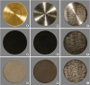 | Figure 2Gross view of titanium discs. (A) M surface without instrumentation, (B) M surface instrumented with a nylon brush, (C) M surface instrumented with a metal brush, (D) SA surface without instrumentation, (E) SA surface instrumented with a nylon brush, (F) SA surface instrumented with a metal brush, (G) surface treated with RBM without instrumentation, (H) RBM surface instrumented with a nylon brush, (I) RBM surface instrumented with a metal brush. The changes on the surfaces instrumented with a nylon brush were indistinct (B, E, and H) and the alterations on the surfaces instrumented with a metal brush were obvious (C, F, and I).M: machined, SA: sandblasted and acid-etched, RBM: resorbable blast media.
|
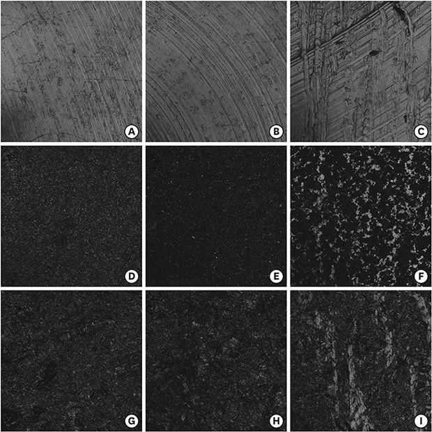
Upon detailed examination of the surfaces with intensity projections with an extended depth of focus (Figure 3), slight changes could be observed on the surfaces instrumented with the nylon brush, which seemed somewhat different compared with the control surfaces. Meanwhile, instrumentation with the metal brush left noticeable changes on all 3 surfaces, which resembled scratches on the M surface and flattening on the SA and RBM surfaces.
 | Figure 3Images from intensity projections with an extended depth of focus obtained by confocal laser microscopy. (A) M surface without instrumentation, (B) M surface instrumented with a nylon brush, (C) M surface instrumented with a metal brush, (D) SA surface without instrumentation, (E) SA surface instrumented with a nylon brush, (F) SA surface instrumented with a metal brush, (G) surface treated with RBM without instrumentation, (H) RBM surface instrumented with a nylon brush, (I) RBM surface instrumented with a metal brush. Instrumentation with the metal brush resulted in scratches on the M surface (C) and flattening of rough surfaces (F and I).M: machined, SA: sandblasted and acid-etched, RBM: resorbable blast media.
|
Changes in parameters of surface properties
The changes in the parameters of surface properties are presented in Table 1. The nylon brush increased average roughness (Ra and Sa) of the M surfaces by more than 2-fold (P<0.001 for Ra and Sa) and increased the Sdr from 6.53% to 20.82% (P=0.001). On the SA surface, the nylon brush decreased Ra and Sa by up to 0.3 µm (P=0.048 for Ra; P=0.001 for Sa) and Sdr from 88.50% to 83.01% (P=0.019). Meanwhile, the Ra and Sa of the RBM surfaces were not affected by the nylon brush.
Table 1
Surface parameter values (mean±standard deviation) measured with confocal microscopy

P values were obtained from one-way analysis of variance for each surface.
M: machined, SA: sandblasted and acid-etched, RBM: resorbable blast media, Ra: arithmetic mean value of a linear profile, Rz: maximum height of a linear profile, Rsk: skewness of the assessed linear profile, Sa: arithmetic mean height of a surface, Sz: maximum height of a surface, Sdr: developed interfacial area ratio, Ssk: skewness of a surface profile, Sku: kurtosis of a surface profile.
a,b,c,d)these letters in parentheses indicate homogenous subsets for each statistically different parameter of each surface according to the post hoc test using the Tukey method.
![]()
The metal brush also increased the Ra and Sa of the M surfaces (P<0.001 for Ra and Sa) and the magnitude of the increase in roughness caused by the metal brush was smaller than that caused by the nylon brush (P=0.034 for Ra; P=0.544 for Sa). The increase in Sdr in the M surfaces showed no significance (P=0.119). The SA surfaces were changed the most by the metal brush. The maximum height (Rz and Sz), Ra, and Sa of the SA surfaces decreased remarkably (P=0.012 for Ra; P=0.009 for Rz; P<0.001 for Sa and Sz), and Sdr also decreased (P<0.001). On the RBM surfaces, the metal brush did not cause changes in the average roughness (Ra and Sa). However, the amount of reduction in Rz, Sz, and Sdr was similar to that of the SA surfaces (P=0.018 for Rz; P=0.004 for Sz; P=0.001 for Sdr).
DISCUSSION
As surface roughness can affect the attachment and/or proliferation of cells and microorganisms, information on surface changes following instrumentation is valuable, especially when the instrument is a newly developed one. The present study evaluated the surface alteration of 3 different types of titanium discs following instrumentation with a recently marketed nylon brush or a metal brush. The effects of various non-metal and metal instruments on titanium surface alterations have been evaluated [9131415161718212627282930313233], and the instruments used in these studies included stainless steel scalers, titanium scalers, plastic scalers, ultrasonic/sonic scalers either with a metal tip or with a plastic tip, stainless steel curettes, titanium curettes, plastic curettes, gold-coated curettes, rubber cups with and without paste, air abrasive polishers, nylon brushes, carbide burs, and diamond burs.
In previously reported studies, non-metal instruments had no effect or increased the roughness of M surfaces, and the roughness of rough surface was unchanged or decreased [91011]. In the present study, instrumentation with a nylon brush increased the average roughness of the M surfaces, decreased the average roughness of the SA surfaces, and had no effect on the RBM surfaces. The M surfaces in the present study had an Ra of up to 0.3 µm, meaning that they could be regarded as smooth. Therefore, any mechanical contact would scratch the surface and increase the surface roughness. The different impact of nylon brush instrumentation on the rough surfaces could be due to the fact that the shape of peaks in the SA and RBM surfaces are different and the peaks on the SA surfaces are more fragile. A similar pattern was reported in the study of Schmage et al. [21], in which instrumentation with uni-tufted nylon brush made average roughness of blasted/acid-etched surface decrease and average roughness of blasted surface increase. On the contrary, Park et al. [9] reported a decrease in average roughness of M surface and SLA surface following instrumentation with an implant brush. These differences may be attributed to the use of prophylaxis paste or dentifrice. Schmage et al. [21] did not use any prophylaxis paste on instrumentation with the nylon brush and reported similar results to the present study while Park et al. [9] brushed M surface with dentifrice for 20 seconds, then washed the surface, and brushed again the surface without dentifrice for 20 seconds, sequentially. It can be presumed that addition of dentifrice acted to decrease the average surface roughness in the study of Park et al. [9], and the effect of a brush alone or with prophylaxis paste/dentifrice should be studied in the future.
The use of the metal brush increased the average roughness of the M disc surfaces, however, the increase was smaller than that seen with the nylon brush. Moreover, the amount of the reduction of the average roughness of the SA surfaces with in response to the metal brush was similar to the findings observed for the nylon brush. This may have resulted from the shape of the instruments, as the metal instrument had a much smaller effective region than the nylon brush. Duarte et al. [31] reported no change following instrumentation with metal curettes/scalers on SLA surfaces, while Park et al. [9] reported a decrease in the average roughness of SLA surfaces. Although the average roughness did not change in the study of Duarte et al. [31], the SLA surface was observed to be flattened out in SEM photomicrographs. From these results, it can be deduced that the surface alteration from metal instruments might be limited to a shallow depth, within 0.5 µm.
In terms of microbial adhesion and surface roughness, it was shown that plaque accumulation decreased until surface roughness reached 0.2 µm [567]. In the present study, the Ra of the M surfaces was 0.26 µm before instrumentation, and it increased to 0.61 µm and 0.47 µm after instrumentation with the nylon brush or metal brush, respectively. Hence, the chance of bacterial adhesion on a M smooth surface becomes greater after instrumentation, even with a nylon brush. Furthermore, the greatest residual plaque biofilm area was observed on titanium surfaces after instrumentation with plastic instruments [32]. Moreover, Bailey et al. [33] showed the possibility of particle dislodgment after instrumentation with plastic instruments, which may affect the titanium surface characteristics and the affinity of cell attachment/proliferation to the surface. Thus, further studies to investigate how the treatment with these 2 types of brushes influence microbial adhesion on the altered surfaces, the efficiency of microbial biofilm removal, and the reestablishment of biocompatibility are needed.
In conclusion, titanium surfaces are altered when instrumented either with a nylon brush or with a metal brush. As a consequence, it is recommended that nylon or metal brushes should be used with caution in order to avoid damaging the implant fixture/abutment surface.
 XML Download