

 PDF
PDF ePub
ePub Citation
 Print
Print


INTRODUCTION
Inflammation is a physiological response that is triggered by noxious stimuli and conditions, such as infection and tissue injury, and it is generally considered beneficial when the response is balanced.1 On the other hand, excessive inflammatory reactions can be harmful and cause a range of inflammatory diseases and clinical syndromes, such as systemic lupus erythematosus,2 asthma,3 and septic shock.4
Acute pancreatitis is one such disease, in which excessive inflammation acts as the trigger mechanism of various symptoms and complications.5 Approximately 80% of patients undergo conservative treatment, such as fasting and fluid resuscitation, but 10–20% show severe clinical symptoms with multiple organ dysfunctions, such as pancreatic necrosis and lung injury.67 In particular, severe and infected necrotizing pancreatitis is associated with a high mortality rate between 30% and 40%, despite intensive care.8910
The high mortality rate of acute pancreatitis is associated with a lack of specific treatments except for fluid resuscitation, antibiotic administration, and palliative interventions for complications.11 On the other hand, the pathogenic mechanisms and pathophysiological changes are unclear. During the early stages of acute pancreatitis, the degree of pancreatic acinar cell injury and inflammation are the key factors that determine the course and prognosis of the disease.12 Necrotic acinar cells not only damage the neighboring cells and tissues directly, but also induce a systemic inflammatory response spreading through the circulation to other tissues and organs.13 This reaction is related to the activation of monocytes, upregulation of cell adhesion molecules and chemokines, release of large amounts of oxygen free radicals, and inhibition of the immune function of lymphocytes.13
Proinflammatory molecules or cytokines, which are released during acute pancreatitis, may be used as markers for a diagnosis or a prediction of the prognosis of acute pancreatitis and can be used as a treatment target. In a previous study, a higher occurrence of apoptosis was observed in lymphocytes treated with the serum from systemic lupus erythematosus patients than the serum from healthy human serum.14 This study hypothesized that the same phenomenon would occur when using the serum from acute pancreatitis patients. Therefore, the degree of apoptosis was examined after treating the cell lines with the serum from patients with acute pancreatitis.
SUBJECTS AND METHODS
1. Study subjects
From June 2016 to August 2018, serum samples were donated from healthy volunteers and acute pancreatitis patients who were admitted to the Eulji Hospital. A diagnosis of acute pancreatitis was made when two of the following three features were noted: persistent abdominal pain characteristic of acute pancreatitis for more than 24 hours; increase in the serum amylase and/or lipase levels to more than three times the upper standard limit; and the presence of a characteristic acute pancreatitis signature detected by a CT scan.11 The resolution of acute pancreatitis was diagnosed based on the improvement of abdominal pain and fever, normalization of serum amylase and lipase, and normalization of leukocytosis and CRP. The attending physician determined the cause of acute pancreatitis based on the patient's history, including alcohol consumption, radiology examination, including the presence of gallstones, and laboratory findings.
2. Ethics statement and serum collection
The Institutional Review Board of Eulji Hospital approved this study (EMCIRB 201509-02). After receiving the participants' consent, serum was collected from healthy volunteers and patients with pancreatitis. For patients with pancreatitis, the blood remaining after a clinical laboratory test was used. Age, gender, BMI, and cigarette smoking habit of the study subjects were recorded and the etiology of acute pancreatitis, the number of days of hospitalization and fasting were also recorded for each acute pancreatitis patient. In the sera from patients with pancreatitis, the blood collected immediately after admission was labeled “active pancreatitis state,” and that collected prior to discharge from hospital was labeled “resolved pancreatitis state.”
3. Cell culture
A skin tissue-derived cell line, BJ, was used to assess the response to the acute pancreatitis patients and healthy volunteers. The cell line was purchased from the Korean Cell Line Bank (Seoul, Korea). The cells were cultured in Dulbecco's modified Eagle's medium (DMEM, Thermo Fisher Scientific, Waltham, MA, USA) supplemented with 1% penicillin-streptomycin (Thermo Fisher Scientific) and 10% bovine serum (Atlas Biologicals, Fort Collins, CO, USA) at 37℃ in an atmosphere containing 5% CO2. For the apoptosis assay, the BJ cells were seeded in 12-well plates (NEST Biotechnology, Jiangsu, China) at a concentration of 3×10⁴ cells/well. After incubation for 24 hours, the BJ cells were treated with DMEM supplemented with 10% human serum from the acute pancreatitis patients (active and resolved) and healthy volunteers to allow the cells to attach to the plate. The apoptosis assay was performed 24 hours after replacing the culture medium.
4. Apoptosis assay
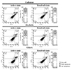
The apoptosis assay was performed on an apoptosis assay kit (Koma Biotech, Seoul, Korea) using annexin V-FITC and propidium iodide (PI). Apoptotic cell staining was performed according to the manufacturer's recommendations. The stained cells were analyzed using an Accuri C6 flow cytometer (Becton Dickinson Biosciences, San Jose, CA, USA) and CFlow sampler program (version 1.0.227.4; Becton Dickinson Biosciences). All experiments were performed in triplicate and repeated on five different days. The dot plots were divided into four quadrants. The live cell quadrant had a negative annexin V and PI (lower left). The early apoptosis quadrant had a positive annexin V and negative PI (lower right). The late apoptosis quadrant had both positive annexin V and PI (upper right).
5. Statistical analysis
The continuous variables are expressed as the mean±SD. Normalization of the apoptosis assay was performed using the resolved state values based on the active state values of pancreatitis caused by each cause to calibrate the deviations of the experiments performed on the other days. A Student's t-test and paired t-test were used to compare the mean differences in the percentage of live and apoptotic cells between the cells treated with the serum from patients with active and resolved pancreatitis conditions. The means, standard errors, and p-values were calculated using GraphPad Prism (version 5.03; GraphPad Software Inc., La Jolla, CA, USA). A p-value less than 0.05 was considered significant.
RESULTS
1. Characteristics of study subjects
Table 1 lists the characteristics of the study subjects. A total of 71 participants were enrolled; among them, 22 were healthy volunteers (control) and 49 were patients with acute pancreatitis (22 gallstone pancreatitis, 16 alcoholic pancreatitis, and 11 pancreatitis due to other causes). Patients with pancreatitis were hospitalized for an average of 6.53 to 14.36 days and kept fasted for an average of 3.60 to 6.46 days during hospitalization.
2. Apoptosis assay
The results of the apoptosis analysis are described as a dot plot (Fig. 1). The mean percentage of live cells in the control group was 78.8±3.5%, whereas the mean percentage of live cells in the active state of acute pancreatitis was 83.3±1.6%. In the case of early apoptosis, the mean percentage in the control group and in the active state of acute pancreatitis was 6.2±0.9% and 6.3±0.8%, respectively. In the case of late apoptosis, the control group and active state of acute pancreatitis group had a mean percentage of 13.1±2.5% and 9.2±1.0%, respectively. No significant difference in apoptosis was observed in the cells treated with the sera from healthy controls and pancreatitis patients.
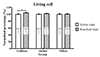
The percentage of live cells in the active state of acute pancreatitis caused by gallstones was 81.9±2.6%, whereas the mean percentage of live cells in the resolved state of acute pancreatitis caused by gallstones was 85.0±1.9% (Fig. 2). The mean percentage of live cells in the active and resolved state of pancreatitis caused by alcohol were 81.9±3.1% and 83.5±3.2%, respectively, and the values for pancreatitis caused by other factors were 83.2±2.9% and 83.4±2.8%, respectively. In the case of early apoptosis, the mean percentage in the active state and resolved state of acute pancreatitis due to gallstones was 6.1±1.2% and 5.3±0.9%, respectively. The percentage for the active state in the alcoholic pancreatitis group was 5.5±1.1% and for the resolved state was 5.7±1.2%. The active and resolved states in pancreatitis from other causes corresponded to 7.3±1.6% and 5.9±0.9%, respectively. In the case of late apoptosis, the active state and resolved state of the acute pancreatitis group due to gallstones was 9.3±1.6% and 7.6±1.4%, respectively. The active and resolved states of the alcoholic pancreatitis group were 10.5±2.0% and 8.4±2.0%, respectively. The corresponding values in the active and resolved states in the acute pancreatitis group due to other factors were 7.7±1.8% and 8.6±1.6%.
3. Comparisons of the cell status after treatment with the serum of active and resolved pancreatitis patients
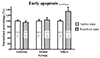
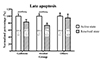
Upon normalization, using the active pancreatitis state of each group as a reference, the live cells of the resolved state were 100.4% to 104.1%, showing a significant increase in the resolved state of gallstone pancreatitis (p=0.0075) (Fig. 3). The normalized percentage of early apoptosis in the resolved state was 94.9% to 133.8%, showing a significant increase in the resolved state of other causes of pancreatitis (p=0.0495) (Fig. 4). In the case of late apoptosis, however, the normalized percentage of the resolved state in the pancreatitis group due to gallstones was 83.2±7.3%. The normalized percentages of the resolved state in alcoholic pancreatitis and the resolved state in the pancreatitis group from other causes were 73.4±6.5% and 94.9±10.9%, respectively; the number of late apoptotic cells in the resolved state of gallstoneand alcohol-induced pancreatitis decreased significantly (p=0.0263 and 0.0266, respectively) (Fig. 5).
DISCUSSION
The inflammatory reaction is a first-line defense mechanism against noxious stimuli and pathogens. In some diseases, however, the inflammatory reaction is itself a cause of disease or an exacerbating factor. Acute pancreatitis is one of the major diseases, in which the overexpression of inflammation is the triggering mechanism. Adequate monitoring and the proper control of inflammation are essential for diagnosis and treatment. Extra-pancreatic inflammation is common in pancreatitis, and several mechanisms have been suggested. In this study, it was hypothesized that the serum from acute pancreatitis patients had apoptosis-inducing factors and that treating the BJ cell line with the patient's serum would increase apoptosis further.
Apoptosis is a key mechanism of homeostasis that usually occurs during the development and aging processes and regulates cell populations in tissues. This can also occur as a defense mechanism as a reaction to an immune response or to cell damage caused by disease or harmful substances. In particular, it responds to the attack of pathogens through the inflammatory reaction and at the same time removes the infected cells, inhibiting the proliferation and diffusion of pathogens. Apoptosis of an infected host cell may be triggered either within the host cell or by external immune cells.151617 Indeed, apoptosis can be induced by circulating immune cells and is affected by the surrounding environment. One of the factors that can affect the cellular behavior in the human body is blood. Blood circulates throughout the body, supplies oxygen and nutrients, and carries the carbon dioxide and waste generated by the cell metabolism. In addition, blood contains cytokines and various proteins secreted by blood cells and cells around the blood vessels. A recent report showed that the blood components affect the apoptosis of cells. Serum-induced cell death rates were different when the sera from cervical cancer patients and controls were used to treat a Jurkat cell line.18 Previous studies documented apoptosis using inflammatory blood. Peripheral blood mononuclear cells were treated with the serum from patients with systemic lupus erythematosus to determine the apoptosis index of the cells. As a result, compared to the control group, the inflammatory serum increased the apoptosis of the cells, which suggests that the underlying cause was the presence of the anti-C1q autoantibody in the inflammatory serum.14 Makino et al.19 reported the apoptosis of endothelial cells caused by high tumor necrosis factor-α levels in the blood of patients with type 2 diabetes. Therefore, apoptosis may be induced by the serum of a person with inflammation or disease.
In the present study, there was no significant difference in the apoptosis of serum-treated cells between the healthy control and patients with pancreatitis. On the other hand, when analyzed according to the cause of pancreatitis, in the gallstone pancreatitis group, the percentage of live cells was significantly higher in the cells treated with the serum from the resolved state pancreatitis patients than in the cells treated with the serum of the active state patients, after normalization. In addition, late apoptosis was reduced significantly in the resolved state of patients with gallstone- and alcohol-induced acute pancreatitis when normalized based on their disease state. These results suggest that there may be apoptosis-promoting factors in the serum of patients with acute pancreatitis.
Several studies have reported changes in the blood component ratio and cytokine concentration in patients with acute pancreatitis. Tumor necrosis factor-α is a well-known cytokine secreted in the early stages of acute pancreatitis2021 that induces apoptosis. In addition, interleukin-6, interleukin-10, and chemokine monocyte chemoattractant protein-1 are also secreted from pancreatic acinar cells and mediate the inflammatory responses and leukocytes recruitment.22 Neutrophil infiltration and macrophage recruitment are also present in acute pancreatitis, as well as in most inflammatory disorders because of the activation of innate immunity.2324 The higher proportion of apoptotic cells after treatment with active pancreatitis serum in this study may be related to the abovementioned substances present in the serum of patients with acute pancreatitis.
This study showed that apoptosis occurred predominantly in a skin tissue-derived cell line, which is less related to systemic inflammation involving the internal organs. This could help better understand the mechanism of systemic inflammation in acute pancreatitis and may be helpful in identifying the appropriate serum markers to diagnose acute pancreatitis and predict the prognosis. If the causes of apoptosis in the serum of acute pancreatitis patients can be identified, they could be the target for treatment. Furthermore, these results can be applied not only to study the causes of apoptosis, but also for biomarker discovery. Many attempts have been made to discover disease biomarkers using blood samples. On the other hand, despite the considerable efforts, the number of biomarkers discovered is limited. The classification of the experimental group and the quality of the sample are essential for solving this issue. A difference in the degree of serum cell death was observed between the active and resolved conditions. In addition, a significant difference was also noted with respect to the causes of acute pancreatitis. This suggests a difference in terms of the blood composition. Therefore, applying the findings of this study to evaluate the prognostic values of the samples prior to an analysis of the serum diagnostic marker will help identify diagnostic markers of the disease.
A limitation of this study was that no evaluation of the clinical outcomes of acute pancreatitis was performed. Moreover, the identity of the substances responsible for triggering apoptosis was not clarified. Future work will examine which molecules present in the serum of patients with acute pancreatitis can trigger apoptosis.
In conclusion, this study suggests that the blood of inflammatory patients may contain apoptosis-inducing factors. These molecules, which are present in the serum of patients with acute pancreatitis, are responsible for the pathophysiological mechanism and once identified, can be targeted for treatment development. Furthermore, differential screening of the blood components through an apoptosis assay may be helpful in selecting samples for biomarker discovery.


 XML Download