

 PDF
PDF ePub
ePub Citation
 Print
Print


INTRODUCTION
The cisterna chyli, a dilated lymphatic sac in the retrocrural space, is usually located to the right of the aorta. Although the cisterna chyli can be located to the left of the aorta, this normal anatomic variant has been rarely reported. We report a case of a left-sided cisterna chyli, which was incidentally detected on the computed tomography (CT) and magnetic resonance (MR) images.
CASE REPORT
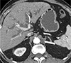
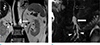
A 76-year-old man was referred to our hospital for jaundice and itching of the whole body. Laboratory workup showed an elevated serum level of total bilirubin (18.7 mg/dL) and carbohydrate antigen 19-9 (1489 U/mL). The differential diagnosis included bile duct obstruction from a malignant tumor, so a CT was performed. Contrast enhanced CT images revealed the Klatskin type II perihilar cholangiocarcinoma, and there were no significantly enlarged regional lymph nodes. However, there was a well-defined left retrocrural lesion that measured 1.2 × 1.4 × 2.5 cm (Fig. 1). It showed near-water attenuation without contrast enhancement and sat between the T12 and L1 vertebral level. MR imaging with MR cholangiopancreatography (MRCP) obtained for further workup of cholangiocarcinoma using 3.0 Tesla system. Respiratory-triggered coronal T2-weighted single-shot fast spine-echo image and breath-hold three-dimensional gradient and spin-echo (GRASE) MRCP image revealed that the left retrocrural lesion appeared as an elongated structure with uniform T2 high signal of its contents, similar to the cerebrospinal fluid (CSF) (Fig. 2). The lesion was consistent with a left-sided cisterna chyli joined by dilated lumbar lymphatics. The left-sided thoracic duct was continuous with the left-sided cisterna chyli, entering through the aortic hiatus of the diaphragm. On follow-up CT 6 months later, there was no change in the appearance of the left retrocrural lesion.
DISCUSSION
The cisterna chyli is the initial prominent confluence of the thoracic duct, which receives the lumbar and intestinal lymphatic trunks. Classically, the cisterna chyli appears as a retrocrural dilated tubular structure of near-water attenuation that is less than 5 cm in length. It is located anterior to the vertebral bodies between T11 and L2, and usually lies to the right of the aorta (1). Approximately 50% of patients have a dominant right-sided thoracic duct that crosses over to the left around the T5-T6 vertebral bodies and terminates into the confluence of the left subclavian and internal jugular veins (1 2). According to a previous study in Japan, a thoracic duct that ran its entire course along the left side was found in 1.0% of cadavers (1/104) (3). The left-sided cisterna chyli is the least common and rare compared with the bilateral cisterna chyli. To the best of our knowledge, there has been only one reported antemortem case of a left-sided cisterna chyli, which was detected incidentally on CT images during the preoperative workup in a 49-year-old man with gastric cancer (3).
Visualization of the cisterna chyli on the cross-sectional images is not rare. Kiyonaga et al. (4) reported that the cisterna chyli was observed on almost 100% of 1 mm-slice thickness axial and multiplanar reformatted CT images (the mean maximum size, 3.8 × 4.0 × 14.1 mm; range, 1.7–7.8 × 1.3–6.9 × 3.8–30.1 mm). A previous study reported that cisterna chyli with a diameter of 5 mm or more were identified in 15% of the patients (30/200) on MR images (5).
Typical imaging findings of the cisterna chyli are located between the T11 and L2 vertebral level, tubular configuration, near-water attenuation and signal intensity on CT and MR images, and lack of change in size (5). Lee et al. (6) described the characteristics of the lymphatic ducts of giant cisterna chyli on post-Gadolinium-DTPA (diethylenetriamine penta-acetic acid) MR images as showing a fluid-fluid level in 30 minutes delayed images and a uniform enhancement of the cisternal contents in 4 to 5 hours delayed images.
The cisterna chyli can mimic pathologic conditions. An unusually located left-sided cisterna chyli may cause confusion in the differential diagnosis. Retrocrural lymphadenopathy, which is seen as a soft tissue mass with enhancement after contrast material administration, can be distinguished from the cisterna chyli, which exhibits fluid contents (7). Occasionally, a neurogenic tumor with cystic degeneration may present as retrocrural mass. However, the internal heterogeneity due to calcification, hemorrhage or a mass-like configuration suggest a neoplasm (8). Abscesses or hematomas can be detected in retrocrural space (9). Retrocrural abscesses manifest as fluid collections with wall enhancement and associated spondylitis. The retrocrural hematoma could be diagnosed by high density on CT or hemorrhagic signal intensity on MR image and accompanying fracture of the adjacent vertebra. With other clinical significance, if surgery for chylothorax is planned or surgery with the risk of developing chylothorax, the presence of the left-sided cisterna chyli should be mentioned in the radiologic report.
In conclusion, radiologists should be aware of the left-sided cisterna chyli and be aware that this normal anatomic structure could be observed on radiologic images.
 XML Download