Abstract
Portable low-cost magnetic resonance imaging (MRI) systems have the potential to enable “point-of-care” and timely MRI diagnosis, and to make this imaging modality available to routine scans and to people in underdeveloped countries and areas. With simplicity, no maintenance, no power consumption, and low cost, permanent magnets/magnet arrays/magnet assemblies are attractive to be used as a source of static magnetic field to realize the portability and to lower the cost for an MRI scanner. However, when taking the canonical Fourier imaging approach and using linear gradient fields, homogeneous fields are required in a scanner, resulting in the facts that either a bulky magnet/magnet array is needed, or the imaging volume is too small to image an organ if the magnet/magnet array is scaled down to a portable size. Recently, with the progress on image reconstruction based on nonlinear gradient field, static field patterns without spatial linearity can be used as spatial encoding magnetic fields (SEMs) to encode MRI signals for imaging. As a result, the requirements for the homogeneity of the static field can be relaxed, which allows permanent magnets/magnet arrays with reduced sizes, reduced weight to image a bigger volume covering organs such as a head. It offers opportunities of constructing a truly portable low-cost MRI scanner. For this exciting potential application, permanent magnets/magnet arrays have attracted increased attention recently. A magnet/magnet array is strongly associated with the imaging volume of an MRI scanner, image reconstruction methods, and RF excitation and RF coils, etc. through field patterns and field homogeneity. This paper offers a review of permanent magnets and magnet arrays of different kinds, especially those that can be used for spatial encoding towards the development of a portable and low-cost MRI system. It is aimed to familiarize the readers with relevant knowledge, literature, and the latest updates of the development on permanent magnets and magnet arrays for MRI. Perspectives on and challenges of using a permanent magnet/magnet array to supply a patterned static magnetic field, which does not have spatial linearity nor high field homogeneity, for image reconstruction in a portable setup are discussed.
Go to : 
References
1. http://www.hitachimed.com/products/mri/RefurbishedMRISystems/Altaire
2. http://www.fonar.com/fonar360.htm
3. https://www.healthcare.siemens.com/magnetic-resonance-imaging/0-35-to-1-5t-mri-scanner/magnetom-c/features
4. https://www.healthcare.siemens.com/magnetic-resonance-imaging/0-35-to-1-5t-mri-scanner/magnetom-aera
5. https://www.healthcare.siemens.com/magnetic-resonance-imaging/0-35-to-1-5t-mri-scanner/magnetom-c/features
6. http://www.fonar.com/standup.htm
7. https://www.paramedmedicalsystems.com/sarat-immagini/mropen_pdf_15.pdf
8. Nishimura DG. Principles of magnetic resonance imaging. Stanford University,. 2010.
9. https://www.esaote.com/dedicated-mri/mri-systems/p/o-scan/
10. Pissanetzky S. Structured coil electromagnets for magnetic resonance imaging and method for fabricating the same. US5382904,. 1995.
11. Vaughan JT, Wang B, Idiyatullin D, et al. Progress toward a portable MRI system for human brain imaging. In 24th ISMRM, Singapore. 2016. 0498.
12. Esparza-Coss E, Cole DM. A low cost MR/permanent magnet prototype. AIP Conference Proceedings. 1998. 440. 119.
13. Sarracanie M, LaPierre CD, Salameh N, Waddington DEJ, Witzel T, Rosen MS. Low-cost high-performance MRI. Sci Rep. 2015; 5:15177.

14. Morgan PS, Conolly S, Mazovski A. Design of uniform field biplanar magnets. In 5th Meeting of ISMRM, Toronto, Canada. 1997. 1447.
15. Hennig J, Welz AM, Schultz G, et al. Parallel imaging in nonbijective, curvilinear magnetic field gradients: a concept study. MAGMA. 2008; 21:5–14.

16. Schultz G, Ullmann P, Lehr H, Welz AM, Hennig J, Zaitsev M. Reconstruction of MRI data encoded with arbitrarily shaped, curvilinear, nonbijective magnetic fields. Magn Reson Med. 2010; 64:1390–1403.

17. Schultz G, Weber H, Gallichan D, et al. Radial imaging with multipolar magnetic encoding fields. IEEE Trans Med Imaging. 2011; 30:2134–2145.

18. Stockmann JP, Galiana G, Tam L, Juchem C, Nixon TW, Constable RT. In vivo O-space imaging with a dedicated 12 cm Z2 insert coil on a human 3T scanner using phase map calibration. Magn Reson Med. 2013; 69:444–455.
19. Stockmann JP, Ciris PA, Galiana G, Tam L, Constable RT. O-space imaging: highly efficient parallel imaging using second-order nonlinear fields as encoding gradients with no phase encoding. Magn Reson Med. 2010; 64:447–456.
20. Cooley CZ, Stockmann JP, Armstrong BD, et al. Two-dimensional imaging in a lightweight portable MRI scanner without gradient coils. Magn Reson Med. 2015; 73:872–883.

21. Ren ZH, Luo W, Su J, Huang SY. Magnet array for a portable magnetic resonance imaging system in RF and wireless technologies for biomedical and healthcare applications (IMWS-BIO), 2015 IEEE MTT-S 2015 International Microwave Workshop Series. Taiwan. 2015. 92–95.
22. Ren ZH, Mu WC, Huang SY. A new yokeless permanent magnet array with high field strength and high field homogeneity for low-field portable MRI system. In Joint Annual Meeting ISMRM-ESMRMB, Paris, France. 2018. 1743.
23. Leupold HA, Potenziani E, Tilak AS. Adjustable multi-tesla permanent magnet field sources. IEEE Trans Magn. 1993; 29:2902–2904.

24. Sagawa M, Fujimura S, Togawa N, Yamamoto H, Matsuura Y. New material for permanent magnets on a base of Nd and Fe (invited). J Appl Phys. 1984; 55:2083–2087.

25. Glover PM, Aptaker PS, Bowler JR, Ciampi E, McDonald PJ. A novel gradient permanent magnet for profiling of planar films and coatings. J Magn Reson. 1999; 139:90–97.
26. Doughty D, McDonald PJ. Drying of coatings and other applications with GARField. In Stapf S, Han S, eds. NMR in chemical engineering. Weinheim: Wiley-VCH. 2006. 89–107.
27. Bennett G, Gorce JP, Keddie JL, McDonald PJ, Berglind H. Magnetic resonance profiling studies of the drying of film-forming aqueous dispersions and glue layers. Magn Reson Imaging. 2003; 21:235–241.

28. Wright SM, Brown DG, Porter JR, et al. A desktop magnetic resonance imaging system. MAGMA. 2002; 13:177–185.

29. Nagata A, Kose K, Terada Y. Development of an outdoor MRI system for measuring flow in a living tree. J Magn Reson. 2016; 265:129–138.

30. Terada Y, Kono S, Uchiumi T, et al. Improved reliability in skeletal age assessment using a pediatric hand MR scanner with a 0.3T permanent magnet. Magn Reson Med Sci. 2014; 13:215–219.

31. Chang WH, Chen JH, Hwang LP. Single-sided mobile NMR with a Halbach magnet. Magn Reson Imaging. 2006; 24:1095–1102.

32. Moresi G, Magin R. Miniature permanent magnet for tabletop NMR. Conc Magn Reson. 2003; B19:35–43.

33. Zhu ZQ, Howe D. Halbach permanent magnet machines and applications: a review. IEE Proc Elec Power Appl. 2001; 148:299–308.

34. Abele MG. Structures of permanent magnets. New York: John Wiley & Sons Inc.,. 1993.
37. Halbach K. Design of permanent multipole magnets with oriented rare earth cobalt material. Nuclear Instruments and Methods. 1980; 169:1–10.

38. Schneiderman J, Wilensky RL, Weiss A, et al. Diagnosis of thin fibrous cap atheromas by a self-contained intravascular magnetic resonance imaging probe in ex-vivo human aortas and in-situ coronary arteries. J Am Coll Cardiol. 2005; 45:1961–1969.
39. Atallah K, Howe D. The application of Halbach cylinders to brushless AC servo motors. IEEE Trans Magn. 1998; 34:2060–2062.

40. Zhu ZQ, Xia ZP, Atallah K, Jewell GW, Howe D. Analysis of anisotropic bonded NdFeB Halbach cylinders accounting for partial powder alignment. IEEE Trans Magn. 2000; 36:3575–3577.

41. Zhu ZQ, Xia ZP, Atallah K, Jewell GW, Howe D. Powder alignment system for anisotropic bonded NdFeB Halbach cylinders. IEEE Trans Magn. 2000; 36:3349–3352.

42. Raich HP, Blumler P. Design and construction of a dipolar halbach array with a homogeneous field from identical bar magnets: NMR Mandhalas. Concepts Magn Reson. 2004; 23B:16–25.

43. Danieli E, Mauler J, Perlo J, Blümich B, Casanova F. Mobile sensor for high resolution NMR spectroscopy and imaging. J Magn Reson. 2009; 198:80–87.

44. Phuc HD, Poulichet P, Cong TT, Fakri A, Delabie C, Fakri-Bouchet L. Design and construction of light weight portable NMR Halbach magnet. International journal on smart sensing and intelligent systems. International Journal on Smart Sensing and Intelligent Systems (S2IS). 2014; 7:1555–1578.
45. Jackson JA, Burnett LJ, Harmon JF. Remote (inside-out) NMR. III. Detection of nuclear magnetic resonance in a remotely produced region of homogeneous magnetic field. J Magn Reson. 1980; 41:411–421.

46. Kleinberg R, Sezginer A, Griffin DD, Fukuhara M. Novel NMR apparatus for investigating an external sample. J Magn Reson. 1992; 97:466–485.

47. Blank A, Alexandrowicz G, Muchnik L, et al. Miniature self-contained intravascular magnetic resonance (IVMI) probe for clinical applications. Magn Reson Med. 2005; 54:105–112.

48. Manz B, Coy A, Dykstra R, et al. A mobile one-sided NMR sensor with a homogeneous magnetic field: the NMR-MOLE. J Magn Reson. 2006; 183:25–31.

49. Eidmann G, Savelsberg R, Blumler P, Blumich B. The NMR MOUSE, a mibile universal surface explorer. J Magn Reson, Series A. 1996; 122:104–109.
50. Landeghem MV, Danieli E, Perlo J, Blumich B, Casanova F. Low-gradient single-sided NMR sensor for one-shot profiling of human skin. J Magn Reson. 2012; 215:74–84.

51. Marble AE, Mastikhin IV, Colpitts BG, Balcom BJ. An analytical methodology for magnetic field control in unilateral NMR. J Magn Reson. 2005; 174:78–87.

52. Perlo J, Casanova F, Blumich B. Profiles with microscopic resolution by single-sided NMR. J Magn Reson. 2005; 176:64–70.

53. Perlo J, Casanova F, Blümich B. Ex situ NMR in highly homogeneous fields: 1H spectroscopy. Science. 2007; 315:1110–1112.
54. McDaniel P, Cooley C, Stockmann J, Wald L. A 6.3kg single-sided magnet for 3D, point-of-care brain imaging. ISMRM-ESMRMB 2018, Paris, France. 2018. 0943.
55. Nishino E. Permanent magnet array for magnetic medical appliance. Gee Co, London, U.K., Tech. Rep. GB 2112645 A, Jul. 1983.
56. Miyajima T. Permanent magnet apparatus. Japanese Patent 60 210 804 A, Oct. 23,. 1985.
57. Aubert G. Permanent magnet for nuclear magnetic resonance imaging equipment. US5332971A,. 1994.
58. Kaczmarz S. Angeneaherte aufloesung von systemen linearer gleichungen. Bull Acad Polon Sci Lett A. 1937; 35:355–357.
59. Lu D, Huang SY. A TSVD-based approach for flexible spatial encoding strategy in portable magnetic resonance imaging (MRI) system. Special session on Electromagnetics at the Heart of the Magnetic Resonance Imaging (MRI). PIETS 2017 Singapore.
60. Ren ZH, Luo W, Su J, Huang SY. Magnet array for a portable magnetic resonance imaging system in RF and Wireless Technologies for Biomedical and Healthcare Applications (IMWS-BIO), 2015 IEEE MTT-S 2015 International Microwave Workshop Series on. Taiwan. 2015. 92–95.
61. Cooley CZ, Haskell MW, Cauley SF, et al. Design of sparse Halbach magnet arrays for portable mri using a genetic algorithm. IEEE Trans Magn. 2018; 54:5100112.

62. Hugon C, Aguiar PM, Aubert G, Sakellariou D. Design, fabrication and evaluation of a low-cost homogeneous portable permanent magnet for NMR and MRI. Comptes Rendus Chimie. 2010; 13:388–393.

63. Aubert G. Cylindrical permanent magnet with longitudinal induced field. US5014032A,. 1991.
64. Ren ZH, Mu WC, Huang SY. A new yokeless permanent magnet array with high field strength and high field homogeneity for low-field portable MRI system. In Joint Annual Meeting ISMRM-ESMRMB, Paris, France. 2018. 1743.
65. Ren ZH, Mu WC, Huang SY. Design and optimization of a ring-pair permanent magnet array for head imaging in a low-field portable MRI system. IEEE Trans Magn. 2018; 55:5100108.

66. Kim SH, Doose C. Temperature compensation of NdFeB permanent magnets. In Proceedings of the 1997 Particle Accelerator Conference (Cat. No.97CH36167). 1997; 3:3227–3229.
67. Rajagopal KR, Singh B, Singh BP, Vedachalam N. Novel methods of temperature compensation for permanent magnet sensors and actuators. IEEE Trans Magn. 2001; 37:1995–1997.

68. Danieli E, Perlo J, Blumich B, Casanova F. Highly stable and finely tuned magnetic fields generated by permanent magnet assemblies. Phys Rev Lett. 2013; 110:180801.

69. Watkins RD, Barber WD, Frischmann PG. Temperature compensated NMR magnet and method of operation therefor. US6252405B1,. 2001.
70. Schmidt R, Slobozhanyuk A, Belov P, Webb A. Flexible and compact hybrid metasurfaces for enhanced ultra high field in vivo magnetic resonance imaging. Sci Rep. 2017; 10:1678.

71. Motovilova E, Huang SY. Hilbert-curve-based metasurface to enhance sensitivity of radiofrequency coils for 7 T MRI. IEEE Trans Microw Theory Tech. 2019; 67:615–625.
72. Idiyatullin D, Suddarth S, Corum CA, Adriany G, Garwood M. Continuous SWIFT. J Magn Reson. 2012; 220:26–31.

73. Sohn SM, Vaughan JT, Lagore RL, Garwood M, Idiyatullin D. In vivo MR imaging with simultaneous RF transmission and reception. Magn Reson Med. 2016; 76:1932–1938.

74. Chen X. Computational methods for electromagnetic inverse scattering. Wiley-IEEE Press 2018, Appendix C.
75. http://iee.ac.cn/Website/index.php?ChannelID=2195
Go to : 
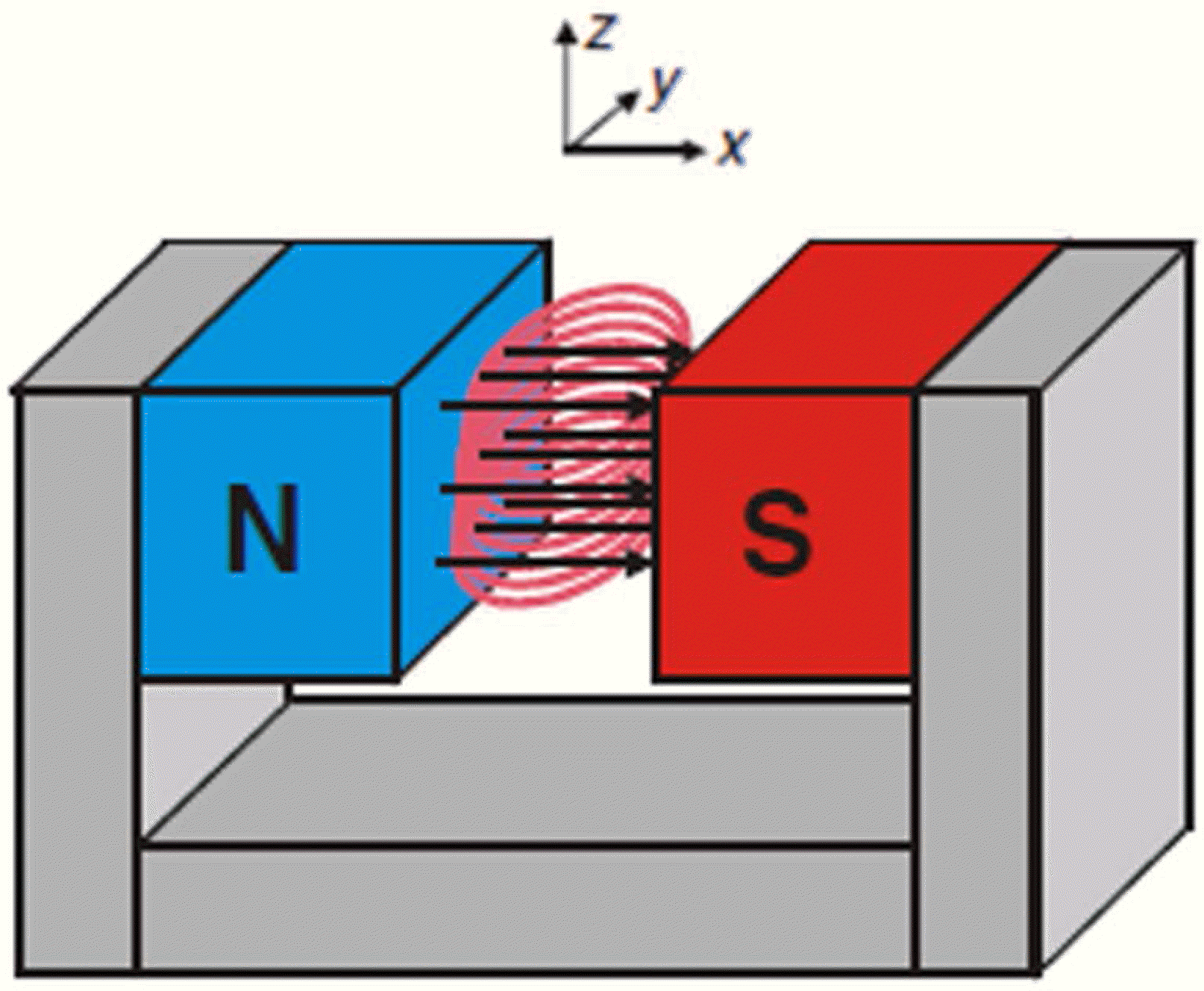
 | Fig. 1.Examples of open MRI (a) Siemens 1.5T MAGNETON Aera, short cylindrical superconducting magnet, 70 cm bore diameter, 137 cm long, system weight of 4.8 tons, minimum room size of 30 m2, FoV of 50 × 50 × 50 cm, gradients 33 mT/m @ 125 T/m/s (4), (b) Siemens 0.35T MAGNETON C, C-shaped dipolar permanent magnet with a vertical magnetic field, bore gap size of 41 cm, 270o accessibility, pole diameter of 137 cm, system weight of 17.6 tons, system dimension of 233 × 206 × 160 cm, minimum room size of 30 m2, FoV of 0.5–40 cm, gradients 24 mT/m @ 55 T/m/s (5), (c) 0.6T UprightTM MRI from Fonar, dipolar electromagnet with a horizontal magnetic field, bore gap size (pole-to-pole) of 46 cm, power requirement of 380–480V, FoV of 6 cm, gradients 12 mT/m, closed-loop water cooling, active and passive shimming, unreported system weight, system dimension, or minimum room size (6), (d) 0.5T PARAmed open MRI, dipolar superconducting magnet using MgB2 with a horizontal magnetic field, cryogen free, low power consumption, bore gap size (pole-to-pole) of 46 cm (7). |
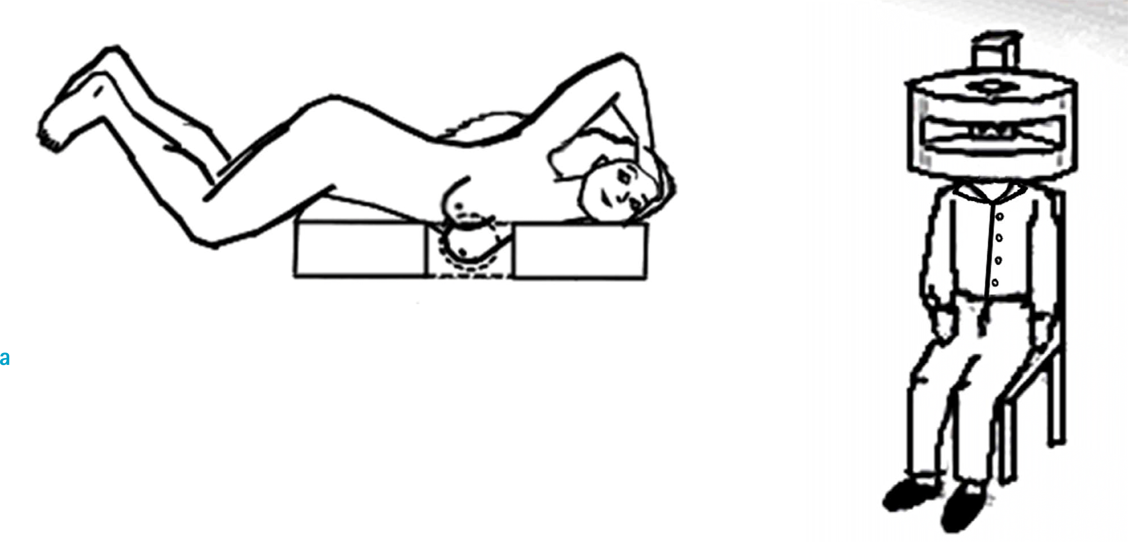 | Fig. 3.The organ specific superconducting magnets (10) (a) A bagel-shaped superconducting magnet for breast imaging (b) a helmet-shaped superconducting magnet for head imaging. |
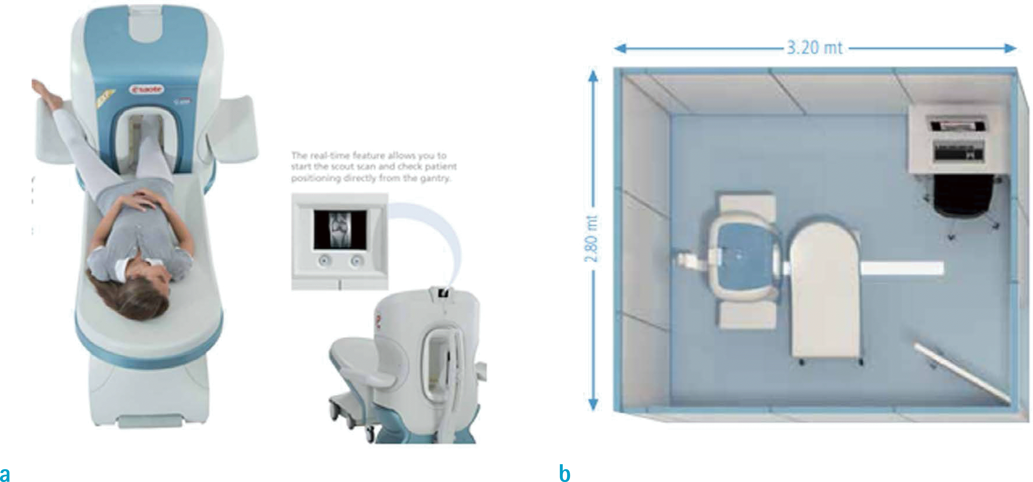
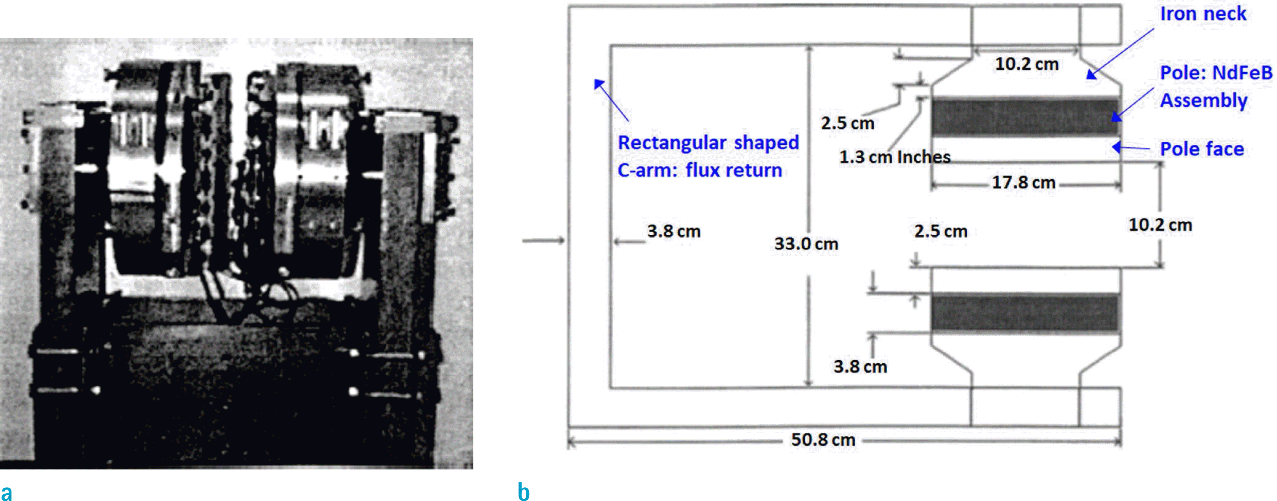 | Fig. 6.A C-shaped table-top permanent magnet array (0.21T) for MRI imaging (12), (a) a photograph, (b) a side view of the system with dimensions. |
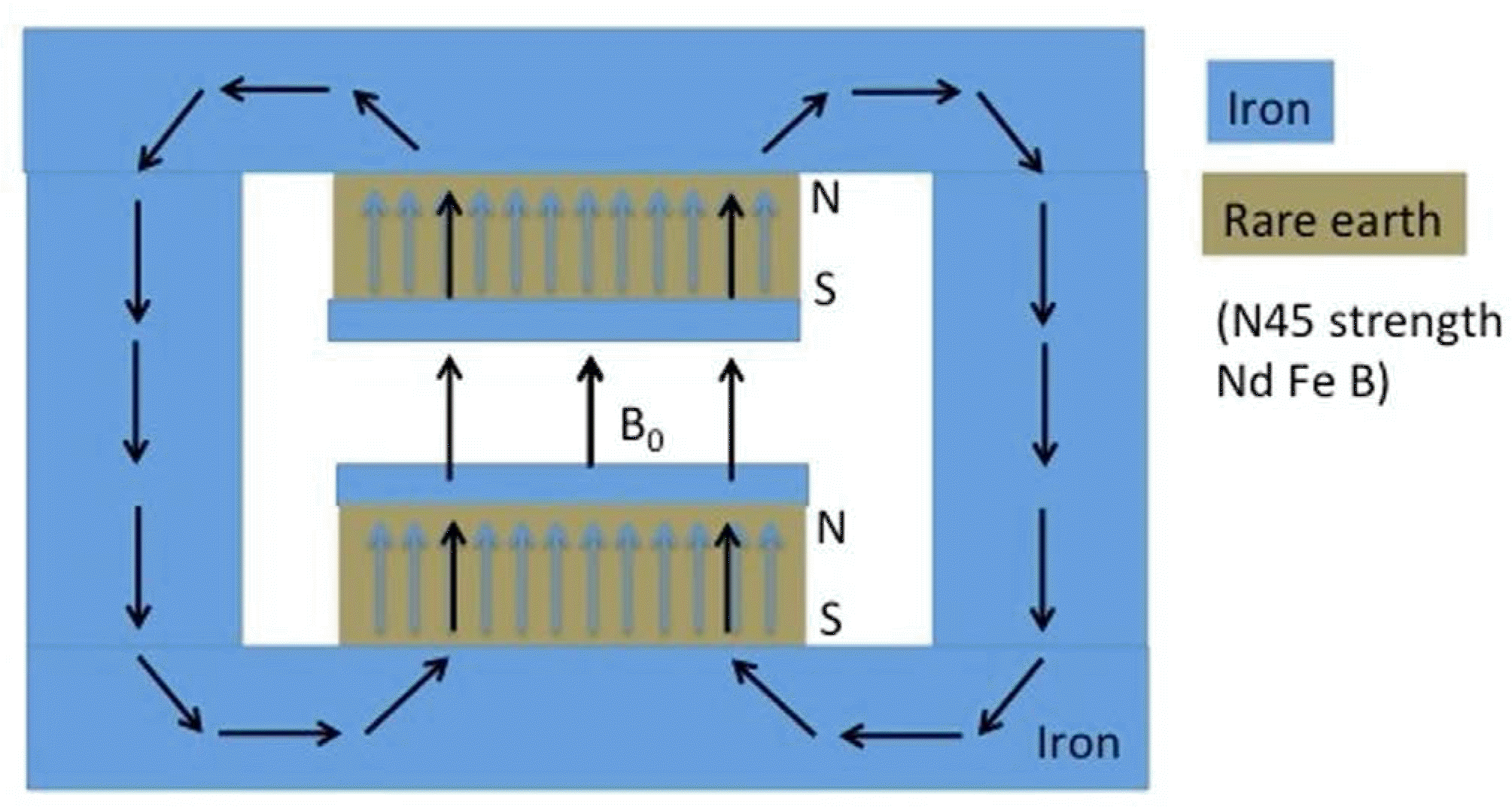 | Fig. 7.The magnet built by the Institute of Electrical Engineering of the Chinese Academy of Sciences in Beijing (75). |
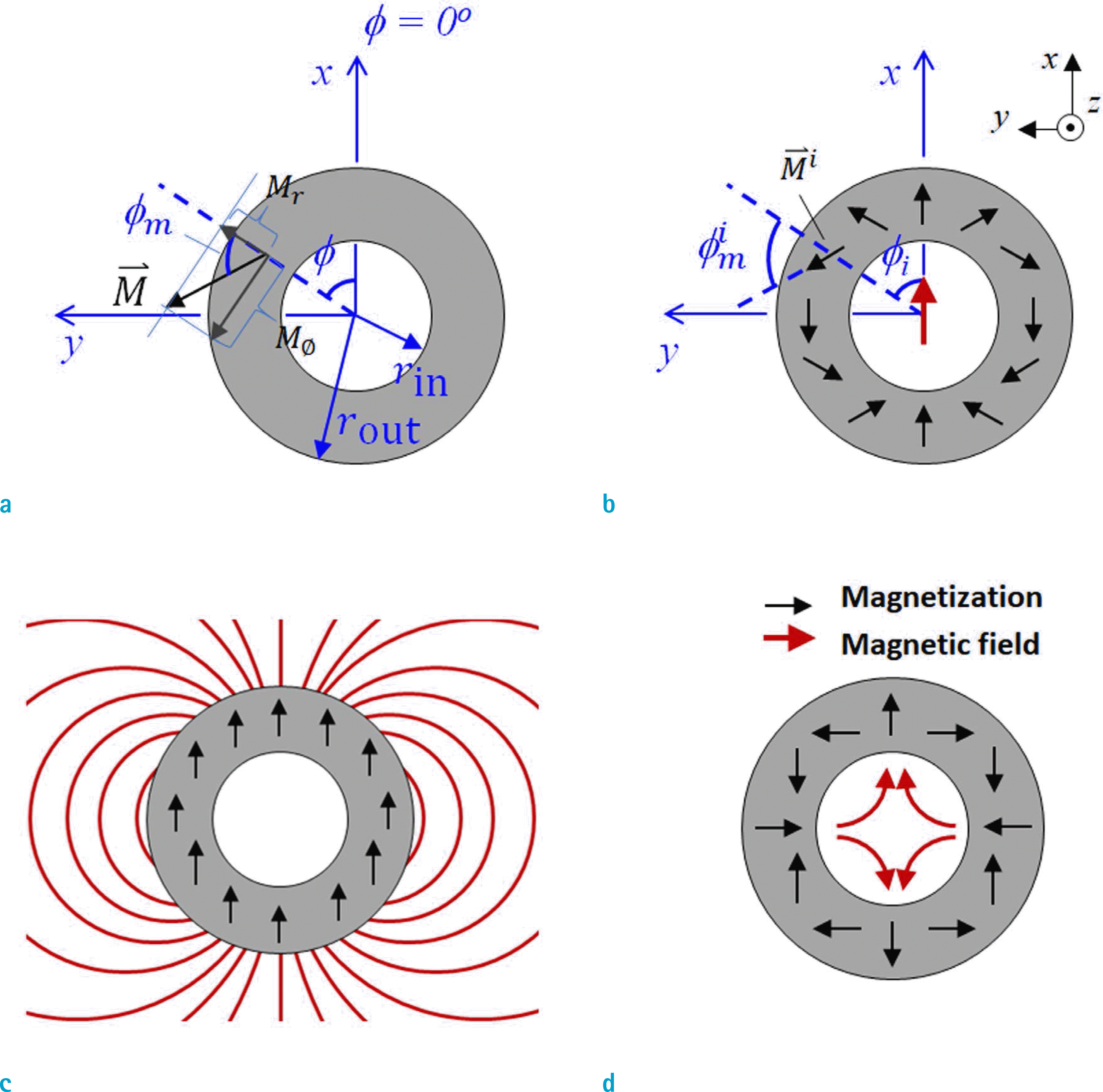 | Fig. 9.Side views of a Halbach cylinder (a) relationship of ⇀ dipolar (n = 1) (d) inner-field, quadrupolar (n = 2). M and angles, (b) inner-field, dipolar (n = 1), (c) outer-field, |
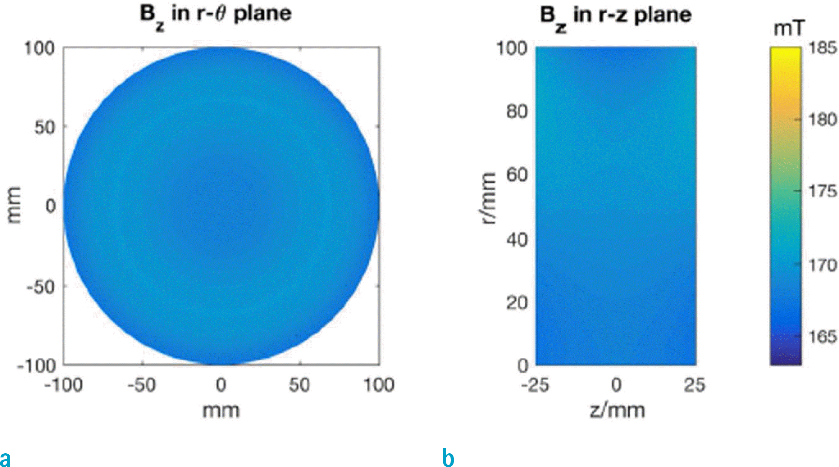 | Fig. 16.The magnetic field generated by the proposed IO ring-pair aggregate in FoV in (a) the rø-plane, (b) the rz-plane (65). |
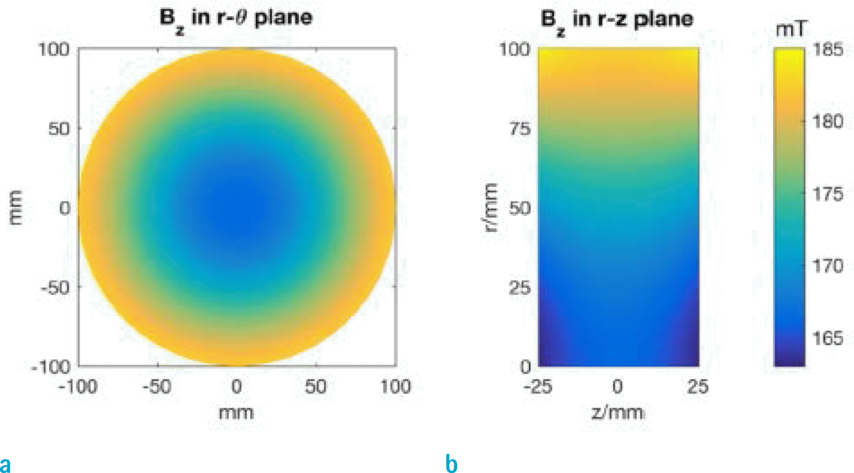 | Fig. 17.The magnetic field generated by the original IO ring pair in FoV in (a) the rø-plane, (b) the rz-plane (65). |


 PDF
PDF ePub
ePub Citation
 Print
Print




 XML Download