

 PDF
PDF ePub
ePub Citation
 Print
Print


Abstract
Purpose
To evaluate the efficacy of 3% diquafosol tetrasodium (DQ) after laser-assisted in-situ keratomileusis (LASIK).
Methods
This prospective randomized study included 150 eyes in 75 patients who were scheduled for LASIK. The patients in the 3% diquafosol tetrasodium (DQ) group (37 patients, 74 eyes) were instructed to apply one drop of DQ, six times daily post-op, while the patients in the 0.3% sodium hyaluronate (HA) group (38 patients, 76 eyes) were instructed to apply one drop of HA, six times daily post-op. A Schirmer test, tear film break-up time (BUT), corneal and conjunctival fluorescein staining score (FLSS), and ocular surface disease index (OSDI) were evaluated pre-op and at 1, 4, and 12 weeks post-op while the tear osmolarity was evaluated pre-op and at 4 and 12 weeks post-op.
Results
There was no significant difference between the two groups regarding Schirmer test results or tear osmolarity and conjunctival FLSS. The BUT was significantly higher in the DQ group at 1 week and 12 weeks post-op. The corneal FLSS was significantly lower in the DQ group at 1 week, 4 weeks and 12 weeks post-op. The OSDI was significantly lower in the DQ group at 1 week post-op.
Figures and Tables
 | Figure 1Cornea and conjunctival fluorescein stain grading. The National Eye Institute/Industry grading system for grading fluorescein staining divides the corneal and conjunctival surfaces to help measure fluorescein uptake. A standardized grading system of 0 to 3 is used for each of the five areas on each cornea and each of the six areas on each conjunctiva.
|
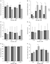 | Figure 2Changes in Tear film break-up time (A), corneal fluorescein staining score (B), conjunctival fluorescein staining score (C), Schirmer test (D), tear osmolarity (E), and ocular surface disease index (F) in the 3% diquafosol tetrasodium and 0.3% sodium hyaluronate group before and after laser-assisted in-situ keratomileusis. ‘DQ’ is 3% diquafosol tetrasodium group, ‘HA’ is 0.3% sodium hyaluronate group. LASIK = laser-assisted in-situ keratomileusis. *Repeat measures analysis of variance test, p < 0.05 between 3% diquafosol tetrasodium and 0.3% sodium hyaluronate group.
|
References
1. Vroman DT, Sandoval HP, Fernández de Castro LE, et al. Effect of hinge location on corneal sensation and dry eye after laser in situ keratomileusis for myopia. J Cataract Refract Surg. 2005; 31:1881–1887.

2. Mian SI, Li AY, Dutta S, et al. Dry eyes and corneal sensation after laser in situ keratomileusis with femtosecond laser flap creation effect of hinge position, hinge angle, and flap thickness. J Cataract Refract Surg. 2009; 35:2092–2098.
3. Shin SY, Lee YJ. Conjunctival changes induced by LASIK suction ring in a rabbit model. Ophthalmic Res. 2006; 38:343–349.
4. Rodriguez AE, Rodriguez-Prats JL, Hamdi IM, et al. Comparison of goblet cell density after femtosecond laser and mechanical microkeratome in LASIK. Invest Ophthalmol Vis Sci. 2007; 48:2570–2575.
5. Jabbur NS, Sakatani K, O'Brien TP. Survey of complications and recommendations for management in dissatisfied patients seeking a consultation after refractive surgery. J Cataract Refract Surg. 2004; 30:1867–1874.
6. Hammond MD, Madigan WP Jr, Bower KS. Refractive surgery in the United States Army, 2000-2003. Ophthalmology. 2005; 112:184–190.
7. De Paiva CS, Chen Z, Koch DD, et al. The incidence and risk factors for developing dry eye after myopic LASIK. Am J Ophthalmol. 2006; 141:438–445.
8. Ambrósio R Jr, Tervo T, Wilson SE. LASIK-associated dry eye and neurotrophic epitheliopathy: pathophysiology and strategies for prevention and treatment. J Refract Surg. 2008; 24:396–407.
9. Nejima R, Miyata K, Tanabe T, et al. Corneal barrier function, tear film stability, and corneal sensation after photorefractive keratectomy and laser in situ keratomileusis. Am J Ophthalmol. 2005; 139:64–71.
10. Linna TU, Vesaluoma MH, Pérez-Santonja JJ, et al. Effect of myopic LASIK on corneal sensitivity and morphology of subbasal nerves. Invest Ophthalmol Vis Sci. 2000; 41:393–397.
11. Yokoi N, Kato H, Kinoshita S. Facilitation of tear fluid secretion by 3% diquafosol ophthalmic solution in normal human eyes. Am J Ophthalmol. 2014; 157:85–92.
12. Fujihara T, Murakami T, Fujita H, et al. Improvement of corneal barrier function by the P2Y(2) agonist INS365 in a rat dry eye model. Invest Ophthalmol Vis Sci. 2001; 42:96–100.
13. Lee JB, Ryu CH, Kim JH, et al. Comparison of tear secretion and tear film instability after photorefractive keratectomy and laser in situ keratomileusis. J Cataract Refract Surg. 2000; 26:1326–1331.
14. Chan TC, Ye C, Chan KP, et al. Evaluation of point-of-care test for elevated tear matrix metalloproteinase 9 in post-LASIK dry eyes. Br J Ophthalmol. 2016; 100:1188–1191.
15. Chao C, Stapleton F, Zhou X, et al. Structural and functional changes in corneal innervation after laser in situ keratomileusis and their relationship with dry eye. Graefes Arch Clin Exp Ophthalmol. 2015; 253:2029–2039.
16. Rodriguez-Prats JL, Hamdi IM, Rodriguez AE, et al. Effect of suction ring application during LASIK on goblet cell density. J Refract Surg. 2007; 23:559–562.
17. Donnenfeld ED, Solomon K, Perry HD, et al. The effect of hinge position on corneal sensation and dry eye after LASIK. Ophthalmology. 2003; 110:1023–1029.
18. Toda I, Ide T, Fukumoto T, et al. Combination therapy with diquafosol tetrasodium and sodium hyaluronate in patients with dry eye after laser in situ keratomileusis. Am J Ophthalmol. 2014; 157:616–622.
19. Fujihara T, Murakami T, Nagano T, et al. INS365 suppresses loss of corneal epithelial integrity by secretion of mucin-like glycoprotein in a rabbit short-term dry eye model. J Ocul Pharmacol Ther. 2002; 18:363–370.
20. Jung HH, Kang YS, Sung MS, Yoon KC. Clinical efficacy of topical 3% diquafosol tetrasodium in short tear film break-up time dry eye. J Korean Ophthalmol Soc. 2015; 56:339–344.
21. Yeon DY, Kang BR, Eom YS, et al. The effect of 3% diquafosol tetrasodium on corneal wetting property and mucin-5AC concentration in rabbits. J Korean Ophthalmol Soc. 2016; 57:208–213.
22. Terakado K, Yogo T, Kohara Y, et al. Conjunctival expression of the P2Y2 receptor and the effects of 3% diquafosol ophthalmic solution in dogs. Vet J. 2014; 202:48–52.
23. Ryan DS, Bower KS, Sia RK, et al. Goblet cell response after photorefractive keratectomy and laser in situ keratomileusis. J Cataract Refract Surg. 2016; 42:1181–1189.
24. Takamura E, Tsubota K, Watanabe H, et al. A randomised, double-masked comparison study of diquafosol versus sodium hyaluronate ophthalmic solutions in dry eye patients. Br J Ophthalmol. 2012; 96:1310–1315.
25. Hwang HS, Sung YM, Lee WS, Kim EC. Additive effect of preservative-free sodium hyaluronate 0.1% in treatment of dry eye syndrome with diquafosol 3% eye drops. Cornea. 2014; 33:935–941.
26. Kamiya K, Nakanishi M, Ishii R, et al. Clinical evaluation of the additive effect of diquafosol tetrasodium on sodium hyaluronate monotherapy in patients with dry eye syndrome: a prospective, randomized, multicenter study. Eye (Lond). 2012; 26:1363–1368.
 XML Download