

 PDF
PDF ePub
ePub Citation
 Print
Print


Abstract
Case summary
A 7.6-year-old female showed numerous small whitish-yellow flecks in the perimacular area and retinal periphery. Dark adapted 0.01 electroretinography (ERG) and dark adapted 3.0 ERG were profoundly reduced. At 26 months after the first visit, the best-corrected visual acuities were 1.0 right eye and 0.9 left eye. There were no pigmented lesions, atrophic lesions, or vascular abnormalities in the retina. Humphrey and Goldmann visual field tests were performed, but neither of the tests revealed any scotomas or other visual field defect. The number and size of characteristic numerous small whitish-yellow retinal flecks seemed almost unchanged. In spectral domain-optical coherence tomography (SD-OCT), the subretinal hyper-reflective lesions spanned the retinal pigment epithelium and the external limiting membrane. ERG showed improved dark adapted responses (dark adapted 0.01 ERG and dark adapted 3.0 ERG) after prolonged dark adaptation (2.5 hours). No family member showed any abnormal findings.
Figures and Tables
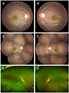 | Figure 1Fundus photography of this patient. (A, B) At the initial visit. There are many yellow whitish spots on the equator and periphery of retina of both eyes. (C, D) Twenty six months after the initial visit. The spots seemed almost unchanged. (E, F) Ultrawide field fundus photography at 26 months after the initial visit. The size of spots was smaller on the inferior retina compared to superior, nasal and temporal retina.
|
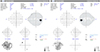 | Figure 2Visual filed test at 26 months after the initial visit. The results of both eyes show generalized reduction of sensitivity. GHT = glaucoma Hemifield test; VFI = visual field index; MD = mean deviation; PSD = pattern standard deviation.
|
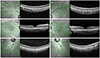 | Figure 3Spectral domain-optical coherence tomography at 26 months after the initial visit. Fovea seemed to be normal in both eyes (C, D), but hyper-reflective lesions were observed between retinal pigment epithelium layer and external limiting membrane on superior and inferior area to the fovea (A, B). The size of hyper-reflective lesions was smaller on the inferior area (A, B, E, F).
|
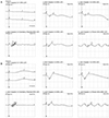 | Figure 4Electroretinography of right eye at 26 months after the initial visit by usual protocol (30 minutes dark adaptation) (A). Scotopic response substantially decreased, but photopic response was normal. After 150 minutes dark adaptation, scotopic response was recovered up to normal range (B). ERG = electroretinography.
|
References
1. Sergouniotis PI, Sohn EH, Li Z, et al. Phenotypic variability in RDH5 retinopathy (Fundus Albipunctatus). Ophthalmology. 2011; 118:1661–1670.

2. Dryja TP. Molecular genetics of Oguchi disease, fundus albipunctatus, and other forms of stationary night blindness: LVII Edward Jackson Memorial Lecture. Am J Ophthalmol. 2000; 130:547–563.
3. Skorczyk-Werner A, Pawłowski P, Michalczuk M, et al. Fundus albipunctatus: review of the literature and report of a novel RDH5 gene mutation affecting the invariant tyrosine (p.Tyr175Phe). J Appl Genet. 2015; 56:317–327.
4. Niwa Y, Kondo M, Ueno S, et al. Cone and rod dysfunction in fundus albipunctatus with RDH5 mutation: an electrophysiological study. Invest Ophthalmol Vis Sci. 2005; 46:1480–1485.
5. Sekiya K, Nakazawa M, Ohguro H, et al. Long-term fundus changes due to fundus albipunctatus associated with mutations in the RDH5 gene. Arch Ophthalmol. 2003; 121:1057–1059.
6. Yamamoto H, Simon A, Eriksson U, et al. Mutations in the gene encoding 11-cis retinol dehydrogenase cause delayed dark adaptation and fundus albipunctatus. Nat Genet. 1999; 22:188–191.
7. Schatz P, Preising M, Lorenz B, et al. Fundus albipunctatus associated with compound heterozygous mutations in RPE65. Ophthalmology. 2011; 118:888–894.
8. Hirose E, Inoue Y, Morimura H, et al. Mutations in the 11-cis retinol dehydrogenase gene in Japanese patients with Fundus albipunctatus. Invest Ophthalmol Vis Sci. 2000; 41:3933–3935.
9. Farjo KM, Moiseyev G, Takahashi Y, et al. The 11-cis-retinol dehydrogenase activity of RDH10 and its interaction with visual cycle proteins. Invest Ophthalmol Vis Sci. 2009; 50:5089–5097.
10. Makiyama Y, Ooto S, Hangai M, et al. Cone abnormalities in fundus albipunctatus associated with RDH5 mutations assessed using adaptive optics scanning laser ophthalmoscopy. Am J Ophthalmol. 2014; 157:558–570.
11. Walia S, Fishman GA, Kapur R. Flecked-retina syndromes. Ophthalmic Genet. 2009; 30:69–75.
12. Ergun E, Hermann B, Wirtitsch M, et al. Assessment of central visual function in Stargardt's disease/fundus flavimaculatus with ultrahigh-resolution optical coherence tomography. Invest Ophthalmol Vis Sci. 2005; 46:310–316.
13. Zaharova E, Sherman J. The use of SD-OCT in the differential diagnosis of dots, spots and other white retinal lesions. Eye Brain. 2011; 3:69–80.
14. Fishman GA, Roberts MF, Derlacki DJ, et al. Novel mutations in the cellular retinaldehyde-binding protein gene (RLBP1) associated with retinitis punctata albescens: evidence of interfamilial genetic heterogeneity and fundus changes in heterozygotes. Arch Ophthalmol. 2004; 122:70–75.
15. Rotenstreich Y, Harats D, Shaish A. Treatment of a retinal dystrophy, fundus albipunctatus, with oral 9-cis-{beta}-carotene. Br J Ophthalmol. 2010; 94:616–621.
 XML Download