Abstract
Purpose
To evaluate the effects of surgery depending on the follow-up duration after superior oblique tuck was performed as the first surgery in unilateral superior oblique palsy patients.
Methods
Sixteen patients who were followed-up for a minimum of 3 months were retrospectively evaluated. The vertical deviation, abnormal head posture, superior oblique underaction, and inferior oblique overaction were evaluated before and at 3, 6, and 12 months after the surgery and at the last follow-up. The angle between the center of the optic disc and fovea (disc-fovea angle) was measured using fundus photography to investigate changes in ocular torsion.
Results
The mean follow-up period was 24.9 ± 21.9 months and the mean tuck was 11.4 ± 4.0 mm. Vertical deviation <7 prism diopters in the primary position was observed in 53.9% of patients at 3 months postoperatively, 50.0% at 6 months, 83.3% at 12 months, and 62.5% at the last follow-up (p = 0.55). Head posture was improved in 66.7% of patients at 3 months, 71.4% at 6 months, 50% at 12 months, and 80% at the last follow-up after surgery (p = 0.73). Ocular torsion was decreased in 37.5% of patients at 3 months postoperatively, 66.7% at 6 months, 75% at 12 months, and 80.0% at the last follow-up (p = 0.11). Superior oblique underaction was improved in 100%, 77.8%, 60%, and 75% of the patients and inferior oblique overaction was improved in 100%, 88.9%, 85.7%, and 81.3% of the patients at postoperative month 3, 6, and 12, and at the last follow-up, respectively.
Figures and Tables
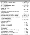
Table 2
Vertical deviations and foveal torsions during the follow-up of superior oblique tuck surgery in unilateral SOP patients

References
1. Helveston EM, Ellis FD. Superior oblique tuck for superior oblique palsy. Aust J Ophthalmol. 1983; 11:215–220.
3. Durnian JM, Marsh IB. Superior oblique tuck: its success as a single muscle treatment for selected cases of superior oblique palsy. Strabismus. 2011; 19:133–137.

4. Nejad M, Thacker N, Velez FG, et al. Surgical results of patients with unilateral superior oblique palsy presenting with large hypertropias. J Pediatr Ophthalmol Strabismus. 2013; 50:44–52.

5. Helveston EM, Mora JS, Lipsky SN, et al. Surgical treatment of superior oblique palsy. Trans Am Ophthalmol Soc. 1996; 94:315–328.

6. Toosi SH, von Noorden GK. Effect of isolated inferior oblique muscle myectomy in the management of superior oblique muscle palsy. Am J Ophthalmol. 1979; 88:602–608.

7. Knapp P, Moore S. Diagnosis and surgical options in superior oblique surgery. Int Ophthalmol Clin. 1976; 16:137–149.

9. Morris RJ, Scott WE, Keech RV. Superior oblique tuck surgery in the management of superior oblique palsies. J Pediatr Ophthalmol Strabismus. 1992; 29:337–346. discussion 347-8.
10. Bhola R, Velez FG, Rosenbaum AL. Isolated superior oblique tucking: an effective procedure for superior oblique palsy with profound superior oblique underaction. J AAPOS. 2005; 9:243–249.

11. Rosenbaum AL, Santiago AP. chap. 35. Clinical strabismus management: principles and surgical management. Superior oblique procedures. Philadelphia: WB Saunders Company;1999.
12. von Noorden GK, Murray E, Wong SY. Superior oblique paralysis. A review of 270 cases. Arch Ophthalmol. 1986; 104:1771–1776.
13. Gräf M, Lorenz B, Eckstein A, Esser J. Superior oblique tucking with versus without additional inferior oblique recession for acquired trochlear nerve palsy. Graefes Arch Clin Exp Ophthalmol. 2010; 248:223–229.

14. Moon HS, Seo SW, Lee JH. Effect of superior oblique tuck surgery in the patients with unilateral superior oblique palsy. J Korean Ophthalmol Soc. 2000; 41:1968–1973.
15. Ducca BL, Souza-Dias CR, Lui AC, Goldchmit M. Measurement of ocular torsion variation following superior oblique tenectomy. Arq Bras Oftalmol. 2014; 77:364–367.

16. von Noorden GK, Campos EC. chap. 20. Binocular vision and ocular motility: theory and management of strabismus. Paralytic strabismus. 6th ed. St. Louis: Mosby;2002.
17. Arici C, Oguz V. The effect of surgical treatment of superior oblique muscle palsy on ocular torsion. J AAPOS. 2012; 16:21–25.

18. Lee J, Suh SY, Choung HK, Kim SJ. Inferior oblique weakening surgery on ocular torsion in congenital superior oblique palsy. Int J Ophthalmol. 2015; 8:569–573.


 PDF
PDF ePub
ePub Citation
 Print
Print


 XML Download