

 PDF
PDF ePub
ePub Citation
 Print
Print


Abstract
Purpose
To evaluate the changes in anterior chamber depth (ACD) and refractive error after combined phacovitrectomy with posterior capsulotomy using a vitrectomy probe.
Methods
In 20 eyes of 20 patients who underwent combined phacovitrectomy with posterior capsulotomy using a vitrectomy probe, the ACD was measured with Scheimpflug imaging (Pentacam®, OCULUS Optikgeräte GmbH, Wetzlar, Germany) preoperatively and postoperatively. We compared the preoperative desired refraction and postoperative refraction using an autokeratorefractometor.
Results
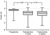
The preoperative ACD was 2.58 ± 0.248 mm; the ACD significantly increased in 1 month postoperatively to 3.65 ± 0.475 mm (p < 0.001), and it was maintained as 3.70 ± 0.452 mm (p = 0.213) at 3 months postoperatively. The preoperative target spherical equivalent was −0.60 ± 0.809 diopters (D). Myopic shifting was noticed at 1 month postoperatively as −1.45 ± 1.252 D, and it changed between 1 month and 3 months postoperatively (−1.48 ± 1.235 D at 3 months postoperatively was not statistically significant). There was no increased intraocular pressure or intraocular lens-related complication.
Conclusions
Phacovitrectomy with posterior capsulotomy using a vitrectomy probe might be a useful way to stabilize the axial position of an intraocular lens without constriction of the capsular bag. However, using this procedure, the surgeon must consider the possibility of myopic shifting in the postoperative refractive error.
Figures and Tables
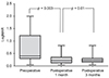 | Figure 1Changes of best corrected visual acuity (BCVA) logarithm of minimal angle of resolution in postoperative 1 month and 3 months. BCVA increased statistically significant in postoperative 1 month, and it maintained in postoperative 3 months. LogMAR = logarithm of minimal angle of resolution.
|
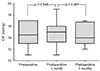 | Figure 2Changes of intraocular pressure (IOP) in postoperative 1 month and 3 months. IOP has no significant differences between preoperative and postoperative 1 month and 3 months.
|
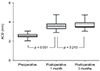 | Figure 3Changes of anterior chamber depth (ACD) in postoperative 1 month and 3 months. ACD showed statistically increased in postoperative 1 month and it maintained until 3 months.
|
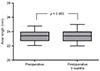 | Figure 4Changes of axial length in postoperative 3 months. Axial length showed no changes in postoperative 3 months compared with preoperative.
|
 | Figure 5Changes of refractory error in postoperative 1 month and 3 months. Postoperative 1 month and 3 months revealed statistically significant myopic shifting compared with preoperative target spherical equivalent.
|
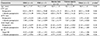
Table 2
Comparison of CST, ACD, axial length and SE in preoperative and postoperative 3 months between diagnoses
![]()

References
1. Steel DH. Phacovitrectomy: expanding indications. J Cataract Refract Surg. 2007; 33:933–936.

2. Muselier A, Dugas B, Burelle X, et al. Macular hole surgery and cataract extraction: combined vs consecutive surgery. Am J Ophthalmol. 2010; 150:387–391.
3. Manvikar SR, Allen D, Steel DH. Optical biometry in combined phacovitrectomy. J Cataract Refract Surg. 2009; 35:64–69.
4. Kim YK, Woo SJ, Hyon JY, et al. Refractive outcomes of combined phacovitrectomy and delayed cataract surgery in retinal detachment. Can J Ophthalmol. 2015; 50:360–366.
5. Pinarci EY, Bayar SA, Sizmaz S, et al. Anterior segment complications after phacovitrectomy in diabetic and nondiabetic patients. Eur J Ophthalmol. 2013; 23:223–229.
6. Chang MA, Parides MK, Chang S, Braunstein RE. Outcome of phacoemulsification after pars plana vitrectomy. Ophthalmology. 2002; 109:948–954.
7. Pinter SM, Sugar A. Phacoemulsification in eyes with past pars plana vitrectomy: case-control study. J Cataract Refract Surg. 1999; 25:556–561.
8. Ling R, Simcock P, McCoombes J, Shaw S. Presbyopic phacovitrectomy. Br J Ophthalmol. 2003; 87:1333–1335.
9. Mochizuki Y, Kubota T, Hata Y, et al. Surgical results of combined pars plana vitrectomy, phacoemulsification, and intraocular lens implantation. Eur J Ophthalmol. 2006; 16:279–286.
10. Toda J, Kato S, Oshika T, Sugita G. Posterior capsule opacification after combined cataract surgery and vitrectomy. J Cataract Refract Surg. 2007; 33:104–107.
11. Roh JH, Sohn HJ, Lee DY, et al. Comparison of posterior capsular opacification between a combined procedure and a sequential procedure of pars plana vitrectomy and cataract surgery. Ophthalmologica. 2010; 224:42–46.
12. Gimbel HV. Posterior capsulorhexis with optic capture in pediatric cataract and intraocular lens surgery. Ophthalmology. 1996; 103:1871–1875.
13. Gimbel HV. Posterior continuous curvilinear capsulorhexis and optic capture of the intraocular lens to prevent secondary opacification in pediatric cataract surgery. J Cataract Refract Surg. 1997; 23 Suppl 1:652–656.
14. Stifter E, Menapace R, Luksch A, et al. Anterior chamber depth and change in axial intraocular lens position after cataract surgery with primary posterior capsulorhexis and posterior optic buttonholing. J Cataract Refract Surg. 2008; 34:749–754.
15. Fajgenbaum MAP, Neffendorf JE, Wong RS, et al. Intraoperative and postoperative complications in phacovitrectomy for epiretinal membrane and macular hole: a clinical audit of 1,000 consecutive eyes. Retina. 2018; 38:1865–1872.
16. Rogers S, Madhusudhana KC, Kang HK, et al. Combined phacovitrectomy for macular hole: long-term results. Ophthalmic Surg Lasers Imaging. 2007; 38:452–456.
17. Karahan E, Er D, Kaynak S. An overview of Nd:YAG laser capsulotomy. Med Hypothesis Discov Innov Ophthalmol. 2014; 3:45–50.
18. Sato S, Inoue M, Kobayashi S, et al. Primary posterior capsulotomy using a 25-gauge vitreous cutter in vitrectomy combined with cataract surgery. J Cataract Refract Surg. 2010; 36:2–5.
19. Ehmann D, García R. Investigating a possible cause of the myopic shift after combined cataract extraction, intraocular lens implantation, and vitrectomy for treatment of a macular hole. Can J Ophthalmol. 2009; 44:594–597.
 XML Download