

 PDF
PDF ePub
ePub Citation
 Print
Print


Abstract
Purpose
To evaluate the repeatability and reproducibility of inferior tear meniscus measurements using two different spectral domain-optical coherence tomography (OCT), and to compare the inter-device agreements between these devices.
Methods
Two examiners evaluated the tear meniscus depth (TMD) and tear meniscus height (TMH) of 20 eyes in 20 normal subjects using Cirrus OCT and Spectralis OCT with the examiners calculating the TMD and TMH. We analyzed intra-examiner repeatability, inter-examiner reproducibility, and inter-device agreement.
Results
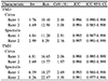
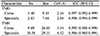
The average TMD measurements using the Cirrus OCT and Spectralis OCT devices were 151.25 ± 41.53 µm and 139.10 ± 40.56 µm by examiner 1, respectively, and 152.03 ± 42.77 µm and 138.35 ± 39.70 µm by examiner 2, respectively. The TMHs were 291.90 ± 100.19 µm and 245.43 ± 66.44 µm by examiner 1, respectively, and 288.25 ± 98.72 µm and 244.23 ± 60.69 µm by examiner 2, respectively. The TMDs and TMHs measured using these OCT devices were not statistically significant for intra-examiner and inter-examiner measurements (all, p > 0.05). These devices showed high repeatability (intraclass correlation coefficient ≥ 0.991) for intra-examiner TMD and TMH measurements and the inter-examiner coefficient of variation ranged from 2.04% to 4.32%. The 95% limits of agreement between the two devices were −66.13 to 91.95 µm for TMD and −127.18 to 217.68 µm for TMH.
Figures and Tables
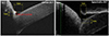 | Figure 1Tear meniscus depth (TMD) and tear meniscus height (TMH). Measurement of TMD and TMH in the Cirrus optical coherence tomography (OCT) (A) and Spectralis OCT (B).
|
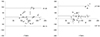 | Figure 2Bland-Altman plots. Bland-Altman plots of the tear meniscus depth tear meniscus depth and tear meniscus height tear meniscus height difference between Cirrus optical coherence tomography (OCT) and Spectralis OCT. TMD = tear meniscus depth; TMH = tear meniscus height.
|
Table 1
Measurement of tear meniscus depth and tear meniscus height using Cirrus OCT and Spectralis OCT
![]()
Notes
References
1. The definition and classification of dry eye disease: report of the Definition and Classification Subcommittee of the International Dry Eye WorkShop (2007). Ocul Surf. 2007; 5:75–92.
2. Lin PY, Tsai SY, Cheng CY, et al. Prevalence of dry eye among an elderly Chinese population in Taiwan: the Shihpai Eye Study. Ophthalmology. 2003; 110:1096–1101.
3. McCarty CA, Bansal AK, Livingston PM, et al. The epidemiology of dry eye in Melbourne, Australia. Ophthalmology. 1998; 105:1114–1119.
4. Kallarackal GU, Ansari EA, Amos N, et al. A comparative study to assess the clinical use of fluorescein meniscus time (FMT) with tear break up time (TBUT) and Schirmer's tests (ST) in the diagnosis of dry eyes. Eye. 2002; 16:594–600.

5. Senchyna M, Wax MB. Quantitative assessment of tear production: a review of methods and utility in dry eye drug discovery. J Ocul Biol Dis Infor. 2008; 1:1–6.
6. Mishima S, Gasset A, Klyce SD Jr, Baum JL. Determination of tear volume and tear flow. Invest Ophthalmol. 1966; 5:264–276.
7. Yokoi N, Bron AJ, Tiffany JM, et al. Relationship between tear volume and tear meniscus curvature. Arch Ophthalmol. 2004; 122:1265–1269.
8. Mainstone JC, Bruce AS, Golding TR. Tear meniscus measurement in the diagnosis of dry eye. Curr Eye Res. 1996; 15:653–661.
9. Lim KJ, Lee JH. Measurement of the tear meniscus height using 0.25% fluorescein sodium. Korean J Ophthalmol. 1991; 5:34–36.
10. Ibrahim OM, Dogru M, Ward SK, et al. The efficacy, sensitivity, and specificity of strip meniscometry in conjunction with tear function tests in the assessment of tear meniscus. Invest Ophthalmol Vis Sci. 2011; 52:2194–2198.
11. Raj A, Dhasmana R, Nagpal RC. Anterior segment optical coherence tomography for tear meniscus evaluation and its correlation with other tear variables in healthy individuals. J Clin Diagn Res. 2016; 10:NC01-4.
12. Chan HH, Zhao Y, Tun TA, Tong L. Repeatability of tear meniscus evaluation using spectral-domain Cirrus(R) HD-OCT and time-domain Visante(R) OCT. Cont Lens Anterior Eye. 2015; 38:368–372.
13. Arriola-Villalobos P, Fernández-Vigo JI, Díaz-Valle D, et al. Assessment of lower tear meniscus measurements obtained with keratograph and agreement with fourier-domain optical-coherence tomography. Br J Ophthalmol. 2015; 99:1120–1125.
14. Vaz S, Falkmer T, Passmore AE, et al. The case for using the repeatability coefficient when calculating test-retest reliability. PLoS One. 2013; 8:e73990.
15. Kong KA. Statistical methods: reliability assessment and method comparison. Ewha Med J. 2017; 40:9–16.
16. Jones LW, Rahman S, Leech R, et al. Determination of inferior tear meniscus height and inferior tear meniscus volume using optical coherence tomography. Invest Ophthalmol Vis Sci. 2004; 45:144.
17. Bitton E, Keech A, Simpson T, Jones L. Variability of the analysis of the tear meniscus height by optical coherence tomography. Optom Vis Sci. 2007; 84:903–908.
18. Arriola-Villalobos P, Fernández-Vigo JI, Díaz-Valle D, et al. Lower tear meniscus measurements using a new anterior segment swept-source optical coherence tomography and agreement with fourier-domain optical coherence tomography. Cornea. 2016; 36:183–188.
19. Zhou S, Li Y, Lu AT, et al. Reproducibility of tear meniscus measurement by Fourier-domain optical coherence tomography: a pilot study. Ophthalmic Surg Lasers Imaging. 2009; 40:442–447.
 XML Download