

 PDF
PDF ePub
ePub Citation
 Print
Print


Abstract
Purpose
To compare corneal astigmatism, keratometry and corneal higher order aberrations between the light emitting diode corneal topography analyzer and Scheimpflug Imager.
Methods
This prospective study involved 45 patients (45 eyes) who visited Seoul St. Mary's hospital before cataract surgery from June 7, 2017, to August 2, 2017. For each eye, keratometry, astigmatism and its axis of cornea, higher-order aberrations were evaluated with a Scheimpflug Imager (Pentacam HR®, Oculus, Wetzlar, Germany) and a color-LED corneal topographer (Cassini®, i-Optics, Den Haag, The Netherlands).
Results
Astigmatism magnitude measured using Cassini® and Pentacam® showed no statistically differences but anterior and total astigmatic axes were significantly different, as measured by the two devices (p < 0.05). Anterior and total mean keratometry were statistically significantly different, as measured by the two devices (p < 0.05). J0 and J45 vectors of anterior and total cornea were statistically different (p < 0.05). In addition, Cassini® and Pentacam® showed discrepancies between total corneal astigmatism, total J0 and J45 vectors. Corneal anterior spherical aberration, vertical and horizontal coma, and oblique and horizontal trefoil aberrations were not statistically different between the two devices.
Go to : 
References
1. Ferreira TB, Ribeiro FJ. A novel color-LED corneal topographer to assess astigmatism in pseudophakic eyes. Clin Ophthalmol. 2016; 10:1521–9.

2. Lombardo M, Lombardo G. Wave aberration of human eyes and new descriptors of image optical quality and visual performance. J Cataract Refract Surg. 2010; 36:313–31.
3. Artal P, Berrio E, Guirao A, Piers P. Contribution of the cornea and internal surfaces to the change of ocular aberrations with age. J Opt Soc Am A Opt Image Sci Vis. 2002; 19:137–43.
4. Bellucci R, Morselli S, Piers P. Comparison of wavefront abdominals and optical quality of eyes implanted with five different abdominal lenses. J Refract Surg. 2004; 20:297–306.
5. Ventura BV, Wang L, Ali SF, et al. Comparison of corneal power, astigmatism, and wavefront aberration measurements obtained by a point-source color light-emitting diode-based topographer, a Placido-disk topographer, and a combined Placido and dual Scheimpflug device. J Cataract Refract Surg. 2015; 41:1658–71.
6. Kanellopoulos AJ, Asimellis G. Color light-emitting diode abdominal topography: validation of keratometric repeatability in a large sample of wide cylindrical-range corneas. Clin Ophthalmol. 2015; 9:245–52.
7. Barkana Y, Gerber Y, Elbaz U, et al. Central corneal thickness measurement with the Pentacam Scheimpflug system, optical low-coherence reflectometry pachymeter, and ultrasound pachymetry. J Cataract Refract Surg. 2005; 31:1729–35.
8. Koch DD, Ali SF, Weikert MP, et al. Contribution of posterior abdominal astigmatism to total corneal astigmatism. J Cataract Refract Surg. 2012; 38:2080–7.
9. Kanellopoulos AJ, Asimellis G. Distribution and repeatability of corneal astigmatism measurements (magnitude and axis) evaluated with color light emitting diode reflection topography. Cornea. 2015; 34:937–44.
10. Klijn S, Reus NJ, van der Sommen CM, Sicam VA. Accuracy of total corneal astigmatism measurements with a scheimpflug imager and a color light-emitting diode corneal topographer. Am J Ophthalmol. 2016; 167:72–8.
11. Davison JA, Potvin R. Refractive cylinder outcomes after abdominal toric intraocular lens cylinder power using total corneal abdominal power. Clin Ophthalmol. 2015; 9:1511–7.
12. Klijn S, Reus NJ, Sicam VA. Evaluation of keratometry with a abdominal color-LED corneal topographer. J Refract Surg. 2015; 31:249–56.
13. Mejía-Barbosa Y, Malacara-Hernández D. A review of methods for measuring corneal topography. Optom Vis Sci. 2001; 78:240–53.
14. Hoffmann PC, Abraham M, Hirnschall N, Findl O. Prediction of residual astigmatism after cataract surgery using swept source fourier domain optical coherence tomography. Current Eye Res. 2014; 39:1178–86.
15. Elliott M, Simpson T, Richter D, Fonn D. Repeatability and abdominal of automated refraction: a comparison of the Nikon NRK-8000, the Nidek AR-1000, and subjective refraction. Optom Vis Sci. 1997; 74:434–8.
16. Choi YJ, Kang NH, Jun RM. Comparison of corneal higher-order aberrations measured with two instruments using scheimpflug camera system. J Korean Ophthalmol Soc. 2015; 56:1497–504.
17. Shin JY, Lee MY, Chung SH. Comparison of keratometry and abdominal higher order aberrations between scout videokeratoscope and pentacam scheimpflug camera. J Korean Ophthalmol Soc. 2014; 55:1758–64.
Go to :
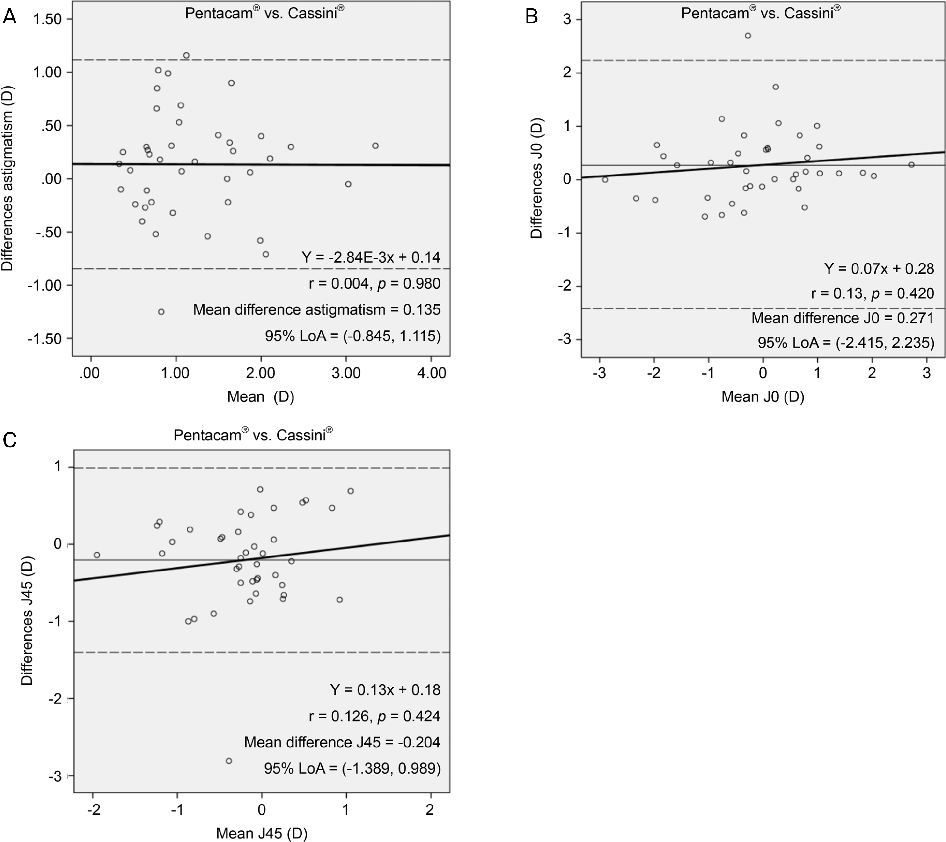 | Figure 1.Bland-Altman plots showing the agreement between total astigmatism magnitude, J0 and, J45 obtained by the 2 devices (Cassini® and Pentacam®). (A-C) graphs plot the differences against the average of the two measurements, with 95% limits of agreement and mean difference. (A) Total astigmatism magnitude (B) Total J0 (C) Total J45. The dotted lines show the 95% LoA. The bold line shows the regression line. D = diopters. |
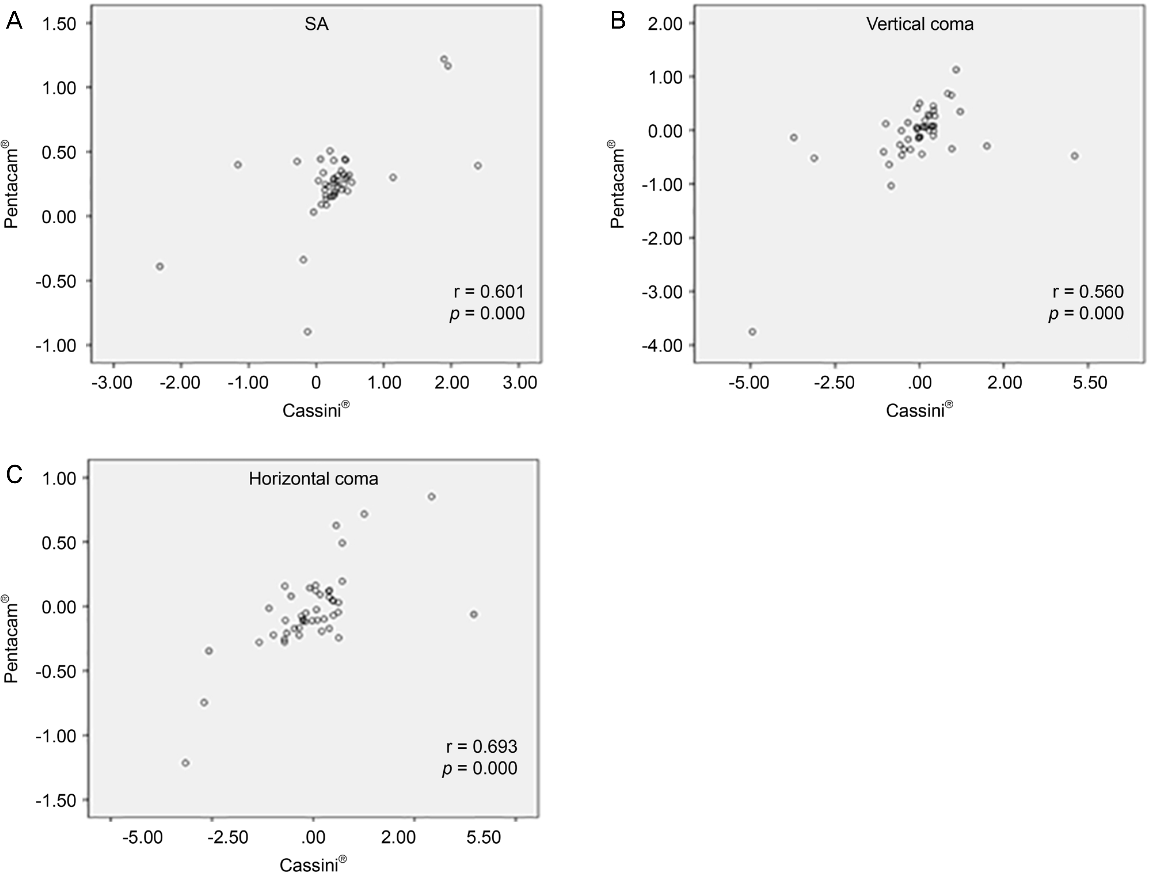 | Figure 2.Correlations of measurements of the (A) spherical aberration, (B) vertical coma, (C) horizontal coma between Pentacam® and Cassini®. SA = spherical aberration; r = Pearson correlation coefficient. * Statistically significant (p < 0.05, based on t-test). |
Table 1.
Demographics of subjects
| Characteristic | Value |
|---|---|
| Number of subjects (eyes) | 45 (45) |
| Age (years) | 59.95 ± 14.82 |
| Male:female | 22:23 |
| UCVA (logMAR) | 0.62 ± 0.31 |
| BCVA (logMAR) | 0.33 ± 0.29 |
Table 2.
Comparison of corneal astigmatic magnitude and axes between the two devices
| Technique | Astigmatism (D) | Axis (D) |
|---|---|---|
| Pentacam® anterior | 1.33 ± 1.19 | 99.65 ± 47.83 |
| Cassini® anterior | 1.33 ± 4.50 | 88.36 ± 46.84* |
| Pentacam® posterior | –0.33 ± 0.21 | 87.82 ± 19.28 |
| Cassini® posterior | –0.36 ± 0.24 | 93.22 ± 32.04 |
| Pentacam® TRP | 1.45 ± 1.25 | 107.42 ± 53.60 |
| Cassini® TCA | 1.43 ± 1.52 | 89.80 ± 50.45* |
Table 3.
Difference of astigmatism magnitude, axes and keratometry between the two devices
| Variable |
Pentacam®-Cassini® difference |
|||
|---|---|---|---|---|
|
Anterior |
Total |
|||
| Mean ± SD | p-value* | Mean ± SD | p-value* | |
| Ast (D) | –0.04 ± 0.30 | 0.378 | 0.13 ± 0.50 | 0.092 |
| Axis° | 11.29 ± 38.59 | 0.032 | 17.62 ± 63.83 | 0.032 |
| Mean K (D) | 0.21 ± 0.63 | 0.019 | 0.83 ± 0.66 | 0.000 |
| J0 (D) | 0.17 ± 0.52 | 0.045 | 0.27 ± 0.66 | 0.012 |
| J45 (D) | –0.17 ± 0.52 | 0.039 | –0.20 ± 0.62 | 0.043 |
Table 4.
The comparison of anterior corneal aberrations between Pentacam® and Cassini® (n = 45)
| Variable | Pentacam® (μ m) | Cassini® (μ m) | p-value * |
|---|---|---|---|
| SA Z (4, 0) | 0.26 ± 0.31 | 0.29 ± 0.69 | 0.206 |
| Vertical coma Z (3, −1) | –0.09 ± 0.68 | –0.05 ± 1.39 | 0.619 |
| Horizontal coma Z (3, 1) | –0.04 ± 0.33 | –0.02 ± 0.47 | 0.955 |
| Oblique trefoil Z (3, −3) | –0.01 ± 0.24 | 0.20 ± 1.52 | 0.843 |
| Horizontal trefoil Z (3, 3) | –0.02 ± 0.28 | –0.44 ± 2.21 | 0.687 |
 XML Download