

 PDF
PDF ePub
ePub Citation
 Print
Print


Introduction
The terpenoid compounds are the hotspots in natural products researches for their complexly structural diversities and excellent multiple biological activities, especially in diterpenoid compounds, such as taxol, ginkgolides and triptolides. There are more than 100 different types of carbon skeletons in the diterpenoid structures and more than 10,000 diterpenoid compounds obtained from the plant origins.1 Isopimane compounds are featuring with a polyhydrophenanthrene core diterpenoid skeletons. The isopimane diterpenoid compounds from the endophytic fungi was relatively few in number, such as 14α,16-epoxy-18-norisopimar-7-en-4α-ol, 16-O-sulfo-18-norisopimar-7-en-4α, 16-diol and 9-deoxy-hymatoxin A from Xylaria sp.,2 smardaesidins A–G and sphaeropsidins A–F from Smardaea sp.,3 aspewentins D–H from Aspergillus wentii,4 and six isopimarane glucosides from Paraconiothyrium sp. MY-42,5 and the above metabolites showed different biological activities. During our chemistry structural diversities survey of endophytic fungi from the endangered plants in Three Gorges area, two isopimarane derivatives together with other three compounds were obtained from the endophytic fungus Xylaralyce sp. of Distylium chinense. Herein we described the isolation and structure elucidation as well as the inhibitory activity against brine shrimp of five secondary metabolites from the plant endophytic fungus Xylaralyce sp. (HM-1).
Experimental
General experimental procedures
UV obtained on a SCINCO Spectrometer, and IR spectra were recorded on Nicoler Auatar Spectrometer series FT360 spectrophotometer. 1D and 2D NMR spectra were recorded on a Bruker Ultrashield-400MHz NMR spectrometer (Switzerland). Mass spectra were obtained on an Agilent 6210 ESI/TOFMS spectrometer (USA) or HRMS and on EI MS Thermofisher ISQ spectrometer. Semi-preparative HPLC was performed on a Dionex Ultra-3000 (USA) and Waters1525 (USA) using a Cosmosil C-18 column (10 µm × 20 mm × 250 mm and 5 µm × 4.6 mm × 250 mm) and Daicel chiral column (5 µm × 4.6 mm × 250 mm). CD spectra were measured on a qCD spectrometer (APL England). Silica gel GF254 (10 – 40 µm) and silica gel (200 – 300 mesh) were obtained from Qingdao Marine Chemical Factory (Qingdao, People's Republic of China). Fractions were monitored by TLC, and the spots were visualized under ultraviolet lamp at 254 nm and in iodine cylinder.
Isolation and identification of the plant-associated fungus
The healthy leaves of Distylium chinense were collected in the Huanghua town in Yichang and washed by running water, then immersed in the 5% – 10% NaClO solution for 5 min and 75% ethanol solution for 3 min, finally, washed by aseptic water three times and removed the aseptic water by the aseptic filter papers. The sterilized leaves were cut into small pieces with 0.5 × 0.5 cm2, and those pieces were placed on the solid medium (70 g bran + 140 g rice + 300 mL tap water) in the room temperature. After the microbes on the solid medium were enough for purification, the growing fungi were repeatedly purified by Streak method, until to obtain the single colonies. The pure single colonies were deposited on the slants under 4 ℃. Totally, twelve plant-associated fungi were obtained from the leaves of Distylium chinense, and those fungi were fermented in 200 mL PDB liquid medium in 500 mL Erlenmeyer flasks. The fungus Xylariales sp. was selected to further chemical constituent investigation according to the results of brine shrimp inhibiting bioactivity assays and chemical structural diversity analysis by HPLC-DAD. The Xylariales sp. was cultured in 70 L PDB liquid medium with 1000 mL and 3000 mL-Erlenmeyer flasks on the electronic oscillator under room temperature with the speed of 120 rpm/min for 15 days. The plant samples were identified as Distylium chinense by Prof. Yu-Bing Wang, and the samples were disposed in Hubei Key Laboratory of Natural Product Research and Development (China Three Gorges University) with the voucher specimen NO. 20160618B. During the identification of the fungus HM-1, we found that the 18S rDNA sequence results shared 99.8% similarity to the Xylariales sp., thus the fungus of HM-1 was identified as Xylariales sp.
Extraction and isolation of compounds
The broth and mycelium were collected by 8 layers of gauze, and the 60 L broth was extracted with 20 L ethyl acetate for five times, then the extract was condensed to 7.2 g crude under vacuum. Then the residues were dissolved in 50 mL methanol and degreased by 30 mL n-hexane for three times to obtain 6.0 g methanol residues, which was subjected to reverse phase silica gel column chromatography and eluted from 20% to 100% methanol/water (in volume ratio). The 80% methanol/water eluted part (400 mg) was performed on positive phase silica gel column chromatography and eluted with 10%, 15%, 20% and 25% ethyl acetate/light petroleum ether to four subfractional parts, finally the four part were carried out to semipreparative HPLC to achieve compounds 1 – 5.
MTT assay
Hep G2 cells were cultured in RPMI 1640 medium (HyClone) supplemented with 10% FBS (HyClone). The cells were maintained in 5% CO2 at 37 ℃. The 3-(4,5-dimethylthiazol-2-yl)2,5-diphenyl-2H-tetrazoliumbromide (MTT, Sigma) colorimetric assay was used to evaluate cell proliferation in the presence of different chemicals. The cells were seeded in 96-well culture plates and treated with vehicleor desired concentrations of chemicals for a further 24 h. After treatment, cells were incubated at 37 ℃ with MTT (10 µL/ well, 5 mg/mL) for 4 h, and the cell growth response to the chemicals was determined by measuring the absorbance at 570 nm on a plate reader. Three replicates were used for each treatment. In the cytotoxic activity in vitro experiment, Mitomycin C was employed as positive control for cytotoxic activity assay, and the IC50 values of mitomycin C for Hep G2 cells was 0.5 µmol/mL.
Antimicrobial assay
Antimicrobial assay against plant-pathogenic Erwinia carotovora subsp. Carotovora (Jones) Bersey et al was carried out using the well diffusion method; streptomycin + amphotericin B in the ratio of 1:1 was used as positive control for the antimicrobial assay, and the positive control MIC50 value was 5.8 µmol/mL.
Brine shrimp inhibiting assay
The eggs of brine shrimp (Artemia salina) were purchased from Qingdao Haina Baichuan Biological Engineering Limited Company (Qingdao, China), and were incubated in artificial sea water. The artificial sea water contained 7.5 g bay salt sine very liter of water, then was boiled and filtrated for the culture mediums of brine shrimp (Artemia salina). The 15 mg of eggs of brine shrimp were suspended in the 300 mL artificial sea water medium in a 500 mL flask for 48 h pumping in air and water, bathing at 25 ℃. The hatchability of brine shrimp was between 85% and 88%. The 1 mg of compounds 1 – 5 were dissolved in 200 µL DMSO. 10 µL, 5 µL and 2 µL of compounds 1 – 5 were dropped into three wells containing 990 µL, 995 µL and 998 µL medium on a 24 wells plate, respectively. Each concentration sample was provided with three parallel samples. There was a DMSO control group and a medium blank group in a 24-well plate. There were 20 brine shrimps in every well. The 24-well plate containing the samples was incubated for 24 h at 25 ℃. The SDS (sodium dodecyl sulfate) was employed as positive control and its inhibiting ratio was 95% for brine shrimp and LC50 0.6 µmol/mL.
Results and Discussions
The fungus Xylaralyce sp. (HM-1) was fermented in 60 L the PDB liquid medium for two weeks, and extracted with ethyl acetate for a 7.2 g residues, then the residues were dissolved in 50 mL methanol and degreased by 30 mL n-hexane for three times to obtain 6.0 g methanol residues, which was subjected to reverse phase silica gel column chromatography and eluted from 20% to 100% methanol/water (in volume ratio). The 80% methanol/ water eluted part (400 mg) was performed on positive phase silica gel column chromatography and eluted with 10%, 15%, 20% and 25% ethyl acetate/light petroleum ether to four subfractional parts, finally the four part were carried out to semipreparative HPLC to achieve compounds 1 – 5.
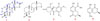
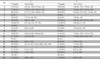
Compound 1 was obtained as colorless crystals, m.p. 168 – 169 ℃. [α]D 25 = −20° (c, 0.1, CH3OH). Its UV spectroscopy displayed the terminal absorbance characteristic at 205 nm. Comprehensive analysis of the ESI-MS, HR-ESI-MS and 1D NMR spectra, the molecular formula was determined as C20H26O4 with the [M+H]+ m/z at 331.1906 (calcd. 331.1909). Twenty carbon atoms resonances were observed in the 1D 13C NMR and DEPT spectra, including three methyl groups at δ 17.9, 21.1 and 24.1, six methene groups at δ 45.3, 33.2, 31.7, 28.1, 18.8 and 17.8, five methine groups at δ 128.9, 88.0, 72.6, 50.9 and 45.1 and six quaternary carbon NMR signals at δ 181.9, 175.7, 138.8, 42.2, 39.1 and 32.9. According to the results of HSQC analysis, the corresponding proton resonances appeared at δ 0.86 (s, 3H), 1.16 (s, 3H) and 1.30 (s, 3H) for methyl groups, at δ 2.51 (d, J = 16.5 Hz, 1H), 2.36 (d, J = 16.5 Hz, 1H), 1.61 (m, 1H), 1.23 (m, 1H), 1.57 (m, 2H), 2.17 (m, 1H), 1.48 (m, 1H), 1.64 (m, 1H), 1.36 (m, 1H) and 1.70 (m,1H), 1.56 (m, 1H) for methenes, at δ 6.27 (dd, J = 2.4, 3.5 Hz, 1H), 4.44 (s, 1H), 4.89 (m, 1H), 1.77 (d, J = 6.0 Hz, 1H) and 2.03 (m, 1H) for methines in compound 1 (Table 1). The comparison of NMR data between compound 1 and xylabisboein A6 suggested that 1 was a diterpenoid structure with an isopimane skeleton.
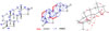
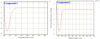
In the 1H-1H COSY, there were cross peaks among δ 2.17 (m, 1H), 1.70 (m, 1H) and 1.23 (m, 1H), and proved that there was substructure of -CH2-CH2-CH2- in compound 1. Subsequently, the correlations of H-7 [δ 6.27 (dd, J = 2.4, 3.5 Hz, 1H)]/H-6 [δ 4.89 (m, 1H)]/H-5 [δ 1.77 (d, J = 6.0 Hz, 1H)] indicated the existence of -CH(R1)-CH(OR2)-CH=C(R3, R4) partial structure in the COSY spectrum, and there was a -CH(R1)-CH2-CH2- structure hinting by the correlations among H-9 [δ 2.03 (m, 1H)], H-11 [δ 1.36 (m, 1H)] and H-12 [δ 1.57 (m, 2H)] (Fig. 2). The above 1H-1H COSY deducing results were proved by the HMBC correlations of H-1/C-3, H-2/C-10, H-3/C-1, C-19, H-5/C-8, C-10, H-7/C-5 and H-12/C-9, C-14. The planar structure of compound 1 was established by the key HMBC correlations of H-3/C-19, H-7/C-5, C-9, H- 14/C-7, C-12, C-15, H-17/C-12, C-13, C-14, C-15, H-18/ C-3, C-4, C-5, C-19, and H-20/C-1, C-5, C-9, C-10 (Fig. 1 and 2). Comparing to reference xylabisboein A, the main difference between them was the carbonyl group at C-16 position in compound 1 replacing the -CH2- group at C-16 position in reference compound. Finally, the relative configurations of 1 were built by the analysis the NOESY spectroscopy, the correlations of H-5/H-9, H-6/ H-9 and H-18 appeared that the four protons of H-5, H-6, H-9 and H-18 lied at the same side in polyhydrophenanthrene core structure, and the correlations of H-7/H-14 and H-20, and H-14/H-17 showed that the proton signals of H-14, H-17 and H-20 located at other side of polyhydrophenanthrene (Fig. 2). The CD spectrum of compound 1 showed negative Cotton effect curve at 210 nm. Comparing to the CD spectrum of compound 1 to that of xylabisboein A,6 the CD spectra of them were almost the same (Fig. 3), thus, the absolute configurations of 1 were determined as 4S, 5R, 9R, 10R, 13R and 14S. Finally, compound 1 was named as xylaroisopimaranin A (1), a new isopimarane derivative.
Compounds 2 – 5 were elucidated as xylabisboein B (2),6 griseofulvin (3),7 5-methylmellein (4),6 and mellein-5-carboxlic acid (5)6 by comparison with their NMR data to the references. The compounds 1 – 5 were evaluated for Hep G2 cells cytotoxicity, antibacterial and inhibiting brine shrimp activity, and the results showed no significant cytotoxicity to Hep G2 cells and no inhibiting Erwinia carotovora sub sp. Carotovora (Jones) Bersey et al, but compounds 1 – 5 displayed brine shrimp inhibiting activities with IC50 value at 10.1, 5.0, 0.52, 18.2, and 25.1 µmol/mL, respectively.
In conclusion, a new isopimarane derivative, xylaroisopimaranin A (1), together with four known compounds xylabisboein B (2), griseofulvin (3), 5-methylmellein (4), and mellein-5-carboxlic acid (5), were isolated from the broth of endophytic fungus Xylaralyce sp., and their structures were elucidated by 1D, 2D NMR, MS and CD spectra. The biological evaluation results showed that the compounds 1–5 displayed no significant Hep G2 cells cytotoxicity and antibacterial activity, but they inhibited the Brine Shrimp with IC50 value at 10.1, 5.0. 52, 18.2, and 25.1 µmol/mL, respectively.
 XML Download