Abstract
Objectives
To report a rare case of a spinal extradural meningioma in a patient with longstanding nonspecific thoracic nocturnal pain
Summary of Literature Review
Meningioma is a frequent intradural extramedullary tumor that is associated with pain, sensory/motor deficits, and sphincter weakness. Spinal meningiomas most commonly occur in the thoracic spine, although they can also be found at other locations.
Materials and Methods
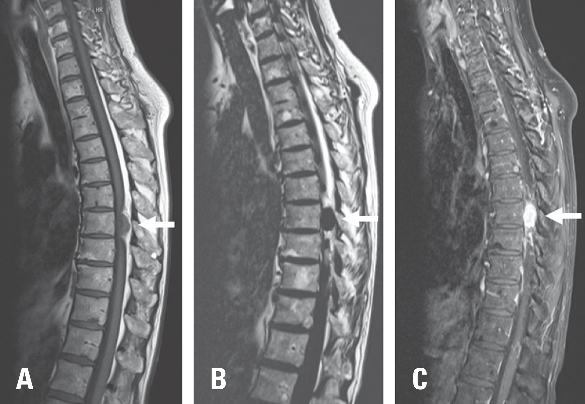
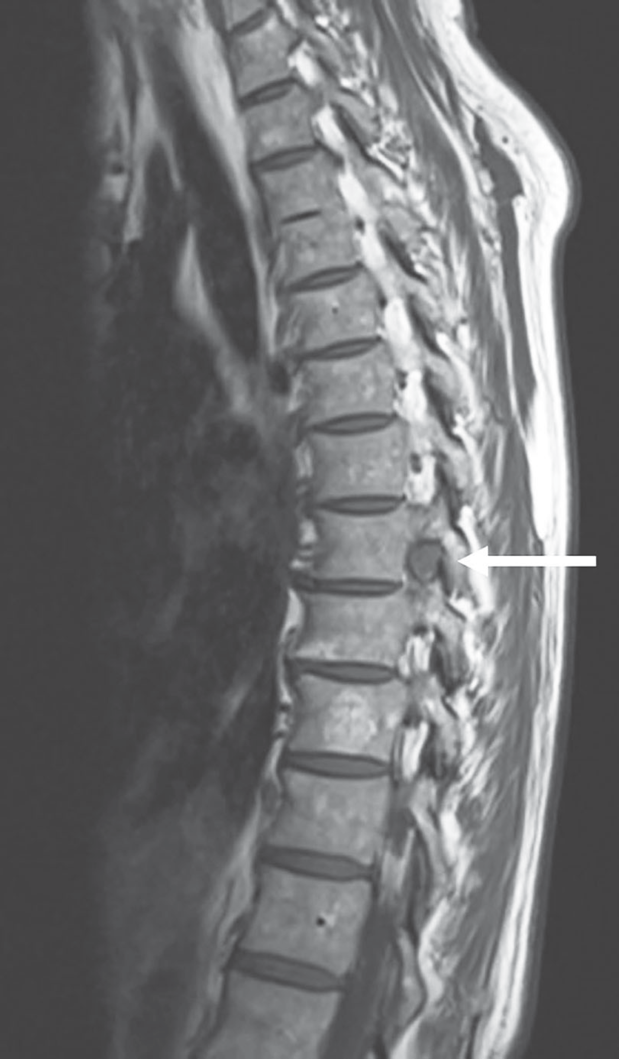
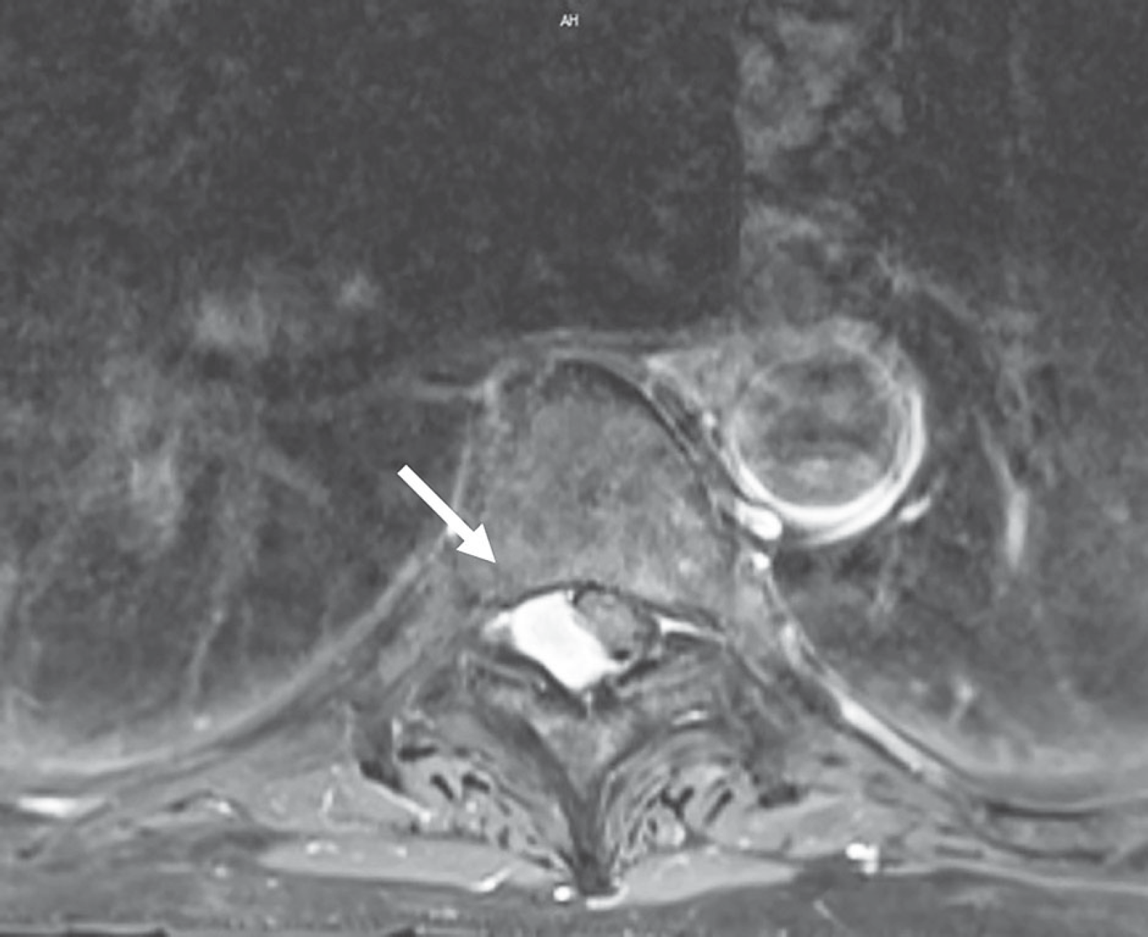
A 65-year-old woman first visited the cardiac and gastrointestinal departments of our institution due to chest pain 2 years previously. No explanation for the complaint could be found in the heart or other organs. On a computed tomography scan of the thorax, a spinal mass was found a few months before the diagnosis. On magnetic resonance imaging, an extramedullary and extradural mass was observed at T7/8.
REFERENCES
1. Calogero JA, Moossy J. Extradural spinal meningiomas: report of four cases. J Neurosurg. 1972 Oct; 37(4):442–7. DOI: 10.3171/jns.1972.37.4.0442.
2. Gottfried ON, Gluf W, Quinones-Hinojosa A, et al. Spi-nal meningiomas: surgical management and outcome. Neurosurg Focus. 2003 Jun 15; 14(6):e2. DOI: 10.3171/foc.2003.14.6.2.

3. Guidetti B. Removal of extramedullary benign spinal cord tumors. Advances and Technical Standards in Neurosurgery. 1974. pp 173-197. DOI: 10.1007/978-3-7091-7099-1_6.
4. Levy WJ Jr, Bay J, Dohn D. Spinal cord meningioma. J Neurosurg. 1982 Dec; 57(6):804–12. DOI: 10.3171/jns.1982.57.6.0804.

5. Powell M. Sir Victor Horsley—an inspiration. BMJ. 2006 Dec 23; 333(7582):1317–9. DOI: 10.1136/bmj.39056.527407.55.

6. Santiago BM, Rodeia P, Cunha E Sa M. Extradural thoracic spinal meningioma. Neurol India. 2009 Jan-Feb; 57(1):98. DOI: 10.4103/0028-3886.48802.

7. Klekamp J, Samii M. Surgical results for spinal meningiomas. Surg Neurol. 1999 Dec; 52(6):552–62. DOI: 10.1016/S0090-3019 (99)00153-6.

8. King AT, Sharr MM, Gullan RW, Bartlett JR. Spinal meningiomas: a 20-year review. Br J Neurosurg. 1998 Dec; 12(6):521–6. DOI: 10.1080/02688699844367.

9. Deyo , Richard A. Diehl, Andrew K. Cancer as a cause of back pain. Journal of General Internal Medicine. 1988; 3(3):230–238. DOI: 10.1007/bf02596337.
10. Herkowitz HN, Garfin SR, Eismont FJ, et al. Rothman-Simeone The Spine E-Book: Expert Consulted. Elsevier Health Sciences;2011.
Fig. 1.
Sagittal T1-weighted (T1W), T2-weighted (T2W), and T1 fat-suppressed and gadolinium-containing contrast-enhanced magnetic resonance imaging scan. (A) T1W image showing an isointense extradural lesion at the T8 level. (B) T2 fat-suppressed image showing a hypointense lesion. (C) T1 gadolinium-containing contrast-enhanced image showing an extradural lesion with some heterogeneity, but mostly a hyperintense signal.


 PDF
PDF Citation
 Print
Print


 XML Download